
The Biology Behind Melancholic Depression: Causes and Risk Factors


Welcome to Mind Care Guide, a compassionate space dedicated to understanding and nurturing your mental well-being. Feeling lost in the fog of melancholic depression? You're not alone. In our featured article, we delve into the biological causes of this specific type of depression, exploring the intricate interplay of factors that can contribute to its development. Are you ready to uncover the science behind the sadness and begin your journey towards understanding and healing? Read on to learn more...
The Biology Behind Melancholic Depression: Causes and Risk Factors | Mind Care Guide
- Understanding Melancholic Depression: A Biological Perspective
- Exploring the Biological Causes of Melancholic Depression
- Risk Factors Contributing to the Biological Basis of Melancholia
- Diagnosing Biologically-Driven Melancholic Depression
- Treatment Approaches Targeting the Biological Aspects of Melancholic Depression
-
Frequently Asked Questions
- What are the primary biological factors implicated in melancholic depression?
- How does genetics contribute to the development of melancholic depression?
- What role do neurotransmitters play in melancholic depression, and which ones are most affected?
- Are there any specific brain regions or structures that are implicated in melancholic depression?
- Can hormonal imbalances directly cause melancholic depression, and if so, which hormones are most relevant?
- Final Reflection: Unraveling the Biology of Melancholic Depression
- Unlock Your Understanding: Take Charge of Your Mental Wellbeing
Understanding Melancholic Depression: A Biological Perspective
What is Melancholic Depression? Defining a Severe Mood Disorder
Melancholic depression is considered a specific and severe subtype of major depressive disorder, characterized by a profound loss of pleasure or interest in all, or almost all, activities (anhedonia) and a lack of reactivity to usually pleasurable stimuli. Unlike other forms of depression where mood might lift slightly in response to positive events, the low mood in melancholia is pervasive and does not improve, even temporarily. Individuals often experience a distinct quality of depressed mood, feeling deeply despondent or empty.
This subtype is also marked by several specific physical symptoms, often worse in the morning. These include significant psychomotor retardation (slowed movements and speech) or agitation (restlessness), substantial weight loss or decreased appetite, and early morning awakening (waking up at least two hours earlier than usual). These pronounced physical manifestations strongly suggest significant underlying biological disruptions, distinguishing it from milder or atypical forms of depression. Understanding the biological causes of melancholic depression is crucial for effective treatment.
Differentiating Melancholic Depression from Other Depressive Disorders
Distinguishing melancholic depression from other depressive presentations is vital because it often indicates a different biological profile and may respond better to specific types of treatment. While many forms of depression involve sadness and loss of interest, melancholia's defining features—pervasive anhedonia, lack of mood reactivity, and prominent physical symptoms like psychomotor changes and early morning awakening—are key differentiators. For instance, atypical depression often involves mood reactivity (mood improves with positive events), increased appetite and weight gain, hypersomnia (excessive sleeping), and a feeling of being weighed down.
The biological underpinnings of melancholic depression appear to involve more pronounced alterations in specific brain circuits and neurochemical systems compared to some other depression types. Research using neuroimaging and biological markers often reveals more significant changes in areas like the HPA axis (stress response system) and certain neurotransmitter pathways in melancholic patients. This distinction highlights why treatments targeting these specific biological mechanisms, such as certain antidepressant classes or brain stimulation therapies, are often particularly effective for this severe subtype, emphasizing the importance of understanding the biological causes of melancholic depression.
Exploring the Biological Causes of Melancholic Depression
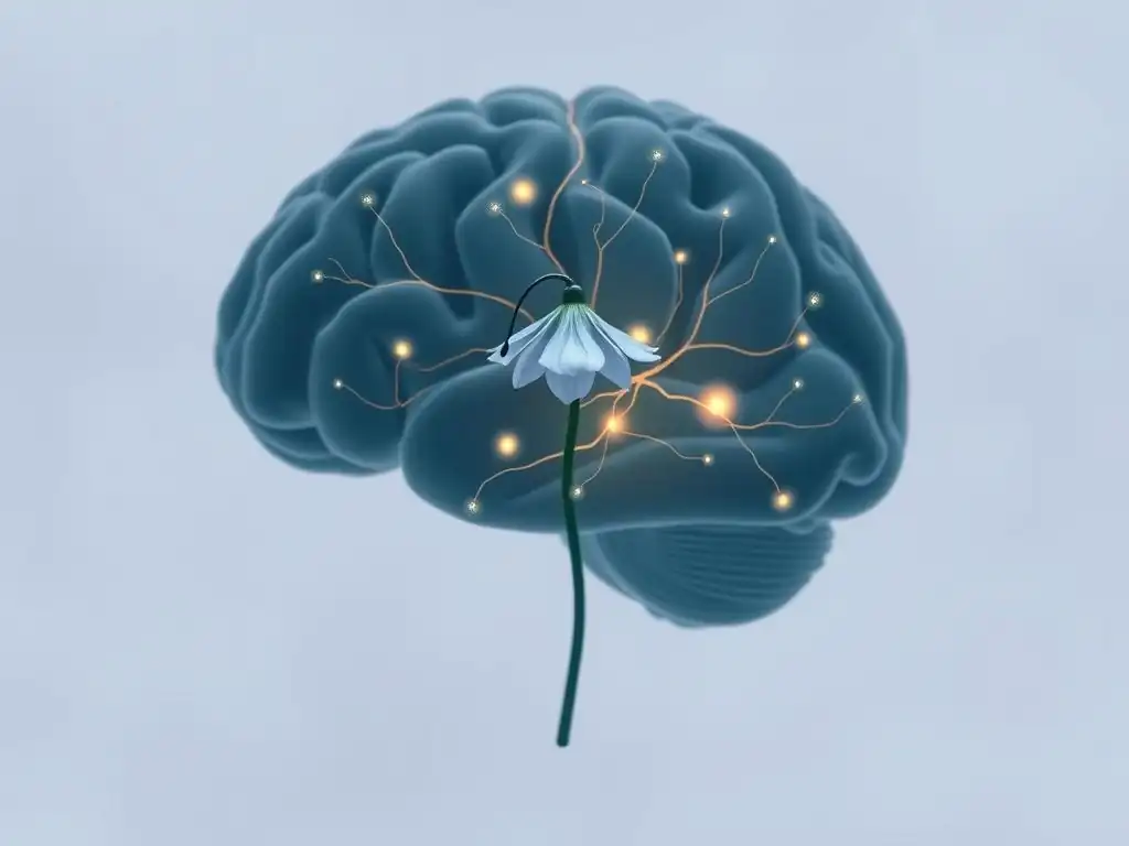
Genetic Predisposition and Melancholic Depression Risk
Genetic factors play a significant role in the vulnerability to developing major depressive disorder, and this influence is particularly strong for severe, recurrent forms like melancholic depression. Studies involving twins and families consistently show a higher concordance rate for depression in identical twins compared to fraternal twins, and a higher prevalence among first-degree relatives of affected individuals. While no single "melancholia gene" has been identified, variations across multiple genes are thought to contribute to an increased biological susceptibility.
Research suggests that having a first-degree relative (parent, sibling) with major depression increases an individual's risk by approximately two to three times compared to the general population. For melancholic depression specifically, this familial risk may be even higher, although precise statistics vary across studies. These genetic influences likely affect the function of neurotransmitters, stress hormone regulation, and brain structure, laying a biological foundation that can be triggered by environmental factors. Exploring these genetic links is a key part of understanding the biological causes of melancholic depression.
Family History and the Likelihood of Developing Melancholic Depression
A strong family history is one of the most significant risk factors for developing melancholic depression. If one parent has a history of major depression, the risk to their child is elevated. If both parents have a history, the risk increases substantially. While estimates vary, some studies suggest the lifetime risk for major depression in individuals with a positive family history can be as high as 20-30%, compared to a general population risk of around 10-15%.
This increased likelihood is not solely due to shared environment but reflects the inheritance of genetic variants that influence brain chemistry, structure, and stress reactivity. For melancholic depression, which is often associated with more pronounced biological markers, the genetic contribution is thought to be substantial. Understanding this familial link helps identify individuals who may have a higher biological predisposition and could benefit from early intervention or monitoring.
Specific Genes Associated with Melancholic Depression Vulnerability
While the genetic picture is complex and involves many genes with small effects, research has identified several candidate genes that may contribute to the biological vulnerability for melancholic depression. Genes involved in serotonin signaling, such as the serotonin transporter gene ( SLC6A4, often referred to as SERT), have been widely studied. Variations in the promoter region of SLC6A4 have been linked to altered serotonin reuptake and increased amygdala reactivity to threat stimuli, potentially contributing to mood dysregulation.
Other genes of interest include those involved in dopamine and norepinephrine pathways ( COMT, MAOA), which are crucial for motivation, reward, and energy levels—areas significantly impacted in melancholia. Genes related to the HPA axis, such as those encoding receptors for cortisol ( NR3C1) or corticotropin-releasing hormone ( CRH), are also investigated, as stress response dysregulation is a prominent feature. While these genetic associations are complex and often depend on interactions with environmental factors, they represent potential pieces of the puzzle regarding the biological causes of melancholic depression.
Neurotransmitter Imbalances in Melancholic Depression
One of the most enduring hypotheses regarding the biological causes of depression, including melancholia, centers on imbalances in key neurotransmitters in the brain. Serotonin, dopamine, and norepinephrine are chemicals that transmit signals between nerve cells and are crucial for regulating mood, motivation, sleep, appetite, and energy levels—all domains profoundly affected in melancholic depression. Disruptions in the synthesis, release, reuptake, or receptor sensitivity of these neurotransmitters are thought to contribute to the symptoms.
While the precise nature of these imbalances is still being fully elucidated, evidence from pharmacological studies supports the neurotransmitter hypothesis. Medications that modulate the levels or activity of these neurotransmitters are often effective treatments, particularly for melancholic depression. However, it's important to note that it's likely not just a simple deficiency but a complex dysregulation involving multiple systems and their interactions. Understanding these potential imbalances is central to exploring the biological causes of melancholic depression.
Serotonin's Role in Regulating Mood and its Impact on Melancholia
Serotonin, often referred to as the "feel-good" neurotransmitter, plays a critical role in regulating mood, sleep, appetite, and social behavior. Low levels or impaired function of serotonin signaling have long been implicated in depression. In melancholic depression, disruptions in serotonin pathways may contribute to the pervasive low mood, sleep disturbances (particularly early morning awakening), and changes in appetite.
Many common antidepressants, such as Selective Serotonin Reuptake Inhibitors (SSRIs), work by increasing the amount of serotonin available in the synaptic cleft, thereby enhancing serotonergic neurotransmission. While not a complete solution for everyone, the efficacy of SSRIs and other serotonergic drugs in treating depression provides evidence for the involvement of this system in the biological causes of melancholic depression.
Dopamine Deficiency and its Connection to Anhedonia in Melancholic Depression
Dopamine is a neurotransmitter critical for reward, motivation, pleasure, and motor control. A hallmark symptom of melancholic depression is severe anhedonia—the inability to experience pleasure. This profound loss of interest and pleasure is strongly linked to dysfunction in the brain's dopamine reward pathways, particularly in areas like the nucleus accumbens and ventral tegmental area.
Evidence suggests that reduced dopamine activity or receptor sensitivity in these circuits contributes significantly to the lack of motivation, loss of interest, and the feeling of emptiness characteristic of melancholia. Some antidepressant medications that also affect dopamine, or augmentation strategies targeting dopamine, can be particularly helpful for anhedonia, further supporting the idea that dopamine dysfunction is a key biological cause of melancholic depression symptoms.
Norepinephrine's Involvement in Energy Levels and Motivation
Norepinephrine is another crucial neurotransmitter involved in the body's "fight or flight" response, arousal, attention, energy levels, and motivation. Individuals with melancholic depression often experience significant fatigue, lack of energy (anergia), and psychomotor retardation—symptoms that align with potential disruptions in norepinephrine signaling.
Dysregulation of norepinephrine pathways, particularly in areas like the locus coeruleus, can impair the brain's ability to maintain alertness, energy, and drive. Antidepressants that target both serotonin and norepinephrine (SNRIs) are often effective in treating melancholic depression, suggesting that addressing norepinephrine imbalances alongside serotonin may be necessary to alleviate the full spectrum of symptoms, including those related to energy and motivation. This further solidifies the role of neurotransmitter imbalances in the biological causes of melancholic depression.
Brain Structure and Function in Melancholic Depression
Neuroimaging studies using techniques like MRI, fMRI, and PET have revealed structural and functional differences in the brains of individuals with depression, with some findings being particularly pronounced in melancholic subtypes. These studies point to alterations in the size, activity, and connectivity of key brain regions involved in mood regulation, cognitive function, and emotional processing. Understanding these brain changes provides crucial insights into the biological causes of melancholic depression.
While these findings are often correlational and it's not always clear whether they are causes or consequences of the illness, they highlight the complex biological basis of the disorder. Specific areas frequently implicated include the prefrontal cortex, hippocampus, and amygdala, which together form crucial circuits for processing emotions, making decisions, and responding to stress.
The Role of the Prefrontal Cortex in Cognitive Function and Mood Regulation
The prefrontal cortex (PFC), particularly the ventromedial and dorsolateral regions, is critical for executive functions, decision-making, regulating emotions, and integrating information from other brain areas. In melancholic depression, studies often show reduced activity or volume in certain areas of the PFC. This dysfunction is thought to contribute to symptoms like impaired concentration, difficulty making decisions, feelings of hopelessness, and the inability to regulate negative emotions.
The PFC's role in top-down control over emotional responses processed in areas like the amygdala is crucial. Impaired PFC function can lead to a diminished capacity to dampen negative feelings or shift attention away from distressing thoughts, contributing to the persistent low mood and rumination seen in melancholia. These observed changes in PFC activity underscore the structural and functional biological causes of melancholic depression.
Hippocampal Volume Reduction and its Impact on Memory and Emotional Processing
The hippocampus is a brain structure vital for learning, memory formation, and regulating the stress response via its connections with the HPA axis. Chronic stress and elevated levels of stress hormones like cortisol can be toxic to hippocampal neurons. Studies have consistently reported smaller hippocampal volume in individuals with recurrent or chronic depression, including melancholic forms, compared to healthy individuals.
Reduced hippocampal volume may contribute to cognitive symptoms like memory problems and concentration difficulties often reported in depression. Furthermore, the hippocampus plays a role in contextualizing emotional memories and regulating the stress response. Its dysfunction could contribute to the difficulty in extinguishing negative emotional responses and the persistent activation of the stress system observed in melancholia, adding another layer to the biological causes of melancholic depression.
Amygdala Activity and the Experience of Negative Emotions in Melancholia
The amygdala is a key region in the brain's emotional processing network, particularly involved in detecting and responding to threats and generating fear and anxiety responses. In melancholic depression, neuroimaging studies often show increased activity in the amygdala, especially in response to negative emotional stimuli. This heightened reactivity is thought to contribute to the increased experience of negative emotions, anxiety, and the pervasive sense of dread or despair characteristic of severe depression.
While the amygdala is hyperactive, the connections between the amygdala and regulatory areas like the PFC may be weakened. This imbalance—an overactive alarm system with insufficient inhibitory control—can lead to a state of heightened emotional distress that is difficult to alleviate. Altered amygdala activity is a significant finding in the biological profile associated with the biological causes of melancholic depression.
The HPA Axis and Stress Response in Melancholic Depression
The Hypothalamic-Pituitary-Adrenal (HPA) axis is the body's central stress response system. When faced with a stressor, the hypothalamus releases corticotropin-releasing hormone (CRH), which signals the pituitary gland to release adrenocorticotropic hormone (ACTH). ACTH then stimulates the adrenal glands to produce cortisol, the primary stress hormone. Normally, a negative feedback loop involving the hippocampus and PFC shuts down this response once the stressor is removed.
However, in a significant subset of individuals with melancholic depression (estimated to be 50-70%), this system is dysregulated. This can manifest as elevated basal cortisol levels, a blunted response of ACTH and cortisol to challenge tests (like the dexamethasone suppression test or DST), and impaired negative feedback. Chronic activation and dysregulation of the HPA axis are considered major biological causes of melancholic depression and contribute to many of its physical and psychological symptoms.
Chronic Stress and its Impact on the HPA Axis
Prolonged or severe stress, particularly early life trauma, can have a lasting impact on the sensitivity and regulation of the HPA axis. Chronic stress can lead to persistent elevation of CRH and cortisol, which can have detrimental effects on brain structures like the hippocampus and PFC, further impairing the negative feedback loop. This creates a vicious cycle where stress dysregulates the HPA axis, leading to higher stress hormone levels, which in turn damage brain regions needed to regulate stress.
This persistent state of physiological stress can contribute to symptoms such as sleep disturbances, changes in appetite, fatigue, and cognitive difficulties often seen in melancholic depression. The link between chronic stress and HPA axis dysfunction is a well-established component of the biological causes of melancholic depression.
Cortisol Dysregulation in Melancholic Depression
Cortisol dysregulation is one of the most consistently observed biological abnormalities in melancholic depression. While some individuals show persistently high cortisol levels throughout the day, others may exhibit a flattened diurnal rhythm (lack of the normal morning peak and evening decline). This abnormal cortisol secretion reflects the underlying HPA axis dysfunction.
High cortisol levels can interfere with neurotransmitter function, impair neurogenesis (the growth of new brain cells) in the hippocampus, and increase inflammation, all of which are implicated in the pathophysiology of depression. The degree of cortisol dysregulation is often correlated with the severity of depressive symptoms, particularly the vegetative signs like sleep and appetite disturbances, further highlighting its role in the biological causes of melancholic depression.
Risk Factors Contributing to the Biological Basis of Melancholia
Several factors can increase an individual's biological vulnerability to developing melancholic depression by influencing genetics, neurotransmitter systems, brain structure, and stress response. These risk factors often interact, creating a complex web of biological predisposition and environmental triggers. Identifying these factors is crucial for understanding the full picture of the biological causes of melancholic depression.
Early Life Trauma and its Long-Term Effects on Brain Development
Adverse Childhood Experiences (ACEs), such as abuse (physical, emotional, sexual), neglect, household dysfunction (e.g., parental mental illness, substance abuse, divorce), have profound and lasting effects on brain development and stress reactivity. Exposure to trauma during critical periods of neurodevelopment can permanently alter the structure and function of brain regions involved in emotional regulation and stress response, such as the amygdala, hippocampus, and prefrontal cortex.
Studies consistently show a strong correlation between the number and severity of ACEs and the risk of developing major depression later in life, including severe and recurrent forms like melancholia. The biological changes induced by early trauma, particularly the sensitization of the HPA axis, are considered significant contributors to the biological causes of melancholic depression.
Adverse Childhood Experiences (ACEs) and Increased Risk of Melancholic Depression
Research, including large-scale studies like the original ACE study, has demonstrated a dose-response relationship between the number of ACEs experienced and the likelihood of developing numerous health problems in adulthood, including depression. Individuals with four or more ACEs are significantly more likely to experience major depression compared to those with none, with some studies indicating a risk increase of four to five times.
For melancholic depression, which is often characterized by pronounced HPA axis dysfunction and altered brain structure, the link to early trauma is particularly strong. ACEs can disrupt the normal development of stress-buffering systems, leaving individuals biologically more vulnerable to the effects of stress throughout their lives. This increased biological vulnerability is a major risk factor for the development of melancholic depression.
The Impact of Trauma on the HPA Axis and Stress Response
Early life trauma can permanently "program" the HPA axis, leading to a state of chronic hyper-reactivity or dysregulation. Exposure to overwhelming stress during childhood can impair the development of the negative feedback mechanisms that normally shut down the stress response. This results in an exaggerated or prolonged release of stress hormones like cortisol in response to even minor stressors later in life.
This persistent HPA axis dysregulation contributes directly to the biological causes of melancholic depression by interfering with neurotransmitter systems, causing neuroinflammation, and potentially leading to the observed changes in brain structure like hippocampal volume reduction. The biological scars left by early trauma significantly increase the risk profile for this severe form of depression.
Chronic Illnesses and Their Association with Melancholic Depression
Chronic physical illnesses are frequently comorbid with depression, and this association is not merely psychological distress in response to illness. Many chronic conditions involve systemic inflammation, hormonal changes, or direct impacts on the nervous system that can contribute to the biological causes of depression. The biological burden of chronic illness can exacerbate underlying vulnerabilities or directly trigger depressive episodes, including melancholic features.
Examples include cardiovascular disease, diabetes, autoimmune disorders, and neurological conditions. The biological pathways linking these illnesses to depression are complex and involve shared inflammatory processes, oxidative stress, and disruptions in neurochemical balance.
Cardiovascular Disease and its Connection to Melancholic Depression
There is a well-established bidirectional relationship between cardiovascular disease (CVD) and depression. Individuals with CVD, such as heart failure or post-myocardial infarction (heart attack), have significantly higher rates of depression, estimated at 15-20%, compared to the general population. Conversely, depression is a significant risk factor for developing CVD and predicts worse outcomes in patients with existing heart conditions.
The biological links include shared inflammatory pathways (e.g., elevated cytokines), HPA axis dysregulation, and autonomic nervous system imbalance. Chronic inflammation associated with CVD can affect brain function and neurotransmitter systems. This complex interplay of biological factors contributes to the increased risk and severity of depression, potentially including melancholic features, in individuals with heart disease.
Inflammation and its Potential Role in the Development of Melancholia
Growing evidence suggests that chronic low-grade inflammation plays a significant role in the biological causes of depression. Inflammatory cytokines (signaling molecules) like IL-6, TNF-alpha, and CRP are often elevated in individuals with major depression, particularly those with more severe or melancholic forms. These cytokines can cross the blood-brain barrier or be produced by glial cells in the brain.
Once in the brain, inflammatory molecules can interfere with neurotransmitter synthesis and metabolism (e.g., reducing serotonin and dopamine precursors), impair neurogenesis and synaptic plasticity, and activate the HPA axis. This neuroinflammatory state can contribute to symptoms like fatigue, anhedonia, cognitive slowing, and mood disturbances characteristic of melancholia. Inflammation is increasingly recognized as a key biological pathway contributing to the biological causes of melancholic depression.
Hormonal Imbalances and Melancholic Depression
Hormones are powerful chemical messengers that regulate numerous bodily functions, including mood and stress response. Imbalances in various endocrine systems can significantly impact mental health and contribute to the biological causes of depression. The intricate interplay between hormones and neurotransmitters highlights another biological pathway to melancholia.
Conditions affecting the thyroid, adrenal glands, or reproductive hormones are particularly relevant. These hormonal disruptions can directly influence brain chemistry and structure or indirectly through systemic effects.
Thyroid Dysfunction and its Impact on Mood
Thyroid hormones are essential for regulating metabolism, energy levels, and brain function. Both hypothyroidism (underactive thyroid) and hyperthyroidism (overactive thyroid) can cause mood disturbances. Hypothyroidism, characterized by insufficient thyroid hormone production, frequently presents with symptoms that overlap significantly with depression, including fatigue, weight gain, cognitive slowing, and low mood.
While hypothyroidism-related depression might not always fit the melancholic subtype perfectly, the biological disruption caused by insufficient thyroid hormone can certainly contribute to a depressive state that requires specific endocrine treatment. Screening for thyroid dysfunction is a standard part of the diagnostic workup for depression due to this clear biological link.
Menopause and the Risk of Melancholic Depression in Women
Menopause, the transition during which a woman's ovaries stop producing eggs and estrogen levels decline significantly, is associated with an increased risk of depression. Estrogen receptors are found throughout the brain, including regions involved in mood regulation (PFC, hippocampus, amygdala). Estrogen influences the synthesis and activity of neurotransmitters like serotonin, norepinephrine, and dopamine.
The fluctuating and declining estrogen levels during perimenopause and menopause can impact these neurochemical systems, potentially contributing to mood instability and depressive episodes. While not all women experience depression during menopause, those with a history of depression, particularly during periods of hormonal change like postpartum, may be more vulnerable to developing melancholic features during this life stage due to the underlying biological sensitivity.
Sleep Disturbances and Their Biological Consequences
Chronic sleep disturbances, such as insomnia or circadian rhythm disruption, are not merely symptoms of melancholic depression but can also act as significant biological risk factors and contribute to its maintenance. Sleep is crucial for brain health, including the regulation of neurotransmitters, clearance of metabolic waste products, and consolidation of memories and emotional experiences. Persistent lack of quality sleep can profoundly disrupt these processes.
The link between sleep and mood is bidirectional and biologically complex. Poor sleep can exacerbate existing vulnerabilities in stress response systems and neurotransmitter balance, making individuals more susceptible to developing or worsening depressive symptoms, including the severe features of melancholia.
Insomnia and its Impact on Neurotransmitter Function
Chronic insomnia, characterized by difficulty falling or staying asleep, can significantly impact neurotransmitter systems involved in mood regulation. Lack of sufficient sleep can alter the sensitivity of receptors for serotonin, dopamine, and norepinephrine, potentially contributing to the very imbalances thought to underlie melancholic depression.
Furthermore, sleep deprivation can increase activity in the amygdala and decrease connectivity between the amygdala and the prefrontal cortex, impairing the brain's ability to regulate emotional responses. This biological consequence of chronic insomnia can heighten emotional reactivity and contribute to the pervasive low mood and anxiety seen in melancholia.
Circadian Rhythm Disruption and its Link to Mood Disorders
The circadian rhythm is the body's internal biological clock that regulates sleep-wake cycles, hormone secretion, and other physiological processes over a 24-hour period. Disruptions to this rhythm, whether due to shift work, frequent travel across time zones, or irregular sleep patterns, are strongly associated with an increased risk of mood disorders, including depression with melancholic features.
In melancholic depression, a common symptom is early morning awakening, indicating a significant disruption of the circadian rhythm. This disruption is linked to abnormalities in the timing and secretion of hormones like cortisol and melatonin, further impacting sleep, energy levels, and mood. The biological clock dysfunction is considered a significant factor contributing to the biological causes of melancholic depression.
Diagnosing Biologically-Driven Melancholic Depression
Diagnosing melancholic depression primarily relies on a thorough clinical evaluation based on specific symptom criteria. While research into biological markers is ongoing, there are currently no routine biological tests that definitively diagnose melancholic depression or reliably distinguish it from other forms of depression. However, understanding the likely biological underpinnings informs the diagnostic process and guides treatment selection.
Clinical Evaluation and Diagnostic Criteria for Melancholic Depression
The diagnosis of melancholic depression is typically made by a mental health professional based on the presence of specific features outlined in diagnostic manuals like the DSM-5 (Diagnostic and Statistical Manual of Mental Disorders, 5th Edition). These criteria include the core symptoms of a major depressive episode (depressed mood or loss of interest/pleasure) plus the presence of at least three out of six specific melancholic features.
These features are: (1) loss of pleasure in all or almost all activities, (2) lack of reactivity to usually pleasurable stimuli, (3) distinct quality of depressed mood (feels different from grief), (4) depression worse in the morning, (5) marked psychomotor retardation or agitation, and (6) significant weight loss or anorexia. The emphasis on these specific vegetative and anhedonic symptoms helps identify a subgroup of depression likely driven by particular biological mechanisms.
Biomarkers and Potential Biological Tests for Melancholic Depression
Research is actively exploring various biological markers (biomarkers) that could potentially aid in diagnosing or predicting treatment response in melancholic depression. These include markers related to HPA axis function (e.g., cortisol levels in blood, saliva, or urine, or response to the dexamethasone suppression test), inflammatory markers (e.g., C-reactive protein, cytokines), and neuroimaging findings (e.g., specific patterns of activity or volume changes in brain regions).
While some biological tests, like the DST, have shown some association with melancholic features, they are not sufficiently sensitive or specific for routine clinical diagnosis. Future research may identify panels of biomarkers or specific neuroimaging patterns that can objectively support the clinical diagnosis and help personalize treatment based on the specific biological causes of melancholic depression in an individual.
Treatment Approaches Targeting the Biological Aspects of Melancholic Depression
Given the strong biological basis of melancholic depression, treatment approaches often focus on correcting underlying biological dysregulations. This typically involves pharmacological interventions aimed at restoring neurotransmitter balance, brain stimulation therapies for severe and treatment-resistant cases, and lifestyle adjustments that support overall brain health. The goal is to address the specific biological causes of melancholic depression symptoms.
Pharmacological Interventions: Antidepressants and their Mechanisms of Action
Antidepressant medications are a cornerstone of treatment for moderate to severe depression, and certain classes are particularly effective for melancholic depression. These drugs work by modulating the activity of neurotransmitters in the brain, aiming to restore more typical signaling patterns. The choice of antidepressant often depends on the specific neurotransmitter systems thought to be most affected.
SSRIs (Selective Serotonin Reuptake Inhibitors) for Serotonin Regulation
SSRIs are the most commonly prescribed class of antidepressants. They work by blocking the reuptake of serotonin in the synapse, thereby increasing the concentration of serotonin available to bind to receptors. This is thought to enhance serotonergic neurotransmission over time. While effective for many forms of depression, SSRIs may be less effective as a sole treatment for severe melancholic depression compared to other options, particularly if dopamine or norepinephrine dysfunction is prominent.
Examples include fluoxetine (Prozac), sertraline (Zoloft), and escitalopram (Lexapro). They generally have a favorable side effect profile compared to older classes, but it may take several weeks to see significant improvement.
SNRIs (Serotonin-Norepinephrine Reuptake Inhibitors) for Dual-Action Support
SNRIs block the reuptake of both serotonin and norepinephrine, leading to increased levels of both neurotransmitters in the synapse. Because melancholic depression often involves dysfunction in both serotonin and norepinephrine pathways (affecting mood, energy, and motivation), SNRIs can be particularly effective for this subtype.
Examples include venlafaxine (Effexor) and duloxetine (Cymbalta). By targeting both systems, SNRIs may provide broader symptom relief, particularly for symptoms like fatigue, anhedonia, and psychomotor slowing, addressing multiple aspects of the biological causes of melancholic depression.
Tricyclic Antidepressants (TCAs) and Monoamine Oxidase Inhibitors (MAOIs)
Older classes of antidepressants, such as Tricyclic Antidepressants (TCAs) and Monoamine Oxidase Inhibitors (MAOIs), are often highly effective for melancholic depression, sometimes more so than SSRIs or SNRIs, especially in severe cases. TCAs block the reuptake of norepinephrine and serotonin (and sometimes dopamine), while MAOIs prevent the breakdown of monoamine neurotransmitters (serotonin, norepinephrine, dopamine).
These medications can have more significant side effects and require careful monitoring (especially dietary restrictions with MAOIs). However, their potent effects on multiple neurotransmitter systems make them valuable options, particularly when other treatments have failed, highlighting their ability to target the core biological causes of melancholic depression.
Brain Stimulation Therapies for Severe Melancholic Depression
For individuals with severe or treatment-resistant melancholic depression, brain stimulation therapies are often considered. These methods directly modulate brain activity and can be highly effective, particularly when pharmacological treatments are insufficient.
Electroconvulsive Therapy (ECT) and its Impact on Brain Activity
Electroconvulsive Therapy (ECT) involves inducing a brief seizure in the brain under general anesthesia. Despite historical stigma, ECT is considered one of the most effective treatments for severe, life-threatening, or treatment-resistant depression, including melancholic subtypes, with response rates often exceeding 70%. The precise mechanisms are not fully understood, but ECT is thought to cause widespread changes in brain chemistry, including increases in neurotransmitter levels, alterations in receptor sensitivity, release of neurotrophic factors (which support neuron growth), and changes in brain connectivity.
ECT can rapidly alleviate severe symptoms like suicidal ideation, psychomotor retardation, and refusal to eat or drink, making it a critical intervention when a quick response is needed. Its profound impact on brain activity underscores its role in addressing the fundamental biological causes of melancholic depression.
Transcranial Magnetic Stimulation (TMS) as a Non-Invasive Treatment Option
Transcranial Magnetic Stimulation (TMS) is a non-invasive procedure that uses magnetic pulses to stimulate specific areas of the brain, typically the dorsolateral prefrontal cortex (DLPFC), which is often underactive in depression. TMS is usually administered daily for several weeks and is typically used for patients who have not responded to antidepressant medications.
By targeting the DLPFC, TMS aims to restore normal activity in brain circuits involved in mood regulation and cognitive control. While generally less effective than ECT for the most severe, acute melancholic episodes, TMS offers a valuable option for patients with treatment-resistant melancholic depression who prefer a non-invasive approach with fewer systemic side effects than medication or ECT.
The Importance of Lifestyle Factors in Supporting Biological Recovery
While medication and brain stimulation therapies target specific biological causes of melancholic depression, lifestyle factors play a crucial supportive role in promoting overall brain health and resilience. Incorporating healthy habits can enhance the effectiveness of other treatments and support long-term recovery by influencing neurochemistry, inflammation, and stress response.
The Role of Diet and Nutrition in Brain Health
Nutrition significantly impacts brain structure and function. A diet rich in omega-3 fatty acids (found in fish), antioxidants (fruits and vegetables), vitamins (particularly B vitamins and vitamin D), and minerals (like zinc and magnesium) supports neurogenesis, reduces inflammation, and provides the building blocks for neurotransmitters. Conversely, a diet high in processed foods, sugar, and saturated fats can promote inflammation and oxidative stress, potentially worsening biological vulnerability to depression.
Ensuring adequate intake of essential nutrients and avoiding inflammatory foods can help optimize brain chemistry and function, supporting the biological recovery from melancholic depression.
Exercise and its Impact on Neurotransmitter Levels and Mood
Regular physical exercise is a powerful biological intervention for depression. Exercise increases the production of endorphins, which have mood-boosting effects. More importantly, chronic exercise promotes the release of neurotrophic factors, such as Brain-Derived Neurotrophic Factor (BDNF), which supports the growth and survival of neurons, particularly in the hippocampus—an area often affected in melancholia.
Exercise also influences neurotransmitter levels, including serotonin, norepinephrine, and dopamine, and can help regulate the HPA axis and reduce inflammation. Incorporating regular physical activity is a vital component of a comprehensive treatment plan, directly addressing several biological causes of melancholic depression and promoting neurobiological resilience.
Frequently Asked Questions
What are the primary biological factors implicated in melancholic depression?
The main biological factors involve neurotransmitter imbalances, particularly serotonin, norepinephrine, and dopamine. Additionally, genetic predisposition plays a significant role, increasing susceptibility. Hormonal dysregulation, especially in the hypothalamic-pituitary-adrenal (HPA) axis, is also crucial.
How does genetics contribute to the development of melancholic depression?
Genetics can increase the risk of developing melancholic depression. Research suggests that specific gene variants are linked to a higher likelihood. However, genetics alone isn't deterministic; environmental factors also contribute.
What role do neurotransmitters play in melancholic depression, and which ones are most affected?
Neurotransmitters are crucial for mood regulation, and melancholic depression often involves dysfunction in these chemicals. Serotonin, norepinephrine, and dopamine are particularly affected, leading to symptoms like anhedonia and psychomotor retardation.
Are there any specific brain regions or structures that are implicated in melancholic depression?
Yes, specific brain areas are often involved. The prefrontal cortex, amygdala, and hippocampus show altered activity and structure. These changes can impact mood regulation, emotional processing, and cognitive function, all relevant to melancholic symptoms.
Can hormonal imbalances directly cause melancholic depression, and if so, which hormones are most relevant?
Hormonal imbalances can contribute to melancholic depression. Dysregulation of the HPA axis, leading to elevated cortisol levels, is a key factor. Thyroid hormone imbalances are also frequently associated with depressive symptoms and can sometimes trigger melancholic episodes.
Final Reflection: Unraveling the Biology of Melancholic Depression
The biological underpinnings of melancholic depression are not merely academic curiosities; they represent the crucial first step towards effective treatment and prevention. Understanding the intricate interplay of genetics, neurochemistry, and hormonal imbalances is paramount in our fight against this debilitating illness, a fight that affects millions worldwide and demands our continued attention and research.
The impact of this research extends far beyond the clinical setting; it shapes our understanding of human suffering and resilience. The more we learn about the biological mechanisms driving melancholic depression, the more effectively we can challenge the stigma surrounding mental illness and foster empathy and support for those affected. The mind is its own place, and in itself can make a Heaven of Hell, a Hell of Heaven.
– John Milton, Paradise Lost. This quote, while not directly about depression, highlights the powerful internal experience of mental illness and the importance of understanding its biological roots to better navigate its complexities.
You, the reader, now possess a deeper understanding of the biological factors contributing to melancholic depression. This knowledge is not just information; it's a tool. Use this knowledge to advocate for better mental health resources, to challenge stigma, and to approach those struggling with compassion and understanding. Remember, progress in understanding and treating melancholic depression requires continued research, open dialogue, and a commitment to fostering a more supportive and informed society. Let's work together to transform the landscape of mental health care.
Unlock Your Understanding: Take Charge of Your Mental Wellbeing
Thanks for taking the time to delve into the complex world of melancholic depression. We hope this article provided valuable insights into its underlying biology and risk factors. Now, let's turn that knowledge into action.
- Prioritize self-care: Identify one small, achievable step you can take today to nurture your mental health—whether it's a mindful walk, a relaxing bath, or simply journaling your thoughts. This is your starting point.
- Share this information: Help others understand this often-misunderstood condition by sharing this article with someone you care about. Knowledge is power, and spreading awareness makes a difference.
- Explore further resources: Consider searching for support groups or mental health professionals in your area to gain even more personalized support and guidance.
What small act of self-care will you prioritize today, and how can you share this valuable information to make a positive impact?