
Can Brain Scans Reveal Your Mood Disorder? Neuroimaging Insights

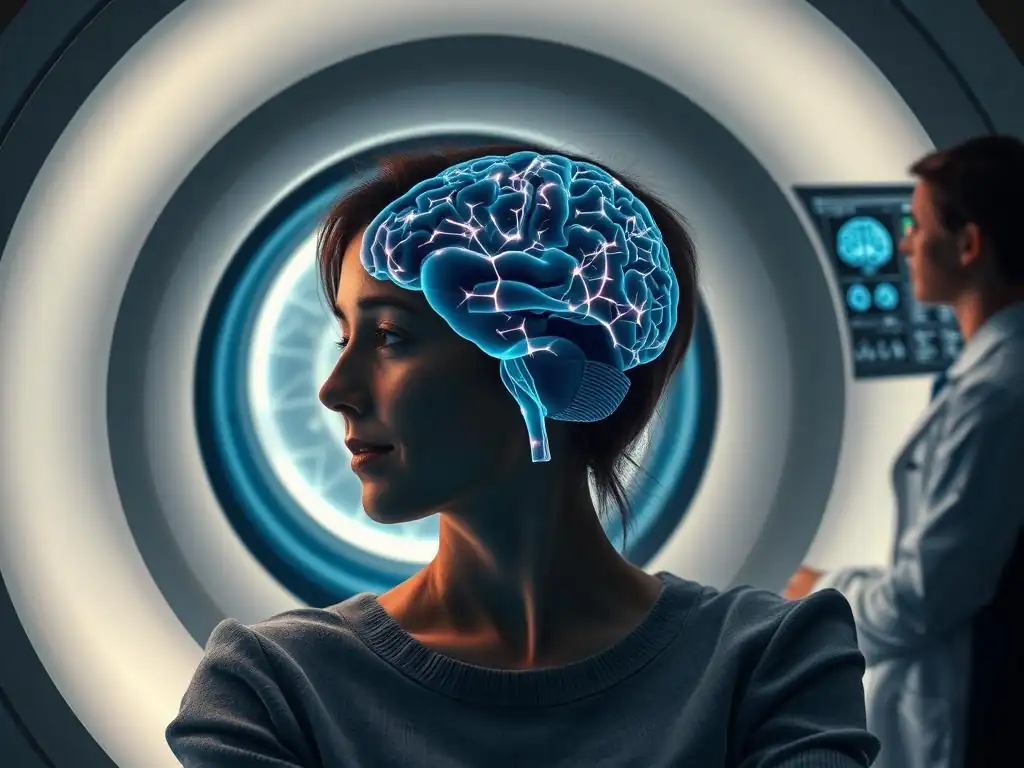
Welcome to Mind Care Guide, your compassionate companion on the journey to mental well-being.
Feeling lost in the labyrinth of mood disorders? You're not alone. Perhaps you're wondering if there's a more objective way to understand what's happening within. Could a brain scan actually reveal the secrets behind your mood? Dive into our in-depth exploration of "Brain scans mood disorder diagnosis" to uncover the fascinating potential – and the limitations – of neuroimaging in understanding and diagnosing these complex conditions. Let's explore together and shed light on this important topic.
Can Brain Scans Reveal Your Mood Disorder? Neuroimaging Insights
- Introduction: Exploring the Role of Brain Scans in Mood Disorder Assessment
- Understanding Mood Disorders: A Neuropsychological Perspective
- Neuroimaging Techniques for Mood Disorder Research and Clinical Applications
-
Brain Scans and the Diagnosis of Specific Mood Disorders
- Brain Imaging in Major Depressive Disorder (MDD): Altered Activity in Prefrontal Cortex and Amygdala
- Neuroimaging Findings in Bipolar Disorder: Identifying Neural Correlates of Mania and Depression
- Examining Brain Activity in Persistent Depressive Disorder (Dysthymia): Chronic Changes in Reward Processing
- The Limitations of Brain Scans in Mood Disorder Diagnosis
- Integrating Brain Scan Data with Clinical Assessment: A Holistic Approach
- Future Directions in Neuroimaging and Mood Disorder Research
- Conclusion: The Promise and Challenges of Brain Scans in Understanding Mood Disorders
-
Frequently Asked Questions
- Can brain scans definitively diagnose a mood disorder?
- What types of brain scans are used in the context of mood disorder research and investigation?
- How can brain scans assist in understanding mood disorders?
- Are brain scans a routine part of mood disorder diagnosis?
- What are the limitations of using brain scans for mood disorder diagnosis?
- Final Reflection: The Evolving Landscape of Mood Disorder Diagnosis
- Unlock Your Mental Wellness Journey Today!
Introduction: Exploring the Role of Brain Scans in Mood Disorder Assessment
Understanding mood disorders, such as major depressive disorder and bipolar disorder, is a complex endeavor. Traditionally, diagnosis relies heavily on clinical interviews, patient self-reports, and behavioral observations based on established diagnostic criteria. However, as our understanding of the brain's role in these conditions grows, researchers and clinicians are increasingly exploring how advanced technologies, particularly brain scans, might contribute to a more objective and nuanced assessment. The potential for brain scans in mood disorder diagnosis is a topic of significant research interest, aiming to identify biological markers that could complement existing methods.
Neuroimaging techniques offer a window into the living brain, allowing scientists to observe its structure, function, and chemical processes. While brain scans are not currently used as a standalone diagnostic tool for mood disorders in routine clinical practice, they are invaluable in research and hold promise for the future. This article delves into how various brain scanning methods work, what they reveal about the brains of individuals with mood disorders, their current limitations, and the exciting future possibilities for integrating neuroimaging into mental health care to potentially aid in brain scans mood disorder diagnosis.
Understanding Mood Disorders: A Neuropsychological Perspective
Defining Mood Disorders: Diagnostic Criteria and Subtypes
Mood disorders are a category of mental health conditions characterized by significant disturbances in emotional regulation. These disturbances can manifest as prolonged periods of excessive sadness (depression), excessive elation (mania), or fluctuating states between these extremes. The Diagnostic and Statistical Manual of Mental Disorders (DSM), currently in its fifth edition (DSM-5), provides the standardized criteria used by clinicians to diagnose these conditions.
Key diagnostic criteria involve specific symptom clusters and duration requirements. For instance, a diagnosis of Major Depressive Disorder requires experiencing at least five symptoms, including depressed mood or loss of interest/pleasure, for a period of at least two weeks. Bipolar Disorder, on the other hand, is defined by the occurrence of manic or hypomanic episodes, often alternating with major depressive episodes.
Subtypes like Bipolar I (involving full manic episodes) and Bipolar II (involving hypomanic but not full manic episodes) highlight the spectrum of these conditions. Persistent Depressive Disorder (Dysthymia) is characterized by chronic, low-grade depressive symptoms lasting for at least two years.
The Biological Basis of Mood Disorders: Neurotransmitters, Genetics, and Brain Circuits
Research consistently points to a complex interplay of biological factors underlying mood disorders. Neurotransmitters, chemical messengers in the brain, are believed to play a significant role. Monoamines like serotonin, norepinephrine, and dopamine are particularly implicated, with theories suggesting imbalances or dysregulation in these systems contribute to symptoms. For example, many antidepressant medications target the reuptake of serotonin and norepinephrine, aiming to increase their availability in synaptic clefts.
Beyond neurotransmitters, genetic predisposition is a recognized risk factor. Studies of twins and families indicate a higher likelihood of developing a mood disorder if a close relative has one. While no single "mood disorder gene" has been identified, multiple genes, each with a small effect, likely contribute to vulnerability.
Furthermore, mood disorders are increasingly understood as disorders of brain circuits – interconnected networks of brain regions. Specific circuits involving areas like the prefrontal cortex (involved in decision-making and emotional regulation), the amygdala (processing emotions), the hippocampus (memory and stress response), and the anterior cingulate cortex (ACC) show altered activity and connectivity in individuals with these conditions, providing a neurobiological basis for symptoms and a target for brain scans mood disorder diagnosis research.
Neuroimaging Techniques for Mood Disorder Research and Clinical Applications
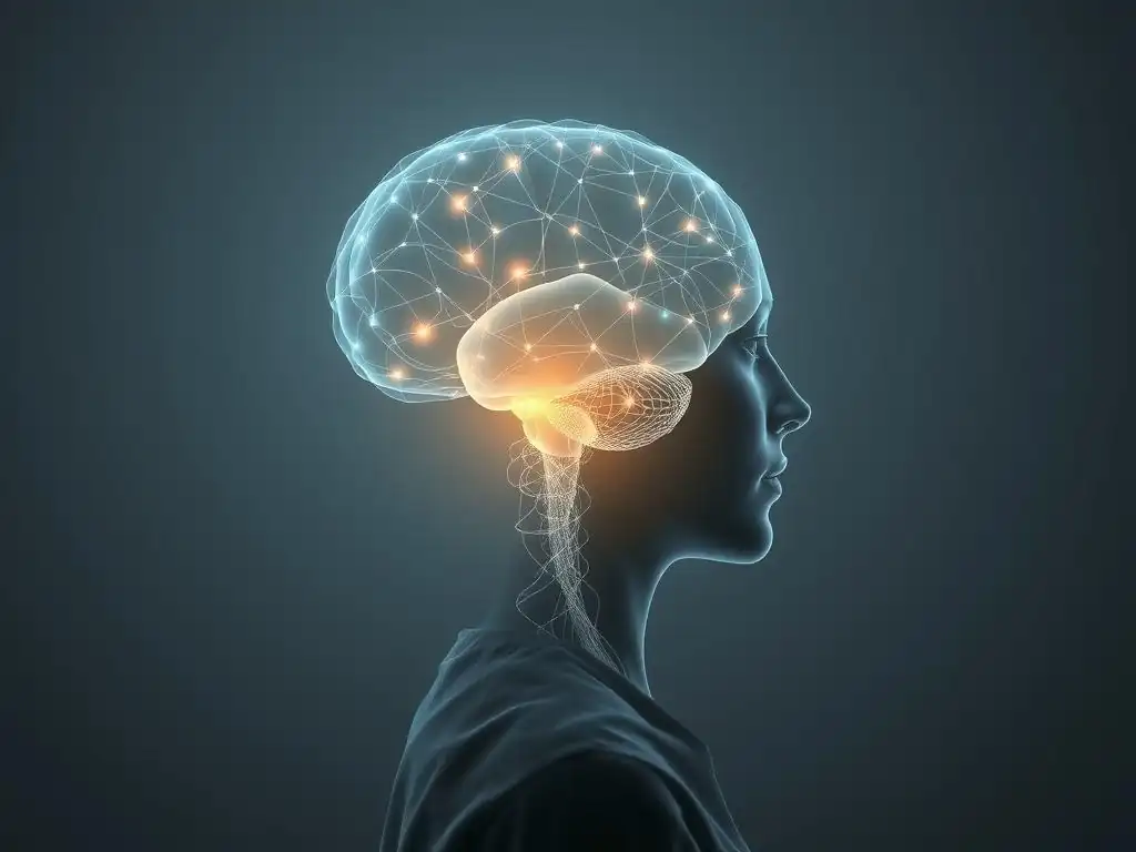
Magnetic Resonance Imaging (MRI): Unveiling Brain Structure and Function
Magnetic Resonance Imaging (MRI) is a powerful non-invasive technique that uses strong magnetic fields and radio waves to create detailed images of the brain. It is widely used in both clinical settings for detecting structural abnormalities (like tumors or lesions) and in research to understand brain differences associated with mental health conditions. MRI provides excellent spatial resolution, allowing researchers to visualize specific brain regions.
Structural MRI: Identifying Volumetric and Morphological Differences
Structural MRI provides static images of brain tissue, allowing researchers to measure the size (volume) and shape (morphology) of different brain regions. Studies using structural MRI in mood disorders have reported findings such as reduced hippocampal volume in individuals with recurrent or long-standing depression, potentially linked to chronic stress exposure. The hippocampus, crucial for memory and stress response, shows volume reductions averaging around 5-10% in some depressed populations compared to controls.
Furthermore, structural MRI research has identified alterations in the prefrontal cortex, specifically areas like the ventromedial prefrontal cortex and anterior cingulate cortex, which are involved in emotional regulation and executive function. While these findings are statistical averages across groups and not diagnostic for individuals, they reveal common patterns of structural differences associated with mood disorders, contributing to the broader picture of the neurobiological underpinnings of these conditions and the potential for structural brain scans mood disorder diagnosis.
Functional MRI (fMRI): Mapping Neural Activity and Connectivity
Functional MRI (fMRI) measures brain activity by detecting changes in blood flow and oxygenation, which are coupled with neural activity. By observing how brain regions "light up" during specific tasks or in a resting state, fMRI allows researchers to map neural activity and connectivity between different brain areas. This technique is invaluable for understanding which brain circuits are involved in processing emotions, regulating mood, and other cognitive functions affected by mood disorders.
In individuals with depression, fMRI studies often show altered activity in the amygdala (hyperactivity in response to negative stimuli) and the prefrontal cortex (hypoactivity in areas involved in cognitive control and reward processing). Connectivity analyses often reveal disrupted networks, such as decreased connectivity within the default mode network (involved in self-referential thought) or altered connections between the amygdala and prefrontal regulatory regions. These functional differences provide insights into the neural basis of symptoms like persistent sadness, anhedonia (loss of pleasure), and rumination, offering functional correlates for brain scans mood disorder diagnosis research.
Positron Emission Tomography (PET): Examining Neurotransmitter Binding and Metabolism
Positron Emission Tomography (PET) is a nuclear imaging technique that uses small amounts of radioactive tracers to visualize and measure metabolic processes or receptor binding in the brain. PET scans can provide quantitative data on aspects like glucose metabolism (an indicator of neural activity) or the density and availability of specific neurotransmitter receptors or transporters (e.g., serotonin transporters, dopamine receptors).
PET studies in mood disorders have contributed significantly to our understanding of neurotransmitter system involvement. For example, PET imaging with appropriate tracers has shown reduced serotonin transporter binding in key brain regions of individuals with depression, potentially indicating fewer sites for serotonin reuptake and thus altered serotonin signaling. Studies examining glucose metabolism have sometimes shown decreased activity in the prefrontal cortex during depressive episodes, consistent with fMRI findings of hypoactivity in these regions. PET offers a unique window into the neurochemical landscape, complementing structural and functional imaging data in the search for biomarkers for brain scans mood disorder diagnosis.
Electroencephalography (EEG) is a non-invasive technique that measures electrical activity in the brain using electrodes placed on the scalp. EEG is excellent at capturing rapid changes in brain activity (high temporal resolution) but has lower spatial resolution compared to MRI or PET. It records brainwaves, which are rhythmic patterns of electrical activity, and can also measure event-related potentials (ERPs), which are characteristic brainwave responses to specific stimuli.
EEG and ERP studies in mood disorders have revealed differences in brain electrical activity associated with emotional processing and cognitive functions. For instance, some studies have reported altered alpha wave asymmetry in the frontal lobes, with relatively less activity in the left frontal region, which has been associated with withdrawal and negative affect in depression.
ERP components related to the processing of emotional stimuli or feedback (like the P300 or reward positivity) also show differences in individuals with mood disorders compared to healthy controls. While less commonly used for direct diagnosis than clinical assessment, EEG provides valuable insights into the dynamic neural processes underlying mood states and symptoms, contributing another modality to the potential for brain scans mood disorder diagnosis research.
Brain Scans and the Diagnosis of Specific Mood Disorders
Brain Imaging in Major Depressive Disorder (MDD): Altered Activity in Prefrontal Cortex and Amygdala
Neuroimaging research on Major Depressive Disorder (MDD) consistently highlights abnormalities in brain regions involved in emotional regulation, reward processing, and cognitive control. Structural MRI often reveals reduced volume in the hippocampus and specific areas of the prefrontal cortex, such as the subgenual anterior cingulate cortex (sgACC). The sgACC, a small region within the prefrontal cortex, has received particular attention, with some studies showing increased activity in this area in severely depressed individuals, which can sometimes normalize with successful treatment.
Functional MRI studies frequently demonstrate altered activity and connectivity in circuits involving the prefrontal cortex and the amygdala. The amygdala, the brain's fear and emotion center, often shows heightened reactivity to negative stimuli in individuals with MDD. Simultaneously, regions of the prefrontal cortex responsible for regulating emotional responses may show decreased activity or connectivity with the amygdala, suggesting a breakdown in the brain's ability to dampen negative emotions. These consistent patterns of altered activity, particularly in the interplay between the prefrontal cortex and amygdala, represent key neurobiological correlates identified through brain scans mood disorder diagnosis research in depression.
Neuroimaging Findings in Bipolar Disorder: Identifying Neural Correlates of Mania and Depression
Bipolar disorder, characterized by distinct periods of elevated mood (mania or hypomania) and depression, also shows characteristic patterns in neuroimaging studies, though findings can vary depending on the current mood state. Structural MRI studies have sometimes reported increased volume in the amygdala or striatum (involved in reward and motivation) in individuals with bipolar disorder, although findings are less consistent than hippocampal changes in MDD.
Functional MRI studies often reveal exaggerated activity in limbic regions like the amygdala and ventral striatum during manic episodes, consistent with heightened emotional reactivity and reward-seeking behavior. Conversely, during depressive episodes, activity patterns may resemble those seen in MDD, with reduced prefrontal cortex activity. The connectivity between prefrontal regulatory regions and limbic areas also appears disrupted in bipolar disorder, potentially contributing to the difficulty in regulating mood swings. Identifying these distinct neural signatures across different mood states is a key goal of brain scans mood disorder diagnosis research in bipolar disorder.
Examining Brain Activity in Persistent Depressive Disorder (Dysthymia): Chronic Changes in Reward Processing
Persistent Depressive Disorder (Dysthymia) involves chronic, low-grade depressive symptoms lasting for years. While less extensively studied with neuroimaging than MDD or Bipolar Disorder, research suggests some overlapping and some distinct neural patterns. Given the chronic nature of the low mood and often present anhedonia (inability to experience pleasure), researchers have focused on brain regions involved in reward processing.
Preliminary studies using fMRI in individuals with Dysthymia have shown altered activity in reward-related circuits, including the ventral striatum and parts of the prefrontal cortex. These areas may show reduced responsiveness to positive stimuli compared to healthy individuals, reflecting the persistent lack of pleasure characteristic of the disorder. While findings are still emerging, understanding these chronic alterations in brain function could help differentiate Dysthymia from episodic MDD and inform therapeutic approaches, contributing to the potential for brain scans mood disorder diagnosis in chronic depression.
The Limitations of Brain Scans in Mood Disorder Diagnosis
Brain Scan Results and Individual Variability: The Influence of Age, Sex, and Genetics
Despite the exciting findings from neuroimaging research, it's crucial to understand that brain scans are not currently used as a definitive diagnostic test for mood disorders in clinical practice. One major limitation is the significant variability in brain structure and function across individuals. Factors such as age, sex, genetic background, life experiences, and even momentary states (like stress levels or sleep deprivation) can influence brain scan results.
While group-level studies can identify average differences between individuals with and without a mood disorder, these differences are often subtle and overlap considerably between groups. This means that an individual's brain scan might fall within the range observed in both healthy individuals and those with a disorder, making it impossible to make a confident diagnosis based solely on the scan.
For example, while average hippocampal volume might be slightly smaller in a group with MDD, many individuals with MDD will have hippocampal volumes within the typical range for healthy people, and some healthy people might have smaller-than-average hippocampi for unrelated reasons.
Ethical Considerations in Neuroimaging for Mental Health: Privacy and Informed Consent
The use of neuroimaging in the context of mental health raises important ethical considerations, particularly regarding privacy and informed consent. Brain scan data is highly personal and potentially revealing, not only about current mental state but also potentially about predispositions or other aspects of cognitive function. Ensuring the secure storage and handling of this sensitive data is paramount to protect patient privacy.
Furthermore, obtaining truly informed consent for brain scanning, especially in individuals who may be experiencing cognitive difficulties due to their mood disorder, requires careful attention. Patients need to fully understand the purpose of the scan (whether for research or potential clinical insight), its limitations (that it's not a diagnostic test), and how their data will be used and protected. There is also an ethical concern about the potential for "neuro-essentialism," where complex mood disorders are reduced solely to brain abnormalities, potentially ignoring the crucial roles of psychological, social, and environmental factors.
The High Cost and Accessibility of Brain Scanning Technology: Disparities in Access to Care
Brain scanning technologies like MRI and PET are expensive, requiring specialized equipment and trained personnel. The cost of acquiring and maintaining these machines, as well as the cost per scan, can be substantial. This high cost limits the widespread accessibility of neuroimaging, particularly in underserved communities or regions with limited healthcare resources.
Relying heavily on expensive brain scans for mood disorder diagnosis or treatment planning could exacerbate existing disparities in mental health care access. While the potential for objective biomarkers is appealing, any future integration of neuroimaging into clinical practice would need to address these cost and accessibility barriers to ensure equitable access to potentially beneficial technologies. Developing more affordable and portable neuroimaging methods or focusing on integrating findings from less expensive techniques like EEG might be crucial steps.
Integrating Brain Scan Data with Clinical Assessment: A Holistic Approach
Combining Neuroimaging with Psychological Evaluations: Improving Diagnostic Accuracy
Given the limitations of brain scans as standalone diagnostic tools, the most promising path forward involves integrating neuroimaging data with traditional clinical assessment methods. A holistic approach that combines objective brain data with subjective reports of symptoms, behavioral observations, psychological testing, and a thorough clinical history is likely to yield the most accurate and comprehensive understanding of an individual's mental health status.
Neuroimaging findings, while not diagnostic on their own, can potentially provide supportive evidence for a diagnosis or help differentiate between conditions that present with similar symptoms. For example, if a patient presents with symptoms that could be either severe depression or a neurological condition like early-stage dementia, a structural MRI might help rule out significant neurodegeneration.
In the future, specific patterns identified through brain scans might serve as "adjunctive" markers, adding weight to a clinical impression formed through interview and psychological evaluation, thereby potentially improving the accuracy of brain scans mood disorder diagnosis when used in concert with other information.
The Role of Brain Scans in Personalized Treatment Planning: Guiding Treatment Selection
Beyond diagnosis, neuroimaging holds significant promise for informing personalized treatment planning. Different individuals with the same mood disorder diagnosis may respond differently to various treatments, such as specific types of psychotherapy or antidepressant medications. Identifying neurobiological markers through brain scans could potentially help predict which treatment is most likely to be effective for a given individual.
For instance, research is exploring whether specific patterns of brain activity or connectivity, as measured by fMRI or EEG, can predict response to different classes of antidepressants or to therapies like transcranial magnetic stimulation (TMS). While this is still largely in the research phase, the goal is to move away from a trial-and-error approach to treatment selection towards a more data-driven, personalized strategy. Using brain scans to identify neurobiological profiles associated with better responses to particular interventions could revolutionize how mood disorders are treated, making care more efficient and effective.
Future Directions in Neuroimaging and Mood Disorder Research
Advancements in Brain Scanning Technology: Higher Resolution and Multi-Modal Imaging
The field of neuroimaging is continuously evolving, with ongoing advancements in technology promising even greater insights into the brain. Future MRI scanners are likely to offer higher spatial and temporal resolution, allowing for the visualization of smaller brain structures and faster brain activity changes. New PET tracers are being developed to target a wider range of neurotransmitter systems and receptors.
Furthermore, there is a growing interest in multi-modal imaging, which combines data from different techniques (e.g., simultaneous fMRI and EEG, or combining structural MRI with PET). This approach leverages the strengths of each modality – for example, the high spatial resolution of MRI with the high temporal resolution of EEG – to provide a more complete picture of brain structure, function, and chemistry simultaneously. These technological leaps will undoubtedly enhance our ability to study the complex neural circuits involved in mood disorders and refine the potential for brain scans mood disorder diagnosis.
Developing Biomarkers for Mood Disorder Prediction and Early Detection
A major goal of future neuroimaging research is the identification of reliable biomarkers for mood disorders. Biomarkers are measurable indicators of a biological state or condition. In the context of mood disorders, researchers are searching for brain imaging patterns that could potentially predict the risk of developing a disorder before symptoms fully manifest, or detect the disorder early in its course.
Identifying predictive biomarkers through brain scans could open up opportunities for early intervention and prevention strategies, potentially mitigating the severity or impact of the disorder. For example, longitudinal studies tracking individuals at high genetic risk for bipolar disorder are using neuroimaging to identify early brain changes that precede the onset of manic or depressive episodes. While the search for definitive biomarkers is challenging due to the complexity and heterogeneity of mood disorders, it remains a key focus for advancing the role of brain scans mood disorder diagnosis and prevention.
Longitudinal Studies: Understanding the Trajectory of Brain Changes in Mood Disorders
Longitudinal studies, which follow the same individuals over extended periods, are crucial for understanding how brain structure and function change over time in individuals with mood disorders. These studies can help clarify whether observed brain differences are causes, consequences, or correlates of the illness. They can also track how the brain responds to treatment or how changes correlate with the course of the disorder (e.g., periods of remission or relapse).
For example, longitudinal MRI studies are tracking changes in hippocampal volume or prefrontal cortex thickness in individuals with recurrent depression to see if these changes progress over time or recover with treatment. Similarly, longitudinal fMRI studies are examining whether patterns of connectivity change as individuals transition between manic and depressive states in bipolar disorder. These studies are essential for building a dynamic understanding of the neurobiological processes underlying mood disorders and for evaluating the long-term utility of brain scans mood disorder diagnosis and monitoring.
Conclusion: The Promise and Challenges of Brain Scans in Understanding Mood Disorders
Neuroimaging techniques have profoundly advanced our understanding of the brain's involvement in mood disorders, revealing complex alterations in structure, function, and neurochemistry across various brain circuits. While these technologies have identified consistent group-level differences in conditions like Major Depressive Disorder, Bipolar Disorder, and Persistent Depressive Disorder, they are not yet precise enough to serve as standalone diagnostic tools for individual patients. The inherent variability in human brains, the influence of numerous non-biological factors, and the high cost and accessibility issues currently limit their routine clinical application for brain scans mood disorder diagnosis.
Emphasizing the Importance of a Comprehensive Diagnostic Approach
Moving forward, the value of brain scans in mood disorder diagnosis lies not in replacing traditional clinical assessment but in complementing it. Integrating objective neuroimaging data with subjective patient reports, clinical interviews, and psychological evaluations offers the most comprehensive approach to understanding and diagnosing these complex conditions. As technology advances and research identifies more specific and reliable neurobiological markers, brain scans may play an increasing role in supporting clinical diagnoses, differentiating subtypes, and guiding personalized treatment decisions, ultimately improving care for individuals living with mood disorders.
Frequently Asked Questions
Can brain scans definitively diagnose a mood disorder?
No, brain scans alone cannot definitively diagnose a mood disorder. They can provide valuable insights but are typically used in conjunction with clinical evaluations and patient history.
What types of brain scans are used in the context of mood disorder research and investigation?
Commonly used brain scans include fMRI (functional Magnetic Resonance Imaging), PET (Positron Emission Tomography), and EEG (Electroencephalography). These scans help visualize brain activity and structural differences.
How can brain scans assist in understanding mood disorders?
Brain scans can help identify abnormal brain activity or structural differences associated with mood disorders like depression or bipolar disorder. This can further understanding of the underlying neurological mechanisms.
Are brain scans a routine part of mood disorder diagnosis?
Brain scans are not a routine part of the diagnostic process for mood disorders in most cases. They are typically used in research settings or when other diagnostic methods are inconclusive.
What are the limitations of using brain scans for mood disorder diagnosis?
Brain scans are expensive and not always accessible. Furthermore, findings can be complex to interpret, and there is significant overlap in brain activity patterns between different mood disorders and even healthy individuals.
Final Reflection: The Evolving Landscape of Mood Disorder Diagnosis
The ability to objectively diagnose mood disorders through brain scans represents a paradigm shift in mental healthcare. While still in its nascent stages, the research presented highlights the potential for neuroimaging to move beyond subjective assessments, offering a more precise and personalized approach to diagnosis and treatment. This holds immense promise for individuals struggling with the often-misunderstood complexities of conditions like depression and bipolar disorder.
The journey towards utilizing brain scans for mood disorder diagnosis is a testament to the power of scientific inquiry and its potential to revolutionize mental health care. The implications are far-reaching, impacting not only diagnostic accuracy but also the development of targeted therapies. As we continue to unravel the intricate neural mechanisms underlying these conditions, we move closer to a future where treatment is tailored to the individual's unique brain signature. The greatest advances in medicine have come from understanding the underlying mechanisms of disease, and this is no less true for mental illness.
Consider the implications of this research. What if accurate, early diagnosis became a reality? What if treatment plans could be personalized based on individual brain patterns, leading to faster recovery and improved outcomes? You have a role to play in this evolution. Stay informed, advocate for research funding, and encourage open conversations about mental health. The future of mood disorder diagnosis is not just a scientific endeavor; it's a collective responsibility, and your engagement is crucial in shaping a more compassionate and effective approach to mental healthcare.
Unlock Your Mental Wellness Journey Today!
Thanks for exploring the fascinating world of neuroimaging and mood disorders with us! We hope you gained valuable insights into how brain scans are revolutionizing mental health understanding. Now it's time to take the next step on your path to well-being.
- Schedule a consultation with a mental health professional: This is your opportunity to discuss your individual needs and explore treatment options tailored specifically to you.
- Share this article with someone you care about: Help spread awareness about the advancements in mood disorder diagnosis and treatment.
- Learn more about neuroimaging research: Explore reputable sources online to delve deeper into this exciting field and stay updated on the latest breakthroughs.
Ready to begin your personal journey towards a brighter, healthier mind? What steps will you take this week to prioritize your mental well-being?