
Unlocking the Brain's Secrets: Essential Imaging Tools in Mental Health Studies

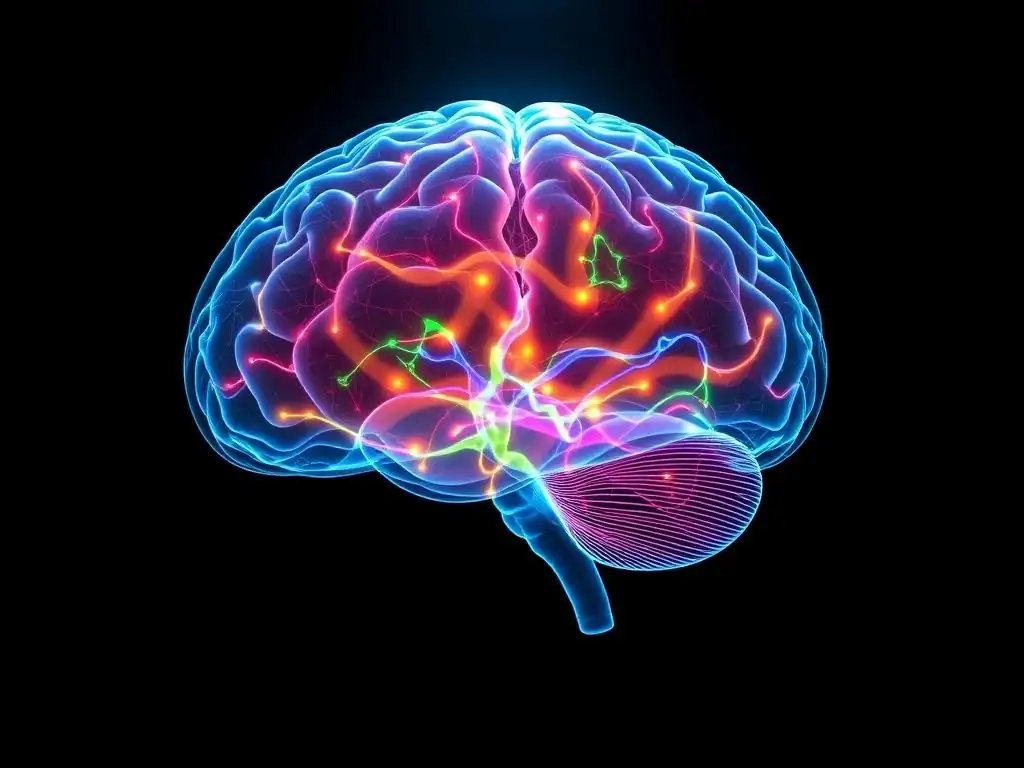
Welcome to Mind Care Guide, a sanctuary for understanding and nurturing your mental well-being.
Feeling lost in the complexities of mental health challenges? You're not alone. Discover how groundbreaking brain imaging mental health research is unveiling the intricate connections within our minds, offering new hope and pathways to recovery. Curious to learn how these powerful tools are revolutionizing our understanding? Let's explore the fascinating world of neuroscience together and unlock the secrets held within.
- Introduction to Brain Imaging in Mental Health Research
- Types of Brain Imaging Techniques Used in Mental Health
- Applications of Brain Imaging in Mental Health Research
- Ethical Considerations in Brain Imaging and Mental Health
- Future Directions in Brain Imaging for Mental Health
- Conclusion: The Power of Brain Imaging in Advancing Mental Health
-
Frequently Asked Questions
- What types of brain imaging are commonly used in mental health research?
- How does brain imaging contribute to understanding mental health disorders?
- Can brain imaging predict the effectiveness of mental health treatments?
- Are there any risks associated with using brain imaging for mental health research?
- How is brain imaging helping advance the field of mental health?
- Final Reflection: The Evolving Landscape of Mental Health Research
- Unlock Your Brain's Potential: Dive Deeper into Mental Health Research!
Introduction to Brain Imaging in Mental Health Research
The Role of Neuroimaging in Understanding Mental Illness
Neuroimaging, a powerful set of techniques that allow us to peer inside the living brain, plays a crucial role in advancing our understanding of mental illnesses. By providing visual representations of brain structure, function, and chemistry, these tools help researchers identify the neural correlates of various disorders. This includes investigating differences in brain size, shape, connectivity, and activity patterns between individuals with mental health conditions and healthy controls.
For example, studies using structural MRI have consistently shown reduced hippocampal volume in patients with major depressive disorder, suggesting a potential biological basis for some symptoms. Furthermore, functional neuroimaging allows us to observe which brain regions are active during specific tasks or emotional states, shedding light on disrupted neural circuits implicated in conditions like anxiety or schizophrenia.
The insights gained from brain imaging are transforming how we conceptualize mental illness, moving beyond purely behavioral descriptions to incorporate biological underpinnings. This biological perspective is vital because it helps to destigmatize conditions by framing them as disorders of the brain, similar to how heart disease is a disorder of the heart. Research using brain imaging in mental health research is actively seeking to map the intricate neural networks involved in complex processes like emotion regulation, decision-making, and social cognition, which are often impaired in mental health conditions.
For instance, fMRI studies in individuals with autism spectrum disorder have revealed altered connectivity patterns, particularly in areas related to social processing, providing tangible evidence of neural differences associated with the condition. This foundational understanding is critical for developing more targeted and effective interventions.
Why Brain Imaging Matters for Mental Health Treatment
Understanding the biological basis of mental health conditions through brain imaging is not just an academic exercise; it has profound implications for treatment. By identifying specific brain abnormalities or dysfunctional circuits associated with a disorder, researchers can develop treatments that target these underlying mechanisms.
For example, knowing that certain areas show altered activity in depression might lead to novel therapeutic approaches like transcranial magnetic stimulation (TMS) or deep brain stimulation (DBS) aimed at modulating activity in those specific regions. Brain imaging mental health research is also instrumental in evaluating how existing treatments, such as psychotherapy or medication, impact brain function and structure.
Moreover, brain imaging holds promise for personalizing mental healthcare. Currently, treatment selection often involves trial and error, which can be a lengthy and frustrating process for patients. Neuroimaging biomarkers could potentially help predict which individuals are most likely to respond to a particular treatment.
For instance, some preliminary studies using fMRI have explored whether patterns of brain activity before treatment can predict response to antidepressant medication or cognitive behavioral therapy in patients with depression. While still largely experimental, this area of brain imaging mental health research is rapidly evolving, aiming to move towards a future where treatment decisions are guided by objective biological data, leading to more efficient and effective care for individuals struggling with mental health challenges.
Types of Brain Imaging Techniques Used in Mental Health
Structural Imaging Techniques
Structural imaging techniques provide detailed pictures of the brain's anatomy. They are used to identify abnormalities in brain size, shape, and tissue integrity, which can be associated with various mental health conditions. These methods essentially create a static map of the brain's physical structure.
Magnetic Resonance Imaging (MRI) in Mental Health Studies
Magnetic Resonance Imaging (MRI) is one of the most widely used structural imaging techniques in brain imaging mental health research. It provides high-resolution images of brain tissue, allowing researchers to measure the volume of different brain regions and detect structural anomalies like lesions or atrophy. The detailed anatomical information obtained from MRI is invaluable for comparing brain structures between healthy individuals and those with mental disorders, helping to identify potential structural markers of illness.
Understanding MRI Principles
MRI works by using strong magnetic fields and radio waves to generate detailed images of the body's internal structures. The process involves aligning the protons within the body's water molecules using a strong magnet. Then, a radiofrequency pulse is applied, which knocks the protons out of alignment. When the pulse is turned off, the protons realign with the magnetic field, emitting energy in the form of radio signals.
These signals are detected by the MRI scanner and processed by a computer to create cross-sectional images of the brain. Different types of tissue emit signals at different rates, allowing for clear differentiation between grey matter, white matter, cerebrospinal fluid, and other structures. The non-invasive nature and excellent soft-tissue contrast make MRI a preferred tool for detailed anatomical studies in brain imaging mental health research.
Applications of MRI in Mental Disorders (e.g., Schizophrenia, Major Depressive Disorder)
MRI has been extensively used to study structural changes associated with numerous mental disorders. In schizophrenia, MRI studies have consistently reported reductions in the volume of certain brain regions, including the hippocampus, amygdala, and prefrontal cortex, compared to healthy controls. Some studies estimate these volume reductions can be subtle, perhaps a few percent difference on average in specific areas across large cohorts.
In major depressive disorder (MDD), research using MRI has frequently shown decreased volume in the hippocampus and anterior cingulate cortex. For example, a meta-analysis of multiple MRI studies might report an average hippocampal volume reduction of around 5-10% in patients with recurrent MDD. These findings contribute to our understanding of the neurobiological underpinnings of these complex conditions and help researchers explore how structural differences might relate to specific symptoms or cognitive impairments.
Diffusion Tensor Imaging (DTI): Mapping White Matter Tracts
Diffusion Tensor Imaging (DTI) is a specialized MRI technique that focuses on the diffusion of water molecules in the brain. Because water diffuses more freely along the direction of white matter tracts (bundles of nerve fibers that connect different brain regions), DTI can be used to map the orientation and integrity of these connections. This provides insights into the brain's wiring and how communication occurs between different areas. Changes in white matter integrity, often measured by metrics like fractional anisotropy (FA), have been implicated in various mental health conditions, suggesting disruptions in neural connectivity.
Limitations of DTI Compared to MRI and fMRI
While DTI is excellent for studying white matter tracts, it has limitations compared to standard structural MRI and functional MRI (fMRI). Standard MRI provides a more comprehensive view of overall brain anatomy, including grey matter structures and gross pathology. DTI primarily focuses on the directional diffusion of water within white matter, offering less detailed information about the structure of grey matter regions like the cortex or subcortical nuclei.
Compared to fMRI, which measures brain activity by detecting changes in blood flow, DTI provides no information about neural activity or function. It gives a picture of the brain's structural connections but not how those connections are being used or how active different brain regions are during a specific task or state. Therefore, DTI is often used in conjunction with MRI and fMRI to gain a more complete picture of brain structure and function in brain imaging mental health research.
Functional Imaging Techniques
Functional imaging techniques allow researchers to measure brain activity, providing insights into which brain regions are involved in specific cognitive, emotional, or behavioral processes. Unlike structural techniques that show the brain's anatomy, functional methods reveal the brain in action.
Functional Magnetic Resonance Imaging (fMRI): Mapping Brain Activity
Functional Magnetic Resonance Imaging (fMRI) is a powerful functional neuroimaging technique that measures brain activity by detecting changes in blood flow. When a brain region becomes more active, it requires more oxygen, leading to an increase in blood flow to that area. fMRI detects the resulting changes in the magnetic properties of oxygenated and deoxygenated blood, known as the BOLD (Blood-Oxygenation-Level Dependent) signal. This allows researchers to create maps of brain activity associated with specific tasks, stimuli, or resting states.
How fMRI Reveals Neural Correlates of Mental Processes
fMRI reveals the neural correlates of mental processes by observing which brain regions show a significant increase in the BOLD signal during a particular cognitive or emotional task compared to a control condition or a resting state. For instance, researchers might ask participants to view fearful faces while in the scanner. Areas like the amygdala, known to be involved in processing fear, would typically show increased activity.
By comparing activity patterns between groups (e.g., individuals with anxiety disorders vs. healthy controls) or within the same individual under different conditions, fMRI helps pinpoint the brain regions and networks involved in specific mental functions and how these might be altered in mental illness. This task-based approach is fundamental to much of the brain imaging mental health research focused on function.
fMRI Applications in Mental Health Research (e.g., Anxiety Disorders, Post-Traumatic Stress Disorder)
fMRI has been widely applied in brain imaging mental health research to study the neural basis of conditions like anxiety disorders and Post-Traumatic Stress Disorder (PTSD). In anxiety disorders, fMRI studies often show heightened activity in the amygdala and altered connectivity within fear circuitry when individuals are presented with anxiety-provoking stimuli. For example, research might demonstrate that the amygdala in individuals with social anxiety disorder shows a significantly larger BOLD response to social rejection cues compared to healthy controls.
In PTSD, fMRI has revealed abnormal activity and connectivity in regions involved in fear processing, emotion regulation (like the prefrontal cortex), and memory (like the hippocampus) during symptom provocation tasks or even during resting state. These findings help researchers understand the neural mechanisms underlying symptoms such as hyperarousal, intrusive memories, and avoidance behaviors.
Positron Emission Tomography (PET) Scans: Exploring Brain Chemistry
Positron Emission Tomography (PET) is a functional imaging technique that provides insights into the metabolic and biochemical activity of the brain. Unlike MRI or fMRI, which primarily focus on structure and blood flow, PET allows researchers to study the distribution and activity of specific molecules, such as neurotransmitters, receptors, or metabolic markers like glucose. This capability makes PET uniquely valuable for investigating the neurochemical imbalances hypothesized to play a role in various mental health conditions.
The Use of Radiotracers in PET Imaging
PET imaging relies on the use of radiotracers, which are molecules labeled with a short-lived radioactive isotope. These radiotracers are injected into the bloodstream and travel to the brain, where they bind to specific targets (e.g., a particular type of neurotransmitter receptor) or are metabolized (e.g., glucose). As the radioactive isotope decays, it emits positrons, which collide with electrons, producing gamma rays.
These gamma rays are detected by the PET scanner, and a computer reconstructs an image showing the distribution and concentration of the radiotracer in different brain regions. By using different radiotracers, researchers can image various aspects of brain chemistry, from glucose metabolism (an indicator of overall neural activity) to the density of specific receptor types.
PET Scans for Studying Neurotransmitter Systems in Mental Illness (e.g., Dopamine, Serotonin)
PET scans are particularly useful for studying neurotransmitter systems that are thought to be dysregulated in mental illness, such as dopamine and serotonin. For instance, PET studies using radiotracers that bind to dopamine receptors have investigated the role of dopamine dysfunction in schizophrenia and addiction. Research might show altered dopamine receptor density or dopamine release in specific brain areas like the striatum in individuals with these conditions.
Similarly, PET imaging with serotonin receptor or transporter radiotracers has been used to explore the involvement of the serotonin system in depression and anxiety disorders. Findings from such studies, like potentially reduced serotonin transporter availability in certain brain regions in depression, contribute significantly to our understanding of the neurochemical basis of these illnesses and inform the development of pharmacological treatments targeting these systems. This neurochemical focus is a key strength of PET in brain imaging mental health research.
Electroencephalography (EEG): Recording Brain Electrical Activity
Electroencephalography (EEG) is a non-invasive technique that measures the electrical activity of the brain using electrodes placed on the scalp. Neurons communicate via electrical signals, and synchronized activity of large populations of neurons generates electrical potentials that can be detected at the scalp surface. EEG records these fluctuations in voltage over time, providing a direct measure of neural activity with excellent temporal resolution (on the order of milliseconds). This allows researchers to capture rapid changes in brain states and responses.
EEG's Role in Diagnosing and Monitoring Mental Health Conditions (e.g., Epilepsy, Sleep Disorders)
While primarily known for its role in diagnosing neurological conditions like epilepsy (where it detects abnormal seizure activity), EEG also has applications in mental health research and clinical practice, particularly in assessing brain states and patterns associated with certain conditions. For example, EEG is a standard tool for diagnosing and studying sleep disorders, which frequently co-occur with mental health conditions like depression and anxiety.
It can identify abnormal sleep architecture or specific sleep-related electrical patterns. In some cases, specific EEG abnormalities or patterns of brain waves (like alpha, beta, theta, delta rhythms) have been associated with different psychiatric conditions, although these are generally not diagnostic markers in the same way they are for epilepsy.
Event-Related Potentials (ERPs) and Cognitive Function
A common application of EEG in mental health research is the study of Event-Related Potentials (ERPs). ERPs are small, voltage fluctuations in the EEG signal that are time-locked to the presentation of a specific stimulus or the execution of a specific event (e.g., seeing a picture, hearing a sound, making a decision). By averaging the EEG signal over many trials, the random background electrical activity is cancelled out, revealing the consistent brain response related to the event.
Different components of ERPs, occurring at specific latencies after the stimulus, are associated with different stages of cognitive processing (e.g., attention, perception, memory, decision-making). Studying ERPs in individuals with mental disorders allows researchers to identify deficits in specific cognitive processes. For instance, studies using ERPs have shown altered P300 components (associated with attention and working memory) in conditions like schizophrenia or ADHD, providing insights into cognitive impairments linked to these disorders.
Magnetoencephalography (MEG): Measuring Magnetic Fields Produced by Brain Activity
Magnetoencephalography (MEG) is another neuroimaging technique that measures the magnetic fields produced by electrical currents within the brain. Similar to EEG, MEG directly measures neuronal activity and offers very high temporal resolution. When neurons fire, the flow of electrical current generates tiny magnetic fields that pass through the skull and scalp relatively undisturbed. MEG sensors, typically housed in a helmet-like device and cooled by liquid helium, detect these weak magnetic fields.
Advantages of MEG Over EEG
MEG offers several advantages over EEG, particularly in terms of spatial resolution and the ability to localize the source of brain activity. While EEG signals are distorted as they pass through the skull and scalp, making it challenging to pinpoint the exact location of the activity source, magnetic fields measured by MEG are less affected by these tissues. This allows MEG to provide more accurate localization of neural activity, especially for activity originating from deeper brain structures.
Furthermore, MEG is particularly sensitive to activity from neurons oriented tangentially to the scalp, which complements EEG's sensitivity to radially oriented activity. This makes MEG a valuable tool for studying rapid neural dynamics with better source localization than EEG, enhancing brain imaging mental health research investigating precise timing and location of neural events.
MEG's Application in Investigating Neural Oscillations in Mental Disorders (e.g., Autism Spectrum Disorder)
MEG is well-suited for studying neural oscillations – rhythmic patterns of electrical activity in the brain that are thought to play a crucial role in coordinating neural processing and communication. Alterations in these oscillations (e.g., in specific frequency bands like alpha, beta, or gamma) have been implicated in various mental disorders. In autism spectrum disorder (ASD), for example, MEG studies have investigated differences in neural oscillations and connectivity patterns, particularly during social or sensory processing tasks.
Research might show altered gamma band activity or reduced coherence (connectivity) between brain regions in individuals with ASD compared to typically developing individuals. These findings contribute to the understanding of how disrupted neural timing and communication might underlie the characteristic symptoms of ASD, such as difficulties with social interaction and sensory sensitivities, providing a powerful tool in brain imaging mental health research for exploring dynamic brain function.
Applications of Brain Imaging in Mental Health Research

Brain imaging techniques offer a diverse toolkit for researchers investigating the complexities of mental illness. Their applications span from identifying biological markers of disorders to understanding the underlying neurobiology and evaluating the impact of treatments.
Identifying Biomarkers for Mental Disorders
One significant application of brain imaging in mental health research is the search for biomarkers – measurable biological indicators that can signal the presence or risk of a disease, or predict treatment response. Structural imaging might identify differences in brain volume or shape associated with a disorder, functional imaging could reveal abnormal patterns of brain activity or connectivity, and PET scans might show altered neurotransmitter system function.
For example, studies have explored whether specific patterns of resting-state functional connectivity, observable with fMRI, could serve as a biomarker for distinguishing between different subtypes of depression or predicting conversion from a high-risk state to psychosis. While no single brain imaging biomarker is currently used for definitive clinical diagnosis of most mental disorders, this area of research is active and holds significant promise for the future of personalized medicine in psychiatry.
Understanding the Neurobiology of Mental Illness
Brain imaging is fundamental to unraveling the complex neurobiological underpinnings of mental illnesses. By mapping brain structures, measuring activity during tasks, and investigating neurochemical systems, researchers can develop and refine models of how disrupted brain circuits and processes contribute to symptoms. For instance, fMRI studies have helped elucidate the role of the default mode network (a set of brain regions active during rest) in conditions like depression and anxiety, showing altered connectivity that might relate to excessive rumination.
PET studies have highlighted the involvement of specific neurotransmitter systems in conditions like obsessive-compulsive disorder (OCD) or substance use disorders. This deepens our understanding beyond simply describing symptoms, moving towards identifying the specific neural mechanisms that go awry, which is crucial for developing more effective, biologically informed interventions.
Evaluating Treatment Effectiveness with Brain Imaging
Brain imaging techniques can also be used to evaluate the effectiveness of different treatments for mental health conditions. By scanning patients before and after a course of therapy (whether medication, psychotherapy, or brain stimulation), researchers can observe whether the treatment leads to measurable changes in brain structure, function, or chemistry. For example, studies using fMRI have shown that successful cognitive behavioral therapy (CBT) for anxiety disorders can lead to changes in activity within fear-related brain circuits.
Similarly, research using structural MRI might investigate whether antidepressant treatment is associated with changes in hippocampal volume over time. This provides objective evidence of a treatment's impact on the brain, helping researchers understand how treatments work at a biological level and potentially identify predictors of treatment response.
Predicting Treatment Response Using Neuroimaging Data
A cutting-edge application of brain imaging in mental health research is the effort to predict how an individual patient will respond to a specific treatment based on their unique brain characteristics. The idea is that pre-treatment brain scans might reveal patterns that indicate whether someone is likely to benefit from a particular medication, type of therapy, or stimulation technique. For example, some preliminary fMRI studies have explored whether the pattern of connectivity in certain brain networks before starting antidepressant medication can predict whether a patient will achieve remission.
While still largely in the research phase and not yet routine in clinical practice, this area of brain imaging mental health research is highly promising. If successful, it could revolutionize mental healthcare by allowing clinicians to select the most appropriate treatment for an individual patient from the outset, avoiding ineffective interventions and speeding recovery.
Ethical Considerations in Brain Imaging and Mental Health
As brain imaging techniques become more sophisticated and widely used in mental health research, important ethical considerations arise. These relate to how data is collected, stored, and used, particularly given the sensitive nature of information about an individual's brain and mental state.
Privacy and Confidentiality of Brain Imaging Data
Brain imaging data is highly personal and potentially identifiable. While scans are typically anonymized by removing direct identifiers like names, the images themselves could potentially be linked back to an individual, especially as databases grow larger and more detailed. Furthermore, as research progresses, it's conceivable that brain scans might reveal information about an individual's predispositions or other health conditions.
Ensuring the robust privacy and confidentiality of brain imaging data is paramount. This requires secure data storage, strict access protocols, and careful consideration of how and where data is shared, particularly in large-scale research initiatives. Researchers and institutions must adhere to strict ethical guidelines and regulations to protect participant information and maintain public trust in brain imaging mental health research.
Informed Consent and Vulnerable Populations
Obtaining truly informed consent from individuals participating in brain imaging studies is critical, especially when working with vulnerable populations who may have impaired cognitive function or decision-making capacity due to their mental health condition. The consent process must clearly explain the nature of the study, the procedures involved (including potential risks, although minimal for techniques like MRI/fMRI), how the data will be used and protected, and the voluntary nature of participation.
For vulnerable populations, additional safeguards are necessary, which may include involving legal guardians or representatives, using simplified language, and ensuring that participants feel no coercion. Ensuring ethical consent procedures is fundamental to responsible brain imaging mental health research.
Responsible Use of Brain Imaging Technology
The increasing capabilities of brain imaging also raise questions about the responsible use of this technology. While brain imaging is a powerful research tool, there is a risk of over-interpreting findings or using them inappropriately. For example, findings about group differences in brain structure or function should not be used to stigmatize individuals or make deterministic claims about individuals based on a single scan.
There are also ethical considerations regarding the potential future use of brain imaging for purposes beyond research and clinical care, such as in legal settings or employment. Researchers and clinicians must communicate findings accurately, avoid neuro-essentialist interpretations (reducing complex individuals to their brain scans), and engage in ongoing dialogue about the societal implications of advances in brain imaging mental health research.
Future Directions in Brain Imaging for Mental Health
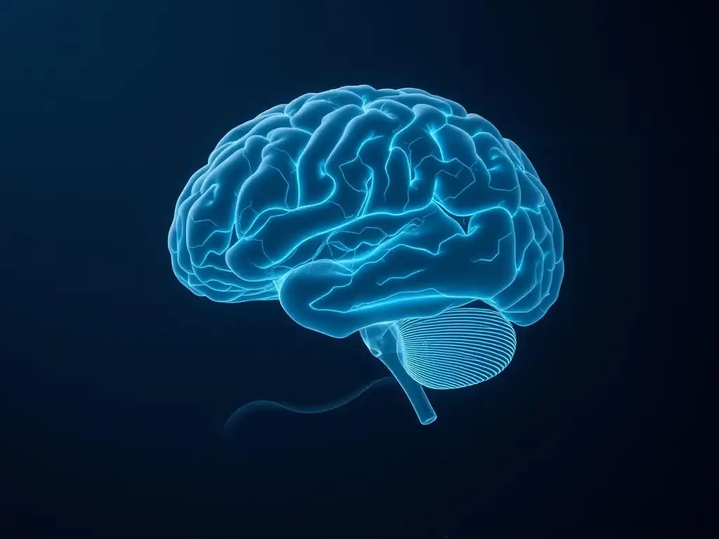
The field of brain imaging is constantly evolving, driven by technological advancements and innovative research approaches. The future holds exciting possibilities for further unlocking the brain's secrets and translating these discoveries into improved mental healthcare.
Advancements in Neuroimaging Technologies (e.g., High-Resolution fMRI)
Technological advancements are continuously pushing the boundaries of what brain imaging can reveal. For instance, the development of higher-field strength MRI scanners (e.g., 7 Tesla and beyond) allows for significantly higher spatial resolution in fMRI, enabling researchers to image activity in smaller brain structures or even within cortical layers. New pulse sequences and analysis methods are improving the sensitivity and specificity of various techniques.
Concurrent EEG-fMRI allows researchers to combine the excellent temporal resolution of EEG with the good spatial resolution of fMRI. These advancements promise more detailed and precise insights into brain structure, function, and connectivity, enabling researchers to investigate neural circuits at a finer scale and potentially identify more subtle abnormalities associated with mental health conditions.
Integrating Brain Imaging with Other Data Sources (e.g., Genetics, Clinical Data, Behavioral Data)
One of the most promising future directions is the integration of brain imaging data with other types of biological and clinical information. Combining neuroimaging data with genetic information, for example, can help researchers understand how genetic variations influence brain structure and function and how this relates to mental illness risk. Integrating imaging data with detailed clinical assessments, behavioral measures, and even data from wearable sensors provides a more comprehensive picture of an individual's health and illness trajectory.
Large-scale initiatives are collecting vast datasets that include brain imaging, genetics, and clinical information from thousands of participants, allowing for powerful analyses to identify complex patterns and interactions. This multi-modal approach is expected to yield deeper insights into the heterogeneity of mental disorders and pave the way for more personalized approaches in brain imaging mental health research.
Personalized Mental Healthcare Through Brain Imaging
Ultimately, a major goal of future brain imaging in mental health research is to contribute to personalized mental healthcare. The hope is that neuroimaging, in combination with other data, can help predict an individual's risk for developing a mental disorder, identify the specific neurobiological profile underlying their symptoms, and forecast their likely response to different treatments. This would allow clinicians to move away from a one-size-fits-all approach towards tailoring interventions to the individual patient.
For example, identifying a specific pattern of brain connectivity might suggest that a patient is more likely to benefit from a particular type of psychotherapy, while another pattern might indicate better response to a specific medication. While significant research is still needed, the vision of using brain imaging data to guide clinical decision-making and optimize treatment outcomes is a powerful driver for the future of the field.
Conclusion: The Power of Brain Imaging in Advancing Mental Health
Brain imaging techniques have revolutionized our ability to study the living brain and have become indispensable tools in mental health research. From mapping the brain's structure with MRI and DTI to capturing its activity with fMRI, PET, EEG, and MEG, these technologies provide unprecedented views into the complex organ underlying our thoughts, feelings, and behaviors. They have significantly advanced our understanding of the neurobiological basis of mental illnesses, helping to identify structural, functional, and chemical alterations associated with conditions like depression, schizophrenia, anxiety disorders, and many others.
Recap of Key Brain Imaging Techniques
We have explored several key brain imaging techniques used in this field. Structural MRI provides detailed anatomical images, useful for measuring brain region volumes. DTI maps white matter tracts, revealing neural connectivity. Functional MRI (fMRI) measures brain activity via blood flow changes, showing which areas are engaged during tasks.
PET scans use radiotracers to explore brain chemistry, particularly neurotransmitter systems. EEG records electrical activity with high temporal precision, useful for studying brain states and event-related potentials (ERPs). Finally, MEG measures magnetic fields from neural activity, offering high temporal resolution and improved source localization over EEG. Each technique offers unique insights, and they are often used in combination to provide a more complete picture in brain imaging mental health research.
The Promise of Neuroimaging for Improved Mental Health Outcomes
The knowledge gained from brain imaging studies has profound implications for improving mental health outcomes. By helping to identify potential biomarkers, understanding the underlying neurobiology, evaluating treatment effectiveness, and holding the promise of predicting treatment response, brain imaging is paving the way for more accurate diagnosis, more targeted treatments, and potentially personalized interventions.
While challenges remain, including ethical considerations and the need for further research to translate findings into routine clinical practice, the power of brain imaging in advancing our understanding and treatment of mental health conditions is undeniable. As technologies continue to evolve and research methods become more sophisticated, brain imaging mental health research will undoubtedly continue to play a vital role in our quest to promote mental well-being and alleviate the burden of mental illness worldwide.
Frequently Asked Questions
What types of brain imaging are commonly used in mental health research?
Common techniques include MRI (Magnetic Resonance Imaging) for structural and functional insights, fMRI (functional MRI) to observe brain activity, and PET (Positron Emission Tomography) to measure metabolic processes.
How does brain imaging contribute to understanding mental health disorders?
It helps identify structural and functional abnormalities in the brain associated with specific disorders, like schizophrenia or depression, aiding in diagnosis and treatment development.
Can brain imaging predict the effectiveness of mental health treatments?
Research is exploring the use of imaging to predict treatment response, allowing for more personalized medicine approaches in psychiatry and mental health care.
Are there any risks associated with using brain imaging for mental health research?
Generally, MRI and fMRI are considered safe, involving no radiation. PET scans use small amounts of radioactive tracers, but exposure is carefully controlled to minimize risk.
How is brain imaging helping advance the field of mental health?
By providing visual representations of brain activity, imaging is revolutionizing our understanding of mental illness and paving the way for novel therapies and improved diagnostic methods.
Final Reflection: The Evolving Landscape of Mental Health Research
The convergence of advanced brain imaging techniques and our understanding of mental illness is revolutionizing the field, offering unprecedented hope for earlier diagnosis and more effective treatments. We are no longer limited to relying solely on subjective patient reports; instead, we can now visualize the intricate neural pathways and structural abnormalities associated with a range of mental health conditions. This shift promises a future where personalized medicine, tailored to the unique brain signature of each individual, becomes a reality.
The journey to understanding the brain's complexities has been long and arduous, but the progress made through brain imaging is truly remarkable. It has not only deepened our scientific understanding but also shifted public perception, fostering greater empathy and reducing stigma surrounding mental illness. The greatest discovery of all time is that a person can change his future by merely changing his attitude.
– Oprah Winfrey. This quote resonates deeply, as the ability to visualize the brain's changes offers a powerful tool for both researchers and patients to understand and actively participate in the journey towards recovery.
Consider the implications of this progress. What if we could identify early warning signs of schizophrenia or depression through brain scans, allowing for timely intervention and preventing debilitating symptoms? What if personalized treatment plans, guided by brain imaging data, could drastically improve treatment outcomes and reduce the burden of mental illness on individuals and society? You have the power to contribute to this future. By staying informed about advancements in brain imaging and supporting research in this vital area, you become a crucial part of unlocking the brain's secrets and building a healthier, more compassionate future for all.
Unlock Your Brain's Potential: Dive Deeper into Mental Health Research!
Thanks for taking the time to explore the fascinating world of brain imaging and mental health research with us! We hope you gained a new appreciation for the tools and techniques scientists use to unravel the complexities of the mind. Ready to take the next step?
- Delve into further research: Explore relevant journals and websites listed in the article's resources section for even deeper understanding. This is your chance to become a true expert!
- Share your thoughts: Leave a comment below – what surprised you most about the methods used in brain imaging studies? Let's start a conversation!
- Challenge yourself: Can you think of innovative ways brain imaging could be used to improve mental health treatments in the future?
What innovative application of brain imaging could revolutionize mental health care tomorrow?