
SPECT vs. PET: Choosing Nuclear Imaging for Mental Health Insights


Welcome to Mind Care Guide, a space dedicated to nurturing your mental well-being.
Are you seeking a deeper understanding of your mental health? Imagine peering into the intricate workings of your brain to unlock crucial insights. This article delves into the fascinating world of Nuclear Imaging Mental Health Assessment, specifically comparing SPECT and PET scans. Which one holds the key to unlocking personalized treatment strategies? Let's explore the science and uncover how these powerful tools can illuminate the path toward improved mental health. Prepare to have your perspective shifted...
SPECT vs. PET: Choosing Nuclear Imaging for Mental Health Insights
- Understanding Nuclear Imaging in Mental Health
- SPECT Imaging for Mental Health: A Detailed Look
- PET Imaging for Mental Health: A Comprehensive Overview
- SPECT vs. PET: A Comparative Analysis for Mental Health Professionals
- Choosing the Right Nuclear Imaging Modality for Mental Health: A Decision Guide
- Future Directions in Nuclear Imaging for Mental Health
- Conclusion: Optimizing Nuclear Imaging for Enhanced Mental Health Insights
-
Frequently Asked Questions
- What exactly is nuclear imaging in the context of mental health assessment?
- How does SPECT differ from PET in nuclear imaging for mental health assessment?
- What mental health conditions might benefit from a nuclear imaging mental health assessment?
- Are there any risks associated with nuclear imaging procedures like SPECT and PET?
- How can nuclear imaging mental health assessment improve patient outcomes?
- Final Reflection: Unveiling the Mind's Landscape Through Nuclear Imaging
- Unlock Deeper Mental Health Insights: Your Next Steps
Understanding Nuclear Imaging in Mental Health
The Role of Brain Imaging Techniques in Mental Health Research
Brain imaging techniques have fundamentally transformed our understanding of mental health conditions. By allowing researchers and clinicians to visualize brain structure and function in living individuals, these tools provide crucial insights into the neural underpinnings of various disorders.
While structural techniques like MRI reveal the anatomy of the brain, functional imaging modalities, including nuclear imaging methods like SPECT and PET, go deeper by illustrating metabolic activity, blood flow, and even the distribution of specific neurochemicals. This functional perspective is particularly valuable for mental health assessment, as many conditions are characterized by disrupted brain activity patterns or neurotransmitter imbalances rather than gross structural abnormalities.
For instance, research utilizing functional brain imaging has identified altered activity in specific brain regions associated with depression, anxiety, and schizophrenia. Studies might show reduced metabolic activity in the prefrontal cortex in individuals with depression or abnormal dopamine signaling in the striatum in those with schizophrenia.
These findings not only enhance our theoretical understanding but also hold potential for improving diagnostic accuracy and guiding treatment strategies. The ability to non-invasively probe the living brain's dynamic processes is a cornerstone of modern neuroscience and its application to mental health, making functional nuclear imaging mental health assessment a vital area of study and clinical potential.
Introduction to SPECT and PET Scans for Mental Health Assessment
Single Photon Emission Computed Tomography (SPECT) and Positron Emission Tomography (PET) are two prominent nuclear imaging techniques used to evaluate brain function. Both methods involve injecting a small amount of a radioactive tracer into the bloodstream. This tracer travels to the brain, and its distribution or binding reflects specific physiological or biochemical processes.
Detectors outside the head then capture the radiation emitted by the tracer, and sophisticated computer algorithms reconstruct images showing the spatial distribution of the tracer within the brain. While the underlying principle is similar – detecting radioactive emissions from a tracer – the type of tracer used and the nature of the emission detected differ significantly, leading to differences in resolution, sensitivity, and the types of biological processes that can be studied.
In the context of mental health, SPECT scans are often used to assess regional cerebral blood flow, which is indirectly related to metabolic activity. Areas with higher blood flow generally indicate greater neuronal activity.
PET scans, on the other hand, can measure metabolic activity (using a tracer like FDG, which tracks glucose metabolism) or, more commonly in research, bind to specific receptors or transporters, allowing for the study of various neurotransmitter systems. Both modalities offer unique windows into brain function, contributing valuable data for the complex task of nuclear imaging mental health assessment, though they are typically used for different specific applications depending on the clinical question or research hypothesis.
SPECT Imaging for Mental Health: A Detailed Look
How SPECT Works: Single Photon Emission Computed Tomography
SPECT imaging relies on tracers that emit single photons as they decay. These tracers are labeled with radioisotopes such as Technetium-99m (⁹⁹mTc), Iodine-123 (¹²³I), or Thallium-201 (²⁰¹Tl). Once injected, the tracer is distributed throughout the body, including the brain. For brain imaging, common tracers like ⁹⁹mTc-HMPAO or ⁹⁹mTc-ECD are lipophilic (fat-soluble) and cross the blood-brain barrier, becoming trapped within brain cells in proportion to regional cerebral blood flow. As the tracer decays, it emits gamma rays (single photons) in random directions.
A SPECT scanner uses gamma cameras that rotate around the patient's head. These cameras are equipped with collimators, lead grids that filter out photons traveling at oblique angles, ensuring that only photons traveling perpendicular to the detector surface are counted. This process allows the system to determine the direction from which the photons originated.
By collecting data from multiple angles as the camera rotates, a computer can reconstruct a 3D image showing the concentration of the tracer in different parts of the brain. This image effectively represents the regional cerebral blood flow at the time the tracer was injected, providing a snapshot of brain activity patterns relevant to nuclear imaging mental health assessment.
SPECT Applications in Mental Health Diagnosis
SPECT imaging offers valuable insights into brain function that can aid in the diagnosis and understanding of certain mental health conditions, particularly those associated with changes in regional blood flow. While not typically used as a standalone diagnostic tool for most psychiatric disorders, it can provide supportive evidence or help differentiate conditions presenting with similar symptoms. For example, SPECT can be useful in evaluating cognitive impairment or suspected organic brain disease, offering a functional perspective alongside structural imaging.
SPECT for Assessing Traumatic Brain Injury
SPECT imaging has proven particularly useful in assessing the functional impact of Traumatic Brain Injury (TBI), even in cases where structural imaging like MRI appears normal. TBI can cause diffuse axonal injury and microvascular damage, leading to subtle but significant alterations in regional cerebral blood flow. A SPECT scan can reveal areas of hypoperfusion (reduced blood flow) in specific brain regions that correlate with the location and severity of injury, as well as the patient's cognitive and behavioral deficits. For instance, studies have shown reduced perfusion in frontal and temporal lobes in individuals with persistent post-concussive symptoms.
While SPECT results in TBI require careful interpretation within the clinical context, they can provide objective evidence of functional impairment that is not always visible on conventional scans. This can be particularly helpful in cases of mild TBI (concussion) or when assessing the long-term consequences of injury. The pattern and extent of hypoperfusion seen on a SPECT scan contribute to a more comprehensive nuclear imaging mental health assessment in the context of brain trauma.
SPECT for Assessing Attention-Deficit/Hyperactivity Disorder (ADHD)
The application of SPECT in diagnosing Attention-Deficit/Hyperactivity Disorder (ADHD) is more controversial and not widely accepted as a standard diagnostic tool by major professional organizations. However, some research and clinical practices explore its potential utility.
Studies using SPECT have often reported findings of reduced cerebral blood flow or metabolic activity, particularly in the prefrontal cortex and basal ganglia, brain regions implicated in executive function, attention, and impulse control, which are core deficits in ADHD. For example, some SPECT studies in groups of individuals with ADHD have suggested a pattern of reduced perfusion in areas like the anterior cingulate cortex or the caudate nucleus compared to control groups.
Despite these findings, the variability in results across studies, lack of standardization, and the fact that similar perfusion patterns can be observed in other conditions mean that SPECT cannot reliably diagnose ADHD in an individual patient. Its role remains primarily in research settings to understand the neurobiology of the disorder or, in some specialized clinical contexts, to potentially support a diagnosis when combined with extensive clinical evaluation. The use of nuclear imaging mental health assessment for ADHD remains an area of ongoing debate and research.
Advantages and Limitations of SPECT in Mental Health
One significant advantage of SPECT imaging is its relative accessibility and lower cost compared to PET scans. SPECT scanners are more widely available in hospitals and imaging centers, and the radioisotopes used generally have longer half-lives (hours), making production and distribution simpler and less expensive. This makes SPECT a more feasible option for clinical nuclear imaging mental health assessment in many settings. Furthermore, SPECT can utilize tracers that measure regional blood flow, which is a fundamental aspect of brain function relevant to many conditions.
However, SPECT has several limitations. The primary limitation is its lower spatial resolution compared to PET, typically ranging from 7-10 mm. This means it is less able to distinguish fine details or small structures in the brain. Additionally, SPECT tracers for studying specific receptors or transporters are less developed and available compared to PET tracers.
The sensitivity of SPECT is also lower than PET, requiring a higher dose of radiation to achieve comparable image quality. While the radiation dose is generally considered safe, it is a factor to consider. These limitations mean that SPECT is less suited for detailed neuroreceptor mapping or studying subtle metabolic changes compared to its PET counterpart.
PET Imaging for Mental Health: A Comprehensive Overview
How PET Works: Positron Emission Tomography
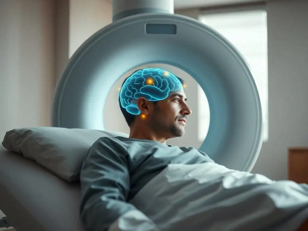
PET imaging utilizes tracers labeled with isotopes that emit positrons, such as Carbon-11 (¹¹C), Fluorine-18 (¹⁸F), Nitrogen-13 (¹³N), or Oxygen-15 (¹⁵O). When a positron is emitted from the nucleus, it travels a short distance (typically less than 1 mm in tissue) before annihilating with an electron. This annihilation event produces two gamma rays that are emitted simultaneously in nearly opposite directions (180 degrees apart). A PET scanner consists of a ring of detectors surrounding the patient's head.
When the two gamma rays from an annihilation event are detected by detectors on opposite sides of the ring within a very narrow time window, the scanner registers a "coincidence event." By recording millions of these coincidence events, the system can determine the lines along which the annihilation events occurred.
Sophisticated computer algorithms then reconstruct a 3D image showing the distribution of the tracer within the brain. The high sensitivity of detecting two simultaneous photons and the ability to use a wider range of biologically active tracers make PET a powerful tool for probing specific molecular processes, crucial for advanced nuclear imaging mental health assessment and research.
PET Applications in Mental Health Research and Clinical Practice
PET imaging is a powerful tool for both mental health research and, in specific instances, clinical practice, largely due to its ability to quantify molecular targets like receptors and transporters. It allows researchers to study the living brain's neurochemistry in detail, providing insights into the mechanisms underlying various disorders. While less commonly used for routine diagnosis than structural imaging, PET plays a vital role in advancing our understanding of the neurobiological basis of psychiatric illnesses and evaluating potential therapeutic targets.
PET for Studying Neurotransmitter Activity in the Striatum
The striatum, a key brain region involved in reward, motivation, and motor control, is heavily modulated by dopamine and other neurotransmitters. PET imaging is extensively used to study neurotransmitter activity in the striatum, particularly the dopamine system, which is implicated in conditions like schizophrenia, addiction, and Parkinson's disease (which can have psychiatric comorbidities). For example, PET tracers like [¹¹C]raclopride or [¹⁸F]fallypride bind to dopamine D2/D3 receptors, allowing researchers to quantify receptor availability.
Studies using these tracers have shown altered dopamine receptor binding or dopamine release in the striatum of individuals with schizophrenia (e.g., increased dopamine synthesis capacity or release) or substance use disorders (e.g., reduced D2 receptor availability in chronic stimulant users). This ability to directly image and quantify neurotransmitter systems makes PET an indispensable tool for understanding the neurochemical basis of psychiatric disorders and for developing targeted treatments, forming a critical component of advanced nuclear imaging mental health assessment in research settings.
PET for Investigating Dopamine Transporter Availability in Mental Disorders
The dopamine transporter (DAT) is responsible for reabsorbing dopamine from the synaptic cleft, regulating the duration of dopamine signaling. Alterations in DAT availability have been implicated in several neurological and psychiatric conditions. PET imaging, using tracers like [¹¹C]WIN 35,428 or [¹²³I]ioflupane (which can also be used with SPECT, often referred to as DaTscan), allows for the quantification of DAT density, particularly in the striatum.
While most commonly used in the diagnosis of Parkinsonian syndromes, PET studies of DAT have also been conducted in other conditions. For instance, some research has explored DAT availability in ADHD, though findings have been inconsistent. Studies in substance use disorders have shown complex relationships between drug use and DAT levels. The ability to measure DAT availability provides valuable information about the integrity of the dopaminergic system, offering insights into conditions where dopamine dysregulation is suspected. This specific application highlights the molecular sensitivity of PET as a nuclear imaging mental health assessment tool.
Advantages and Limitations of PET in Mental Health
A major advantage of PET imaging is its superior spatial resolution compared to SPECT, typically ranging from 4-6 mm, allowing for clearer visualization of smaller brain structures. More importantly, PET offers significantly higher sensitivity and a much wider array of radiotracers available or under development that can target specific receptors, transporters, enzymes, or metabolic pathways. This molecular specificity allows researchers to directly study the neurochemical imbalances believed to underlie many mental health conditions, providing a deeper level of insight into disease mechanisms.
However, PET imaging is considerably more expensive than SPECT. The radioisotopes used in PET often have very short half-lives (e.g., ¹¹C has a half-life of ~20 minutes, ¹⁸F has a half-life of ~110 minutes), requiring an on-site cyclotron to produce them or rapid transport from a nearby facility. This logistical challenge and the higher cost of the scanner and tracers limit the availability of PET, particularly for routine clinical nuclear imaging mental health assessment outside of major academic or research centers.
SPECT vs. PET: A Comparative Analysis for Mental Health Professionals
Key Differences in Image Resolution and Sensitivity
When comparing SPECT and PET for nuclear imaging mental health assessment, resolution and sensitivity are critical distinctions. PET generally offers better spatial resolution, allowing for clearer images and the ability to resolve smaller brain structures or subtle changes in tracer distribution. Typical spatial resolution for clinical SPECT is around 7-10 mm, while clinical PET scanners can achieve 4-6 mm, and high-resolution research scanners even better. This difference can be significant when studying fine-grained regional activity or tracer binding.
Furthermore, PET is inherently more sensitive than SPECT. This higher sensitivity stems from the detection of two coincident photons in PET compared to a single photon in SPECT, which reduces background noise and improves signal-to-noise ratio. Higher sensitivity means that PET can detect lower concentrations of the radiotracer, allowing for studies using tracers that bind to less abundant targets or permitting lower injected radiation doses while maintaining image quality. This difference in sensitivity makes PET particularly advantageous for quantitative studies of low-density receptors or transporters.
Cost-Effectiveness Comparison: SPECT vs. PET for Mental Health Evaluations
The cost of performing a nuclear imaging scan is a major factor influencing its accessibility for routine clinical mental health assessment. SPECT scans are significantly less expensive than PET scans. The capital cost of a SPECT scanner is lower, the radioisotopes are less costly to produce and have longer half-lives which simplifies logistics, and the overall procedure is typically less complex to perform and interpret in a basic sense (though interpretation of functional data is always nuanced). A typical SPECT scan might cost several hundred to a couple of thousand dollars, depending on the facility and location.
In contrast, PET scans are substantially more expensive. The necessity of an on-site cyclotron or access to a facility with one for short-lived isotopes adds considerable infrastructure cost. The PET scanner itself is more complex and expensive to purchase and maintain.
The production and delivery of PET radiotracers are also more costly. A typical PET scan, particularly one using complex tracers for receptor binding, can cost several thousand dollars, often significantly more than a SPECT scan. This cost disparity is a primary reason why SPECT is somewhat more available for clinical purposes (like perfusion imaging in dementia workups), while advanced PET for molecular targets is largely confined to research settings or specialized clinical centers for nuclear imaging mental health assessment.
Radiation Exposure Considerations in SPECT and PET Brain Scans
Both SPECT and PET scans involve injecting a radioactive tracer, meaning patients are exposed to a small amount of ionizing radiation. The dose of radiation received depends on the specific radioisotope used, the amount injected, and how quickly it is cleared from the body. Generally, the effective radiation dose from a brain SPECT scan is comparable to or slightly lower than that from a brain PET scan, although this can vary depending on the tracer. For example, a typical brain perfusion SPECT scan might result in an effective dose of around 5-10 millisieverts (mSv).
PET scans, especially those using ¹⁸F-FDG (for glucose metabolism), often result in a similar effective dose, perhaps in the range of 4-8 mSv. However, some PET tracers targeting specific receptors might require different injected activities, potentially altering the dose. Both doses are within levels considered acceptable for diagnostic medical procedures but are not negligible. Clinicians must weigh the potential diagnostic benefit of nuclear imaging mental health assessment against the minimal radiation risk, especially in vulnerable populations or if repeat scans are anticipated. The decision to use either modality always involves a careful risk-benefit analysis.
Availability and Accessibility of SPECT and PET Technology
The availability and accessibility of SPECT and PET technology differ significantly, impacting their widespread use in mental health settings. SPECT scanners are more common in hospitals and general imaging centers globally. Their lower cost, simpler infrastructure requirements (not needing an on-site cyclotron for most common tracers), and the longer shelf-life of SPECT radioisotopes make them more accessible outside of large metropolitan or academic centers. This wider availability makes SPECT a practical tool for blood flow imaging when needed for clinical nuclear imaging mental health assessment, such as in the evaluation of suspected dementia or stroke.
PET scanners, particularly those capable of advanced neuroreceptor imaging, are less widely available. They are typically found in larger hospitals, specialized imaging centers, or research institutions due to their higher cost, the need for a cyclotron or close proximity to one, and the requirement for specialized expertise in administering the tracers and interpreting the complex data. While ¹⁸F-FDG PET for oncology is more common, neuro-PET for specific molecular targets remains less accessible, limiting its routine use in clinical psychiatric practice compared to its significant role in research.
Choosing the Right Nuclear Imaging Modality for Mental Health: A Decision Guide
Factors Influencing the Selection of SPECT or PET
Deciding whether to use SPECT or PET for nuclear imaging mental health assessment is a multi-faceted process guided by several key factors. It's not simply a matter of which technology is "better," but rather which is more appropriate for the specific question being asked, the patient's condition, and the available resources. This decision requires careful consideration by clinicians and researchers to ensure the most relevant and informative data is obtained while minimizing risks and costs.
Specific Clinical Question or Research Objective
The most crucial factor is the specific clinical question or research objective. If the primary goal is to assess regional cerebral blood flow, which can indicate areas of reduced or increased activity, SPECT is often the preferred and sufficient modality due to its accessibility and lower cost. This is useful for evaluating conditions where perfusion deficits are suspected, such as certain types of dementia or TBI.
However, if the objective is to quantify the density or binding of specific neurotransmitter receptors or transporters, investigate metabolic pathways at a molecular level, or achieve higher spatial resolution for smaller brain regions, PET is the modality of choice. PET's diverse range of specific tracers allows for targeted investigation of the neurochemical systems hypothesized to be involved in a particular mental health condition.
Patient Condition and Tolerance
Patient factors also play a role in selecting the imaging modality. Both SPECT and PET scans require the patient to remain still for a period, typically ranging from 20 minutes to over an hour depending on the specific protocol and tracer. Patients with severe anxiety, claustrophobia, or movement disorders may find it challenging to remain still.
The injection procedure for the radiotracer is similar for both. However, the scan duration can sometimes be shorter for certain PET protocols compared to SPECT, potentially benefiting patients who have difficulty staying still for extended periods. The radiation dose, while generally low for both, is also considered, especially in pediatric or pregnant patients, though nuclear imaging mental health assessment is rarely performed in pregnancy.
Available Resources and Expertise
The practical realities of available resources and expertise significantly influence the choice between SPECT and PET. As discussed earlier, PET scanners are less common and more expensive to operate than SPECT scanners. Access to specific radiotracers, particularly for PET, may be limited to centers with an on-site cyclotron or reliable access to radiopharmacies that can synthesize and deliver short-lived isotopes quickly.
Furthermore, interpreting the results of both SPECT and PET scans, especially complex quantitative studies of receptor binding, requires specialized training and expertise in nuclear medicine and potentially neuroradiology or psychiatry. If a facility only has a SPECT scanner and the clinical question can be reasonably addressed with blood flow information, SPECT will be the practical choice for nuclear imaging mental health assessment.
Case Studies: Illust illustrating the Use of SPECT and PET in Mental Health
To better understand how these modalities are applied, let's consider hypothetical but realistic case studies illustrating their use in nuclear imaging mental health assessment. These examples highlight the types of questions each technology is best suited to address and the kind of information they can provide.
Case Study 1: Using SPECT to Evaluate Memory Loss in Dementia
- Scenario: A 72-year-old patient presents with progressive memory loss, difficulty with daily tasks, and changes in behavior. Clinical evaluation and structural MRI are suggestive of dementia, but the specific type (e.g., Alzheimer's vs. vascular dementia) is unclear.
- SPECT Application: A brain perfusion SPECT scan is ordered. The patient receives an injection of a ⁹⁹mTc-based tracer, and images are acquired. The SPECT scan reveals a significant pattern of hypoperfusion (reduced blood flow) predominantly in the parietal and temporal lobes, particularly in the posterior cingulate cortex and precuneus. This pattern of reduced blood flow is highly characteristic of Alzheimer's disease and supports this diagnosis over other potential causes like diffuse vascular disease or frontal lobe dementia, which typically show different perfusion patterns.
This case demonstrates how SPECT, by visualizing regional blood flow, can provide valuable functional information that complements structural imaging and clinical data. The specific pattern of reduced perfusion observed acts as a biomarker supporting a particular diagnostic hypothesis in the context of nuclear imaging mental health assessment for cognitive decline. While not definitive on its own, the SPECT findings contribute significantly to the overall diagnostic picture, guiding further management and treatment decisions.
Case Study 2: Using PET to Investigate Serotonin Receptor Binding in Major Depressive Disorder
- Scenario: A research study aims to investigate whether patients with treatment-resistant Major Depressive Disorder (MDD) have altered serotonin 1A (5-HT1A) receptor availability compared to healthy controls, potentially explaining poor response to SSRI antidepressants.
- PET Application: Participants with MDD and a matched group of healthy controls undergo a PET scan using a radiotracer that binds specifically to 5-HT1A receptors, such as [¹¹C]WAY 100635. After tracer injection, dynamic PET images are acquired over 60-90 minutes. Quantitative analysis is performed to calculate the binding potential (BPND) of the tracer in various brain regions known to have 5-HT1A receptors, such as the prefrontal cortex, amygdala, and raphe nuclei.
The study finds that, on average, participants with treatment-resistant MDD show a statistically significant reduction (e.g., approximately 15-20% lower) in 5-HT1A receptor binding potential in the prefrontal cortex compared to healthy controls. This finding supports the hypothesis that altered serotonin receptor function may contribute to treatment resistance in some individuals with depression. This case illustrates PET's unique capability to quantify specific molecular targets in the living brain, providing insights into the neurochemical basis of mental disorders that are not possible with blood flow imaging alone, showcasing its power in nuclear imaging mental health assessment for research purposes.
Future Directions in Nuclear Imaging for Mental Health
Advancements in SPECT and PET Technology
The field of nuclear imaging is continuously evolving, with ongoing advancements promising to enhance the utility of SPECT and PET in mental health. Hybrid scanners, combining PET or SPECT with MRI (PET/MRI, SPECT/MRI), are becoming more common. These systems allow for simultaneous acquisition of functional nuclear imaging data and high-resolution structural/functional MRI data, providing perfect spatial co-registration and richer, multi-modal information within a single scan session. This is particularly beneficial for mental health research, allowing researchers to link molecular or perfusion changes directly to brain structure and activity patterns measured by fMRI.
Furthermore, there are developments in scanner technology, such as digital PET detectors offering improved resolution and sensitivity, and total-body PET scanners that can image the entire body simultaneously with much higher sensitivity and lower dose. Advances in radiochemistry are also crucial, with efforts focused on developing new, highly specific radiotracers for a wider range of neurotransmitter systems, receptors, and other molecular targets relevant to psychiatric disorders. These technological leaps are set to improve the quality and expand the scope of nuclear imaging mental health assessment.
Potential for Personalized Mental Health Treatment Based on Nuclear Imaging Findings
One of the most exciting future directions for nuclear imaging in mental health is its potential to contribute to personalized treatment approaches. Currently, selecting treatments for many psychiatric conditions relies heavily on trial and error. Nuclear imaging, particularly PET with specific tracers, could potentially provide biomarkers that predict treatment response or help select the most appropriate therapy for an individual patient based on their unique neurobiological profile.
For example, if a PET scan could reliably identify subtypes of depression characterized by different neurochemical abnormalities (e.g., primarily dopaminergic vs. serotonergic dysfunction), clinicians could potentially tailor antidepressant medication choices accordingly. Similarly, imaging specific targets might help determine if a patient is likely to respond to a particular type of psychotherapy that targets a specific brain circuit. While still largely in the research phase, the prospect of using nuclear imaging mental health assessment to move towards precision medicine in psychiatry is a significant area of focus.
Ethical Considerations in Using Nuclear Imaging for Mental Health Assessment
As nuclear imaging techniques become more advanced and potentially more accessible for mental health assessment, several ethical considerations come to the forefront. One key issue is the interpretation and potential misuse of findings. Brain scans can reveal complex patterns that require expert interpretation and should not be used in isolation to diagnose or stigmatize individuals. There is a risk of oversimplifying complex conditions based on imaging findings or using scans in a discriminatory manner (e.g., for employment screening).
Privacy and data security are also paramount, as imaging data is highly personal and potentially sensitive. Ensuring robust protocols for data handling and anonymization is essential, especially in large research studies. Furthermore, the cost and accessibility disparities raise questions of equity – who has access to these advanced diagnostic tools? As the technology progresses, addressing these ethical challenges proactively will be crucial to ensure that nuclear imaging mental health assessment is used responsibly, ethically, and equitably for the benefit of patients.
Conclusion: Optimizing Nuclear Imaging for Enhanced Mental Health Insights
Nuclear imaging, encompassing both SPECT and PET modalities, offers invaluable tools for peering into the functional and molecular landscape of the living brain. While SPECT provides accessible insights into regional cerebral blood flow, useful for assessing conditions like dementia and TBI, PET offers superior resolution and sensitivity, allowing for detailed investigation of specific neurotransmitter systems and metabolic pathways crucial for understanding the neurochemical basis of complex psychiatric disorders. Both techniques play distinct yet complementary roles in nuclear imaging mental health assessment, contributing significantly to both clinical evaluation (in specific cases) and groundbreaking research.
Choosing between SPECT and PET depends critically on the specific clinical question, the desired level of detail (perfusion vs. molecular targets), patient factors, and the availability of resources and expertise. While challenges related to cost, accessibility, and the need for careful interpretation remain, ongoing technological advancements and the development of new radiotracers promise to enhance the power and utility of nuclear imaging in the future. Ultimately, optimizing the use of nuclear imaging techniques holds significant potential to improve our understanding, diagnosis, and personalized treatment of mental health conditions, leading to better outcomes for individuals affected by these challenging illnesses.
Frequently Asked Questions
What exactly is nuclear imaging in the context of mental health assessment?
Nuclear imaging uses radioactive tracers to visualize brain activity. This allows doctors to see how the brain is functioning metabolically and chemically, providing insights that structural imaging (like MRI) cannot.
How does SPECT differ from PET in nuclear imaging for mental health assessment?
SPECT (Single-Photon Emission Computed Tomography) is generally more accessible and less expensive than PET (Positron Emission Tomography). However, PET offers higher resolution images and can detect a wider range of brain activities.
What mental health conditions might benefit from a nuclear imaging mental health assessment?
Conditions like dementia, traumatic brain injury, and certain mood disorders (such as treatment-resistant depression) may benefit. Nuclear imaging can help identify specific brain regions affected by these conditions.
Are there any risks associated with nuclear imaging procedures like SPECT and PET?
The primary risk is exposure to a small amount of radiation. While the dosage is generally considered safe, it's important to discuss potential risks with your doctor, especially if you are pregnant or breastfeeding.
How can nuclear imaging mental health assessment improve patient outcomes?
By providing a more detailed understanding of brain function, nuclear imaging can help doctors make more accurate diagnoses and develop more targeted treatment plans, potentially leading to improved patient outcomes.
Final Reflection: Unveiling the Mind's Landscape Through Nuclear Imaging
The quest for objective biomarkers in mental health is no longer a futuristic fantasy; it's a rapidly evolving reality. SPECT and PET scans, once relegated to the realm of physical medicine, are increasingly proving their value in illuminating the complex neurobiological underpinnings of mental illness. This shift holds immense promise for earlier diagnosis, personalized treatment, and a deeper understanding of the brain's intricate workings.
The journey through the intricacies of SPECT and PET imaging has underscored the profound impact these technologies are having, and will continue to have, on our understanding of mental health. The ability to visualize brain activity and neurotransmitter function offers a level of precision previously unimaginable. This objective data can help challenge ingrained stigmas and facilitate more empathetic and effective interventions. The greatest advances in medicine are made when we can see what we are doing.
This quote, while not directly about mental health imaging, perfectly encapsulates the transformative potential of visualizing the brain's inner workings.
Consider the implications: more accurate diagnoses leading to tailored treatment plans, a reduction in trial-and-error medication approaches, and ultimately, improved patient outcomes. You, the reader, now possess a deeper understanding of the tools available to explore the complexities of the mind. Take this knowledge forward. Advocate for increased access to these technologies, engage in informed discussions about mental health, and remember that the journey towards a healthier mind begins with a deeper understanding of its intricate mechanisms.
Unlock Deeper Mental Health Insights: Your Next Steps
Thanks for exploring the world of SPECT and PET scans with us! You've now armed yourself with valuable knowledge to understand these powerful mental health imaging tools. Ready to take the next step?
- Schedule a consultation: Discuss your specific needs and if a SPECT or PET scan is right for you with a qualified healthcare professional. This is the most important action to take after reading the article.
- Share this knowledge: Help others understand the potential benefits of SPECT and PET scans by sharing this article with friends, family, or support groups who might benefit from this information.
- Explore further: Dive deeper into research papers and studies on SPECT and PET scans for mental health conditions – you might find answers that are pertinent to your situation!
What specific questions do you still have about choosing the right nuclear imaging technique for your mental health journey? Let's discuss!