
The Bipolar Brain: Exploring the Neurobiological Basis of Mania and Depression

Welcome to Mind Care Guide, a sanctuary for understanding and nurturing your mental well-being.
Are you seeking clarity on the intricate workings of the mind, particularly when it comes to mood disorders? This is a safe space to explore. Perhaps you've wondered about the specific biological processes that underlie the dramatic shifts in mood characteristic of bipolar disorder.
In our featured article, "The Bipolar Brain," we delve into the fascinating and complex world of bipolar disorder brain function, offering insights into the neurobiological basis of mania and depression. What key differences exist in the brains of individuals with bipolar disorder, and how do these differences impact their lived experiences? Let's embark on this journey of discovery together.
The Bipolar Brain: Exploring the Neurobiological Basis of Mania and Depression
- Understanding Bipolar Disorder: A Neurological Perspective
- The Neurobiology of Bipolar Disorder: Key Brain Regions Involved
- Brain Chemistry and Bipolar Disorder: Neurotransmitter Imbalances
- Neuroimaging Studies and Bipolar Disorder: Visualizing Brain Activity
- Genetic Factors and Bipolar Brain Function: The Heritability of Bipolar Disorder
- Treatment Strategies Targeting Brain Function in Bipolar Disorder
- The Future of Bipolar Disorder Research: Advances in Understanding the Bipolar Brain
-
Frequently Asked Questions
- What is the primary focus of understanding Bipolar Disorder in terms of brain function?
- How do neurotransmitters like dopamine and serotonin relate to Bipolar Disorder?
- Which specific brain regions are implicated in Bipolar Disorder?
- Can brain imaging techniques help diagnose or understand Bipolar Disorder?
- Is Bipolar Disorder purely a result of brain function, or are other factors involved?
- Final Reflection: Unraveling the Bipolar Brain: A Journey Towards Understanding
- Unlock Your Understanding of Bipolar Disorder
Understanding Bipolar Disorder: A Neurological Perspective
Bipolar Disorder: Defining the Mood Disorder
Bipolar disorder is a complex mental health condition characterized by significant shifts in mood, energy levels, thinking patterns, and behavior. These shifts typically manifest as distinct episodes of elevated or irritable mood (mania or hypomania) and episodes of depressed mood. Unlike typical mood swings, these episodes are often intense, prolonged, and significantly impact a person's ability to function in daily life. Understanding the underlying bipolar disorder brain function is crucial to grasping the nature of this illness.
The diagnostic criteria, such as those outlined in the DSM-5, specify different types of bipolar disorder, including Bipolar I (characterized by at least one manic episode) and Bipolar II (characterized by at least one hypomanic episode and at least one major depressive episode). Cyclothymic disorder involves less severe but more chronic mood fluctuations. These classifications highlight the episodic nature of the disorder, where individuals cycle between different mood states, often with periods of relatively stable mood in between. The severity and frequency of these episodes can vary greatly among individuals.
Prevalence and Impact of Bipolar Disorder on Brain Health
Bipolar disorder affects a significant portion of the global population. Estimates suggest that the lifetime prevalence of Bipolar I and II combined is approximately 1% to 2%, though broader definitions including subthreshold cases can push this figure higher, possibly towards 3-4%. This makes it one of the more common serious mental health conditions. The onset often occurs in late adolescence or early adulthood, though it can appear at any age.
The impact of bipolar disorder on brain health is profound and multifaceted. Research into bipolar disorder brain function reveals alterations in brain structure, connectivity, and neurochemistry. These changes are not merely consequences of the illness but are believed to contribute significantly to its pathophysiology. Chronic or recurrent episodes, particularly manic episodes, may potentially lead to more pronounced structural brain changes over time, although this area is still under active investigation. The disorder can disrupt various cognitive functions, including attention, memory, and executive function, even during periods of remission, underscoring the persistent impact on the brain.
The Neurobiology of Bipolar Disorder: Key Brain Regions Involved
Prefrontal Cortex Dysfunction in Bipolar Disorder
The prefrontal cortex (PFC), located at the front of the brain, is critical for complex cognitive behaviors, decision-making, personality expression, and moderating social behavior. It acts as the brain's control center, integrating information from other brain regions to guide action. In bipolar disorder, studies consistently point to dysfunction within the PFC, which is thought to contribute to impaired emotional regulation, poor judgment, and difficulty with planning characteristic of both manic and depressive states.
Research using neuroimaging techniques like fMRI often shows altered activity in various subregions of the PFC in individuals with bipolar disorder compared to healthy controls. For instance, during manic episodes, certain areas of the PFC may show decreased activity, potentially contributing to disinhibition and impulsivity. Conversely, during depressive episodes, other areas might show abnormal activity patterns. These findings suggest that the communication and processing within the PFC are disrupted, impacting overall bipolar disorder brain function.
The Role of the Dorsolateral Prefrontal Cortex (DLPFC) in Executive Function in Bipolar Disorder
The dorsolateral prefrontal cortex (DLPFC) is a key area within the PFC responsible for executive functions such as planning, working memory, problem-solving, and cognitive flexibility. These are the skills that allow us to manage time, pay attention, switch focus, and remember details. Dysfunction in the DLPFC is strongly implicated in the cognitive deficits observed in bipolar disorder.
Studies have shown reduced activity and volume in the DLPFC in individuals with bipolar disorder, especially during depressive episodes or even during periods of remission. This reduced activity is correlated with impaired performance on tasks requiring executive functions. For example, individuals may struggle with tasks that require holding and manipulating information in their mind (working memory) or switching between different rules or strategies (cognitive flexibility). This suggests that disrupted DLPFC function contributes to the cognitive difficulties that can persist even when mood symptoms are stable, impacting daily functioning and quality of life.
Orbitofrontal Cortex (OFC) and Impulse Control in Bipolar Disorder
The orbitofrontal cortex (OFC), another part of the PFC, plays a crucial role in evaluating the emotional value of outcomes and guiding decision-making, particularly in relation to reward and punishment. It helps us inhibit inappropriate behaviors and make choices that lead to favorable results. Alterations in OFC function are thought to contribute to the impulsivity and risk-taking often seen during manic episodes.
Neuroimaging research indicates that the OFC shows altered activity patterns in bipolar disorder. During mania, there may be changes in OFC activity that lead to an overvaluation of potential rewards and a reduced sensitivity to potential negative consequences. This can manifest as impulsive spending, reckless behavior, or poor decision-making. Conversely, abnormal OFC function might contribute to the difficulty experiencing pleasure (anhedonia) seen in depressive states. Understanding the OFC's role is vital for understanding the affective and behavioral dysregulation in the bipolar disorder brain function.
Amygdala and Emotional Processing in Bipolar Disorder
The amygdala is an almond-shaped structure deep within the temporal lobe that is central to processing emotions, particularly fear and pleasure, and plays a significant role in emotional learning and memory. It is part of the limbic system, which is heavily involved in mood regulation. In bipolar disorder, the amygdala is often found to be hyper-responsive to emotional stimuli, contributing to the intense and volatile emotional experiences characteristic of the illness.
Research using fMRI has frequently shown heightened amygdala activity when individuals with bipolar disorder are presented with emotional faces or stimuli, even subtle ones. This heightened reactivity can contribute to an exaggerated emotional response to everyday events, making individuals more susceptible to mood swings. The amygdala's strong connections to the PFC are also crucial; a breakdown in communication between these regions may impair the PFC's ability to regulate or dampen the amygdala's emotional responses.
Amygdala Hyperactivity During Manic Episodes
During manic episodes, the amygdala often shows elevated levels of activity. This hyperactivity is thought to contribute to the heightened emotional reactivity, irritability, and euphoria experienced during mania. The brain's emotional alarm system seems to be constantly triggered or operating at a higher sensitivity threshold.
This increased amygdala activity during mania can lead to an amplified response to positive or rewarding stimuli, potentially driving the goal-directed behavior, impulsivity, and inflated self-esteem seen in this phase. For example, a mildly positive event might trigger an excessively euphoric response, or a minor frustration could escalate into intense irritability. The persistent over-activation of the amygdala is a key finding in studies exploring bipolar disorder brain function during mania.
Amygdala Hypoactivity During Depressive Episodes
Conversely, during depressive episodes, some studies suggest that the amygdala may show reduced reactivity to positive stimuli, while still potentially remaining hyper-reactive to negative stimuli or displaying altered resting-state activity. This pattern of activity in the amygdala and its connections could contribute to the core symptoms of depression, such as persistent sadness, loss of interest or pleasure (anhedonia), and feelings of hopelessness.
While the picture is complex and findings can vary between studies, a common theme is the dysregulation of amygdala function across mood states. The amygdala does not simply switch "on" or "off" but shows altered patterns of activity and connectivity depending on whether the individual is experiencing mania, depression, or even remission. This highlights the pervasive impact of bipolar disorder on the brain's emotional processing centers.
Hippocampus and Memory Formation in Bipolar Disorder
The hippocampus, another structure within the limbic system, is vital for forming new memories (particularly episodic and spatial memories) and plays a role in regulating the stress response. It is also interconnected with the amygdala and PFC, contributing to the processing and regulation of emotional memories. Changes in the hippocampus are frequently observed in individuals with bipolar disorder, particularly those with more severe or chronic forms of the illness.
Studies using structural MRI have often reported reduced hippocampal volume in individuals with bipolar disorder compared to healthy controls. While the exact cause is not fully understood, factors such as chronic stress (due to frequent mood episodes), elevated cortisol levels, and potentially the effects of medication or the illness itself are thought to contribute. This reduction in volume suggests a structural vulnerability or impact on neuronal health within this critical memory-forming region.
Reduced Hippocampal Volume in Bipolar Disorder
Multiple studies have provided evidence for reduced hippocampal volume in patients with bipolar disorder. Meta-analyses pooling data from numerous studies have estimated that this volume reduction can be in the range of approximately 5% to 10% on average, though individual variability is significant. This finding has been observed in both Bipolar I and Bipolar II disorder, and some research suggests the reduction may be more pronounced in individuals with longer illness duration or a greater number of mood episodes.
The reduced size of the hippocampus is a consistent structural difference observed in the bipolar disorder brain function. While the direct functional consequences are still being fully mapped out, it is believed to contribute to some of the cognitive difficulties experienced by individuals with the disorder, particularly those related to memory and learning.
Impact on Cognitive Function and Emotional Memory
The structural changes in the hippocampus are linked to impairments in cognitive function in bipolar disorder. Given the hippocampus's role in memory, it's not surprising that difficulties with verbal memory, recall of personal events (episodic memory), and learning new information are commonly reported. These memory challenges can impact daily life, academic performance, and occupational function.
Furthermore, the hippocampus's interaction with the amygdala is critical for processing emotional memories. Alterations in hippocampal function could contribute to difficulties in regulating emotional responses based on past experiences or retrieving emotionally neutral memories, potentially favoring the recall of negative or mood-congruent information. This interplay between memory and emotion highlights how changes in the hippocampus contribute to the complex picture of bipolar disorder brain function.
Basal Ganglia and Motor Activity in Bipolar Disorder
The basal ganglia are a group of subcortical structures involved in motor control, procedural learning, and aspects of emotional processing, particularly those related to reward and motivation. They play a key role in selecting and initiating actions, habits, and goal-directed behavior. In bipolar disorder, the basal ganglia have been implicated due to the prominent psychomotor symptoms and alterations in reward processing observed during mood episodes.
Studies have sometimes reported altered volume or activity in regions of the basal ganglia, such as the striatum (which includes the caudate nucleus and putamen) and the globus pallidus, in individuals with bipolar disorder. These changes are thought to contribute to both the excessive goal-directed activity and motor restlessness seen in mania and the psychomotor retardation (slowed movement and thought) characteristic of severe depression. Understanding the basal ganglia's role is crucial for a complete picture of how bipolar disorder brain function is altered.
The Role of the Basal Ganglia in Reward Processing
A key function of the basal ganglia, especially the striatum, is its involvement in the brain's reward system, heavily influenced by dopamine signaling. This system motivates behavior by associating actions with pleasurable outcomes. In bipolar disorder, dysregulation of this reward circuitry is believed to be central to the shift between manic and depressive states.
During mania, the reward system, likely involving increased dopamine signaling in the basal ganglia, may become hypersensitive. This can lead to an exaggerated pursuit of reward, increased motivation, elevated mood, and impulsive behaviors aimed at seeking pleasure or novelty. Conversely, during depression, this reward circuitry may become hypoactive, leading to an inability to experience pleasure (anhedonia), lack of motivation, and withdrawal from rewarding activities. This dysregulation in how the basal ganglia process reward is a significant aspect of bipolar disorder brain function.
Impact on Psychomotor Activity during Mania and Depression
The basal ganglia's role in motor control also directly relates to the psychomotor symptoms of bipolar disorder. During manic episodes, individuals often display increased psychomotor activity, such as restlessness, pacing, rapid speech, and an inability to sit still. This aligns with potential overactivity or dysregulation within basal ganglia circuits that facilitate movement initiation and execution.
Conversely, during severe depressive episodes, psychomotor retardation is common, characterized by slowed movements, speech, and thought processes. This could be linked to reduced activity or altered signaling within the basal ganglia circuits responsible for initiating and executing voluntary movements. The profound changes in motor activity across mood states underscore the involvement of the basal ganglia in the physical manifestations of bipolar disorder brain function.
Brain Chemistry and Bipolar Disorder: Neurotransmitter Imbalances
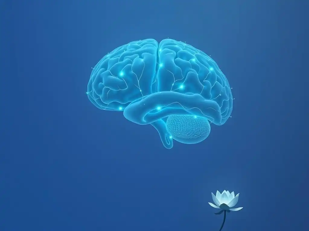
Neurotransmitters are chemical messengers that transmit signals between neurons in the brain. They play a critical role in regulating mood, sleep, appetite, energy levels, and cognitive function. Imbalances or dysregulation in the activity of certain neurotransmitters are widely believed to be central to the pathophysiology of bipolar disorder, contributing significantly to the altered bipolar disorder brain function. While no single neurotransmitter is solely responsible, research has focused on the roles of dopamine, serotonin, glutamate, and GABA.
Dopamine Dysregulation in Bipolar Disorder
Dopamine is a key neurotransmitter involved in reward, motivation, pleasure, and motor control. It is a central player in the brain's reward pathway, originating in the ventral tegmental area (VTA) and projecting to areas like the nucleus accumbens and prefrontal cortex. Dopamine signaling is strongly implicated in the energetic and euphoric aspects of mania.
Research, though complex and not always consistent, often points towards increased dopamine activity or sensitivity during manic episodes. This heightened dopamine signaling is thought to drive the increased energy, goal-directed behavior, euphoria, and impulsivity characteristic of mania. The brain's reward system appears to be operating in overdrive.
Elevated Dopamine Levels During Mania
While directly measuring neurotransmitter levels in the living human brain is challenging, studies using techniques like PET scans have provided insights into dopamine system activity. Some studies suggest increased dopamine synthesis, release, or receptor sensitivity in key brain regions, particularly the striatum, during manic episodes. This surge in dopamine activity is hypothesized to fuel the manic state, promoting intense feelings of reward, motivation, and energy.
The precise mechanisms are still being elucidated, but the prevailing hypothesis is that a functional excess of dopamine contributes significantly to the manic phase of bipolar disorder. This aligns with the fact that drugs that increase dopamine activity can sometimes trigger manic symptoms, while many effective treatments for mania target dopamine receptors.
Dopamine's Role in Reward and Motivation
Dopamine's primary role in the brain's reward and motivation system makes its dysregulation particularly relevant to bipolar disorder. In healthy individuals, dopamine release is associated with pleasurable experiences and reinforces behaviors that lead to rewards. This system is essential for learning and goal pursuit.
In bipolar disorder, the exaggerated or blunted responses within the dopamine reward pathway are thought to underpin the extreme shifts in motivation and pleasure across mood states. During mania, the reward system may be hypersensitive to potential rewards, driving excessive goal-directed behavior and risk-taking. During depression, a hypoactive dopamine system could contribute to anhedonia and lack of motivation. Understanding this dopamine dysregulation is key to understanding the altered bipolar disorder brain function.
Serotonin Imbalances in Bipolar Disorder
Serotonin is another crucial neurotransmitter involved in regulating mood, sleep, appetite, and impulse control. It has widespread projections throughout the brain, influencing many different functions. Serotonin dysfunction has long been associated with mood disorders, particularly depression, and is also implicated in the complex picture of bipolar disorder.
While often linked to depression, serotonin's role in bipolar disorder is more nuanced than simply being "low." Its interaction with other neurotransmitter systems and its influence on mood stability are critical. Abnormalities in serotonin transporter function or receptor sensitivity are areas of active research in understanding bipolar disorder brain function.
Reduced Serotonin Levels and Depression
During the depressive phase of bipolar disorder, research suggests that abnormalities in serotonin signaling, potentially involving reduced availability or function, may contribute to symptoms like persistent sadness, loss of interest, sleep disturbances, and changes in appetite. This aligns with the serotonin hypothesis of depression, although it's recognized that other systems are also involved.
It's important to note that simply increasing serotonin may not be sufficient or appropriate for treating bipolar depression, as antidepressants that primarily target serotonin can sometimes trigger manic or hypomanic episodes in vulnerable individuals. This highlights the complex interplay of neurotransmitters in bipolar disorder.
Serotonin's Role in Mood Regulation and Impulse Control
Beyond its link to depressive symptoms, serotonin is also involved in modulating mood stability and impulse control. Dysregulation in serotonin pathways could contribute to the rapid or extreme shifts in mood seen in some forms of bipolar disorder. Furthermore, serotonin helps regulate inhibitory control, and dysfunction in this system could play a role in the impulsivity observed during manic episodes.
The intricate balance between serotonin and other neurotransmitters, particularly dopamine and norepinephrine, is thought to be essential for maintaining mood stability. Imbalances in this complex network, rather than a simple deficiency or excess of a single chemical, likely contribute to the fluctuating mood states and altered bipolar disorder brain function.
Glutamate and GABA: The Excitatory-Inhibitory Balance in Bipolar Disorder
Glutamate is the brain's primary excitatory neurotransmitter, meaning it increases the likelihood that a neuron will fire. GABA (gamma-aminobutyric acid) is the primary inhibitory neurotransmitter, decreasing the likelihood of neuronal firing. The balance between glutamate and GABA is critical for maintaining healthy brain activity, regulating neuronal excitability, and facilitating processes like learning and memory. Disruptions to this delicate excitatory-inhibitory balance are increasingly recognized as playing a significant role in the pathophysiology of bipolar disorder.
Research suggests altered levels or function of both glutamate and GABA in various brain regions implicated in bipolar disorder, including the prefrontal cortex, hippocampus, and basal ganglia. These imbalances can lead to abnormal patterns of neural activity, potentially contributing to the extreme fluctuations in mood and energy levels. For example, excessive excitation or insufficient inhibition in certain circuits could contribute to the heightened activity seen in mania.
Glutamate's Role in Neural Excitation
Glutamate is involved in virtually all aspects of brain function, including synaptic plasticity, learning, and memory. However, excessive glutamate activity can be neurotoxic and lead to excitotoxicity, potentially contributing to neuronal damage or loss observed in some brain regions in bipolar disorder.
Studies using magnetic resonance spectroscopy (MRS) to measure brain metabolites have sometimes shown altered glutamate levels in specific brain areas in individuals with bipolar disorder. For instance, some findings suggest elevated glutamate or related metabolites in certain regions during mood episodes. This potential dysregulation of the brain's main excitatory system could contribute to the overall state of heightened neural activity hypothesized during mania.
GABA's Role in Neural Inhibition
GABA acts as the brain's main brake pedal, reducing neuronal excitability and counteracting the effects of excitatory neurotransmitters like glutamate. It plays a critical role in calming neural activity and maintaining stability. Deficits in GABAergic signaling could lead to excessive or uncontrolled firing of neurons.
MRS studies have also sometimes indicated altered GABA levels in individuals with bipolar disorder, with some findings suggesting reduced GABA concentrations in certain brain regions. A relative deficiency in inhibition, coupled with potential excess excitation from glutamate, could create an environment of neural instability that contributes to the rapid and extreme shifts in mood characteristic of the illness.
Imbalances in Glutamate and GABA and their relation to Bipolar Brain Function
The interplay between glutamate and GABA is fundamental to understanding the altered bipolar disorder brain function. A disruption in the normal balance between these two neurotransmitters can lead to hyperexcitability in certain brain circuits, potentially contributing to manic symptoms, or hypoactivity in others, contributing to depressive symptoms.
For example, an imbalance favoring excitation in limbic areas like the amygdala could contribute to emotional lability, while an imbalance in prefrontal-subcortical circuits could affect mood regulation and executive function. Many effective treatments for bipolar disorder, such as lithium and valproate, are known to influence glutamate and GABA systems, supporting the importance of this excitatory-inhibitory balance in managing the disorder.
Neuroimaging Studies and Bipolar Disorder: Visualizing Brain Activity
Neuroimaging techniques have revolutionized our understanding of the brain by allowing researchers to visualize its structure and function in living individuals. These tools have been instrumental in identifying key differences in the bipolar disorder brain function compared to healthy brains. By examining structural volumes, neural activity patterns, and connectivity between regions, neuroimaging provides crucial insights into the biological underpinnings of the disorder.
MRI (Magnetic Resonance Imaging) Studies in Bipolar Disorder
Magnetic Resonance Imaging (MRI) uses strong magnetic fields and radio waves to create detailed images of brain structures. Structural MRI allows researchers to measure the volume and shape of different brain regions, while functional MRI (fMRI) measures brain activity by detecting changes in blood flow. Both techniques have provided valuable, albeit sometimes variable, findings in bipolar disorder research.
Structural MRI studies have consistently revealed differences in brain volume in certain areas in individuals with bipolar disorder. While findings can vary depending on illness stage, medication status, and individual characteristics, patterns of altered volume in regions like the prefrontal cortex, hippocampus, and amygdala are frequently reported. fMRI studies have shed light on how these regions function and interact during different mood states and cognitive tasks.
Structural Brain Changes in Bipolar Patients
Numerous structural MRI studies have documented specific differences in the brains of individuals with bipolar disorder. As mentioned earlier, reduced gray matter volume in the prefrontal cortex and hippocampus is a relatively consistent finding, particularly in individuals with longer illness duration or multiple episodes. Some studies also report altered white matter integrity, which affects the connections between brain regions, potentially disrupting communication networks.
While these structural changes are often subtle on an individual level, they are statistically significant when examining groups of patients. They suggest that bipolar disorder is associated with tangible alterations in brain architecture, which likely contribute to the persistent cognitive and emotional dysregulation observed in the illness.
Functional Connectivity Differences
Functional MRI studies often focus on functional connectivity, which measures how correlated the activity is between different brain regions. This provides insights into how well different parts of the brain are communicating with each other. Studies in bipolar disorder have revealed altered functional connectivity within and between key brain networks.
For example, there is evidence of disrupted connectivity within the default mode network (active during rest), the salience network (involved in detecting important stimuli), and the executive control network (involved in planning and problem-solving). Furthermore, connectivity between limbic regions (like the amygdala) and regulatory regions (like the prefrontal cortex) is often found to be altered, potentially explaining the difficulty in regulating intense emotions. These connectivity abnormalities are a key aspect of understanding the complex network dysfunction in bipolar disorder brain function.
PET (Positron Emission Tomography) Studies in Bipolar Disorder
Positron Emission Tomography (PET) is a neuroimaging technique that uses radioactive tracers to visualize metabolic activity or the distribution of specific molecules, such as neurotransmitter receptors or transporters, in the brain. PET studies have been crucial for investigating brain chemistry and energy metabolism in bipolar disorder.
By using different tracers, PET scans can provide insights into aspects of bipolar disorder brain function that MRI cannot, such as regional glucose metabolism (an indicator of neural activity) or the density and availability of dopamine or serotonin transporters and receptors. These studies complement structural and functional MRI findings by providing a biochemical perspective.
Examining Neurotransmitter Activity
PET studies have been particularly valuable in examining the activity of neurotransmitter systems in bipolar disorder. For instance, studies using tracers that bind to dopamine receptors or transporters have provided some evidence suggesting altered dopamine signaling, particularly in the striatum, which may be more active during mania.
Similarly, PET studies investigating serotonin transporters or receptors have contributed to the understanding of serotonin system dysregulation in bipolar disorder, especially in relation to depressive symptoms. While findings can be complex and dependent on the specific tracer and brain region studied, PET helps build a picture of the neurochemical environment underlying mood episodes.
Identifying Metabolic Abnormalities
Beyond neurotransmitters, PET can also measure regional brain metabolism, typically using a tracer that tracks glucose uptake (FDG-PET). Areas of the brain that are more active consume more glucose. Studies using FDG-PET in bipolar disorder have sometimes shown altered metabolic activity in various brain regions depending on the mood state.
During manic episodes, some studies have reported increased metabolic activity in certain subcortical regions, consistent with a state of heightened energy and activity. During depressive episodes, decreased metabolic activity has sometimes been observed in areas like the prefrontal cortex. These metabolic findings provide another layer of evidence for widespread brain dysfunction in bipolar disorder.
EEG (Electroencephalography) Studies in Bipolar Disorder
Electroencephalography (EEG) is a non-invasive technique that measures the brain's electrical activity using electrodes placed on the scalp. EEG provides excellent temporal resolution, meaning it can capture rapid changes in brain activity, which fMRI cannot. Different brain states are associated with distinct patterns of electrical oscillations or brainwaves (e.g., alpha, beta, theta, delta).
EEG studies in bipolar disorder have investigated differences in brainwave patterns, event-related potentials (ERPs – brain responses to specific stimuli), and connectivity measures derived from electrical activity. These studies can help identify potential biomarkers for the disorder or specific mood states, contributing to a dynamic understanding of bipolar disorder brain function.
Analyzing Brainwave Patterns
EEG studies have reported various abnormalities in brainwave patterns in individuals with bipolar disorder, both during mood episodes and in remission. For example, some research suggests altered power or connectivity in specific frequency bands (like theta or alpha) in certain brain regions, such as the frontal lobes. These alterations might reflect underlying differences in neuronal network activity and communication.
Specific patterns might correlate with different mood states; for instance, certain EEG signatures could be more prominent during mania compared to depression. While EEG findings in bipolar disorder are still an area of active research and can be variable, they offer a window into the real-time electrical dynamics of the brain.
Identifying Biomarkers for Bipolar Disorder
One goal of EEG research in bipolar disorder is to identify neurophysiological biomarkers that could help in diagnosis, predicting treatment response, or tracking illness progression. ERPs, which measure the brain's electrical response to a specific event (like seeing a picture or hearing a sound), have shown promise in this regard. For example, altered amplitude or latency of specific ERP components related to emotional processing or cognitive control have been observed in bipolar disorder.
While no definitive EEG biomarker for bipolar disorder is currently used clinically, ongoing research aims to refine these measures. EEG's ability to capture rapid brain activity makes it a valuable tool for understanding the dynamic nature of altered neural circuits in the bipolar disorder brain function.
Genetic Factors and Bipolar Brain Function: The Heritability of Bipolar Disorder
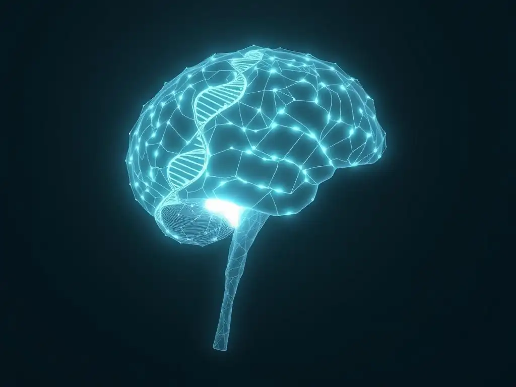
Bipolar disorder is a highly heritable condition, meaning genetic factors play a significant role in an individual's risk of developing the illness. Studies of families, twins, and adopted individuals consistently show a strong genetic component. For instance, the risk of developing bipolar disorder is significantly higher for first-degree relatives of individuals with the disorder (e.g., parents, siblings, children) compared to the general population. This strong genetic influence underscores that differences in bipolar disorder brain function are, to a significant extent, genetically predisposed.
However, it's crucial to understand that bipolar disorder is not caused by a single gene. It is a complex genetic disorder involving the interplay of multiple genes, each contributing a small effect to overall risk. Furthermore, environmental factors also play a critical role, interacting with genetic predispositions to influence whether and how the disorder manifests.
Identifying Genes Associated with Bipolar Disorder
Large-scale genetic studies, such as genome-wide association studies (GWAS), have identified several genes and genetic regions associated with an increased risk of bipolar disorder. These studies compare the genetic makeup of thousands of individuals with bipolar disorder to that of healthy controls to find common genetic variations (like single nucleotide polymorphisms, or SNPs) that occur more frequently in the affected group.
Genes implicated in bipolar disorder are often involved in brain function, including those related to:
- Neurotransmitter signaling (e.g., genes affecting dopamine, serotonin, or glutamate pathways).
- Ion channels (proteins that regulate the flow of ions in neurons, critical for electrical signaling).
- Intracellular signaling pathways (molecular cascades within neurons that respond to external signals).
- Circadian rhythms (the body's internal biological clock, often disrupted in bipolar disorder).
- Immune and inflammatory processes (increasingly linked to psychiatric disorders).
While these genes contribute to risk, no single gene variation is necessary or sufficient to cause bipolar disorder. The collective effect of many small genetic variations, along with environmental factors, determines an individual's susceptibility and influences the specific patterns of altered bipolar disorder brain function.
Gene-Environment Interactions in Bipolar Disorder
The genetic predisposition to bipolar disorder does not exist in isolation; it interacts with environmental factors to shape the final risk and presentation of the illness. This concept of gene-environment interaction is critical to understanding why some individuals with a genetic vulnerability develop the disorder while others do not.
Environmental factors that have been studied in relation to bipolar disorder risk include stressful life events (particularly early-life trauma or severe stress), substance use (like heavy cannabis use), and sleep disruption. For example, an individual carrying certain genetic variants might be more sensitive to the impact of stress on their mood or sleep patterns, and this heightened sensitivity, when combined with significant stress, could trigger a mood episode or the onset of the disorder.
These interactions highlight the complexity of the illness and how genetic vulnerabilities translate into altered bipolar disorder brain function within the context of a person's life experiences.
Treatment Strategies Targeting Brain Function in Bipolar Disorder
Treatment for bipolar disorder aims to stabilize mood, reduce the frequency and severity of episodes, and improve overall functioning and quality of life. Many effective treatments work by directly or indirectly targeting the neurobiological abnormalities underlying the disorder, aiming to restore more typical bipolar disorder brain function. This includes both pharmacological and non-pharmacological approaches.
Pharmacological Interventions for Bipolar Disorder
Medications are the cornerstone of bipolar disorder treatment, particularly for managing acute mood episodes and preventing recurrences. Different classes of medications target different aspects of brain chemistry and function.
Mood Stabilizers: Lithium and Valproate
Mood stabilizers are the primary class of medications used to treat and prevent mood episodes in bipolar disorder. Lithium is one of the oldest and most effective mood stabilizers, particularly for preventing mania and reducing suicide risk. Its exact mechanism of action is complex and not fully understood, but it is known to influence multiple neurotransmitter systems (including serotonin and glutamate), modulate intracellular signaling pathways, and potentially exert neuroprotective effects, helping to stabilize neural activity and protect against the potential neurotoxicity associated with repeated episodes.
Valproate (an anticonvulsant also used as a mood stabilizer) is effective for both manic and depressive episodes and is often used for rapid cycling bipolar disorder. Valproate is known to increase GABA levels in the brain, enhancing inhibitory neurotransmission. It also affects voltage-gated sodium channels and other pathways, contributing to its mood-stabilizing effects by dampening excessive neural activity and helping to re-establish a better excitatory-inhibitory balance, thereby normalizing aspects of bipolar disorder brain function.
Antipsychotics: Mechanism of Action on the Brain
Atypical antipsychotics are frequently used in the treatment of bipolar disorder, particularly for acute mania, mixed episodes, and as adjunctive therapy for bipolar depression and maintenance treatment. These medications block dopamine receptors (specifically D2 receptors) and often also affect serotonin receptors (particularly 5-HT2A receptors).
By blocking dopamine receptors, antipsychotics can help reduce the hyperactivity in dopamine pathways thought to contribute to manic symptoms like elevated energy, goal-directed behavior, and psychosis (if present). Their effects on serotonin and other systems may contribute to their mood-stabilizing properties and efficacy in bipolar depression. They help to modulate neural circuits involved in mood regulation, perception, and motivation, addressing key aspects of altered bipolar disorder brain function.
Antidepressants: Considerations for Use in Bipolar Disorder
Antidepressants are used with caution in bipolar disorder due to the risk of precipitating manic or hypomanic episodes (known as "switching"). When used, they are typically prescribed in combination with a mood stabilizer or antipsychotic to mitigate this risk. Antidepressants primarily target serotonin and/or norepinephrine systems, aiming to increase the availability of these neurotransmitters in the brain.
While they can be effective for treating the depressive phase, their use in bipolar disorder requires careful monitoring. The potential for switching highlights the underlying instability of neurotransmitter systems and mood circuits in the bipolar disorder brain function, where interventions aimed at one system can have cascading effects on the delicate balance between mood states.
Non-Pharmacological Treatments for Bipolar Disorder
In addition to medication, non-pharmacological treatments, particularly psychotherapies and neuromodulation techniques, play an important role in managing bipolar disorder. These approaches can help individuals cope with the disorder, improve functioning, and may also exert effects on brain function.
Cognitive Behavioral Therapy (CBT) and Neural Pathways
Cognitive Behavioral Therapy (CBT) is a psychotherapy that helps individuals identify and change negative thought patterns and behaviors. While it doesn't directly target neurotransmitter levels, CBT is believed to work by influencing neural pathways involved in emotional regulation, cognitive processing, and behavior.
Through techniques like challenging distorted thoughts, developing coping strategies, and practicing behavioral skills, CBT can strengthen the connections between the prefrontal cortex (involved in executive control and regulation) and limbic regions (like the amygdala, involved in emotion). Over time, this can help individuals better regulate their emotional responses and improve cognitive control, potentially leading to more stable patterns of bipolar disorder brain function.
Electroconvulsive Therapy (ECT) and its Impact on Brain Function
Electroconvulsive Therapy (ECT) is a highly effective treatment for severe, treatment-resistant depression and mania, particularly when rapid response is needed. ECT involves inducing a brief seizure under controlled medical conditions. While its precise mechanisms are not fully understood, ECT is known to cause widespread neurobiological changes in the brain.
ECT is thought to affect multiple neurotransmitter systems (including dopamine, serotonin, and glutamate), alter gene expression, increase neurotrophic factors (which support neuronal health and growth), and modify connectivity patterns within brain circuits. These changes appear to help reset or normalize dysfunctional neural activity and connectivity, making ECT a powerful intervention for severely disrupted bipolar disorder brain function.
Transcranial Magnetic Stimulation (TMS) and Neuromodulation
Transcranial Magnetic Stimulation (TMS) is a non-invasive neuromodulation technique that uses magnetic pulses to stimulate specific areas of the brain. It is an FDA-approved treatment for major depressive disorder and is being investigated for use in bipolar depression and other mood states. TMS typically targets the dorsolateral prefrontal cortex (DLPFC), an area known to be hypoactive in depression.
By delivering repetitive magnetic pulses to the DLPFC, TMS aims to modulate its activity and its connections to other brain regions involved in mood regulation, such as the limbic system. The goal is to restore more balanced activity within these circuits. While research on TMS in bipolar disorder is ongoing, it represents a promising approach to directly modulate specific brain regions implicated in altered bipolar disorder brain function.
The Future of Bipolar Disorder Research: Advances in Understanding the Bipolar Brain
Research into bipolar disorder brain function is continually advancing, driven by technological innovations in neuroimaging, genetics, and molecular biology. The future holds promise for a deeper understanding of the disorder's complex neurobiological basis, leading to more precise diagnostic tools and more effective, personalized treatments.
Personalized Medicine and Bipolar Disorder
One major goal in psychiatric research is the development of personalized medicine approaches. Given the significant variability in symptom presentation, course of illness, and treatment response among individuals with bipolar disorder, moving beyond a one-size-fits-all approach is critical. Future research aims to identify biological markers (biomarkers) that can predict who will respond best to specific treatments or who is at highest risk for certain outcomes.
This could involve using neuroimaging data to identify specific patterns of altered brain structure or function, genetic testing to identify relevant risk variants, or analyzing other biological measures. The hope is that by understanding the unique profile of bipolar disorder brain function in an individual, clinicians can select the most effective treatment from the outset, minimizing trial and error and improving outcomes.
Novel Therapeutic Targets for Bipolar Disorder
A deeper understanding of the neurobiological pathways involved in bipolar disorder is leading to the identification of novel targets for therapeutic intervention. Instead of solely relying on medications that broadly affect neurotransmitter systems, future treatments may target specific molecules or pathways that are dysregulated in the illness.
For example, research is exploring the potential of compounds that modulate glutamate receptors, target inflammatory pathways, or influence circadian rhythm genes. Advances in neuromodulation techniques are also opening up possibilities for more targeted interventions to specific brain circuits. Identifying these novel targets based on a sophisticated understanding of bipolar disorder brain function is key to developing more effective and perhaps better-tolerated treatments.
Understanding the Bipolar Brain: Longitudinal Studies and Early Intervention
Longitudinal studies, which follow individuals over extended periods, are crucial for understanding how bipolar disorder develops and progresses over time. These studies can track changes in brain structure, function, and chemistry from the early stages of the illness, or even before onset in high-risk individuals (e.g., children of parents with bipolar disorder).
By studying the brain over the course of the illness, researchers can gain insights into critical periods for intervention and identify early markers of risk or progression. This could pave the way for preventative strategies or early interventions aimed at mitigating the impact of the disorder on the developing brain and potentially altering the long-term trajectory of the illness. Understanding the dynamic nature of the bipolar disorder brain function throughout life is a key area of future research.
Frequently Asked Questions
What is the primary focus of understanding Bipolar Disorder in terms of brain function?
The primary focus lies in understanding the neurobiological basis of the mood swings. This involves examining how brain structures and neurotransmitters contribute to manic and depressive episodes. We want to know what is happening in the brain.
How do neurotransmitters like dopamine and serotonin relate to Bipolar Disorder?
Imbalances in neurotransmitters, particularly dopamine and serotonin, are thought to play a crucial role. During mania, dopamine levels may be elevated, while depression may be linked to decreased serotonin.
Which specific brain regions are implicated in Bipolar Disorder?
Research suggests that several brain regions are involved, including the prefrontal cortex (responsible for decision-making), the amygdala (emotional processing), and the hippocampus (memory).
Can brain imaging techniques help diagnose or understand Bipolar Disorder?
While not a definitive diagnostic tool, brain imaging techniques like MRI and fMRI can reveal structural and functional differences in the brains of individuals with Bipolar Disorder compared to those without. This can help understand underlying neurological differences.
Is Bipolar Disorder purely a result of brain function, or are other factors involved?
While Bipolar Disorder has a strong neurobiological component, it's understood to be a complex condition influenced by a combination of factors. These include genetics, environmental stressors, and life experiences.
Final Reflection: Unraveling the Bipolar Brain: A Journey Towards Understanding
The intricate dance between mania and depression in bipolar disorder is not merely a psychological struggle; it's a deeply rooted neurobiological reality. Understanding the complex interplay of brain regions and neurotransmitters is crucial not only for accurate diagnosis but also for developing more effective and personalized treatments. The implications of this research extend far beyond the individual, impacting families, communities, and the broader understanding of mental health.
The journey of exploring the bipolar brain has profoundly impacted my understanding of mental illness, shifting it from a purely symptomatic perspective to one that acknowledges the profound biological underpinnings. This exploration highlights the urgent need for continued research and destigmatization. The mind is its own place, and in itself can make a Heaven of Hell, a Hell of Heaven.
– John Milton, Paradise Lost. This quote, while not directly about bipolar disorder, speaks to the internal landscape of those experiencing this condition, emphasizing the subjective reality shaped by neurobiological processes. The ongoing research into the brain's intricate mechanisms offers hope for a future where this internal landscape can be better understood and navigated.
Now, consider your own understanding of mental health. How has this exploration of the bipolar brain challenged your preconceptions? What steps can you take, whether through education, advocacy, or simply increased empathy, to contribute to a more compassionate and informed understanding of bipolar disorder and other mental illnesses? Let us work together to build a future where the complexities of the brain are embraced, not feared, and where those living with bipolar disorder find support, understanding, and hope.
Unlock Your Understanding of Bipolar Disorder
Thanks for exploring the complex world of bipolar disorder with us! We hope this article provided valuable insights into the neurobiological mechanisms driving mania and depression. Now, it's time to take the next steps on your journey to understanding and supporting mental wellness.
- Deepen your knowledge: Explore reputable resources like the National Institute of Mental Health (NIMH) website or search for relevant documentaries that delve into the latest research on bipolar disorder. This will provide a broader perspective and allow you to engage with information beyond this article.
- Share your insights: Discuss your key takeaways from this article with friends, family, or online communities focused on mental health. Sharing knowledge helps reduce stigma and fosters support. Perhaps even start a conversation about the challenges and triumphs of managing bipolar disorder.
- Connect with support groups: Find a local or online support group for individuals with bipolar disorder or their loved ones. Connecting with others who understand can offer invaluable support and shared experiences.
What specific aspect of bipolar disorder resonated most with you, and how will you incorporate this new understanding into your life or approach to mental health?