
Beyond the Neuron: Imaging Glial Cells and Other Brain Components in Mental Health

Welcome to Mind Care Guide, a space dedicated to understanding and nurturing your mental well-being.
Feeling overwhelmed by the complexities of mental health? You're not alone. Recent research is revealing fascinating connections between brain structure and mental state. In this article, we delve beyond the neuron to explore the unsung heroes of the brain: glial cells. Could understanding the role of glial cells unlock new avenues for mental health support? Join us as we uncover the latest insights and explore how these often-overlooked cells might hold the key to a brighter, healthier future.
- Beyond the Neuron: Imaging Glial Cells and Other Brain Components in Mental Health
- Brain Imaging Techniques for Visualizing Glial Cells
- Imaging Other Brain Components Relevant to Mental Health
-
Glial Cells and Specific Mental Health Conditions: Imaging Insights
- Glial Cells in Depression: Imaging Evidence and Therapeutic Implications
- Glial Cells in Schizophrenia: Investigating Glial Cell Abnormalities with Brain Imaging
- Glial Cells in Anxiety Disorders: Exploring the Role of Glial Cells in Fear and Stress Responses
- Glial Cells in Neurodevelopmental Disorders: Imaging Studies in Autism and ADHD
- Future Directions in Glial Cell Imaging and Mental Health Research
- Glial Cell-Targeted Therapies and Mental Health Support
-
Frequently Asked Questions
- How are glial cells relevant to mental health?
- What imaging techniques are used to study glial cells in relation to mental health?
- Can glial cells be targeted therapeutically for mental health conditions?
- What other brain components, besides neurons and glial cells, are important for mental well-being?
- Where can I find more information and support related to glial cells and mental health?
- Final Reflection: Unveiling the Unsung Heroes of Mental Health
- Unveil the Secrets of Your Mind: Explore Brain Imaging Further
Beyond the Neuron: Imaging Glial Cells and Other Brain Components in Mental Health
The Importance of Glial Cells: More Than Just Support for Neurons
For decades, neurons were considered the primary functional units of the brain, responsible for transmitting information through electrical and chemical signals. Glial cells, often referred to collectively as "glia," were largely thought to play a passive, supportive role—providing structural integrity, insulation, and nutrient supply to the more dynamic neurons. However, this perspective has dramatically shifted with advancements in neuroscience research. We now understand that glial cells, which outnumber neurons in some brain regions, are active participants in complex brain functions, including synaptic plasticity, neurotransmission regulation, and immune responses within the central nervous system.
There are several main types of glial cells, each with distinct roles. Astrocytes, with their star-like shape, regulate blood flow, maintain the blood-brain barrier, control the chemical environment around synapses, and even participate in synaptic signaling. Oligodendrocytes in the central nervous system (and Schwann cells in the peripheral nervous system) produce myelin, the fatty sheath that insulates neuronal axons and allows for rapid transmission of electrical impulses.
Microglia are the brain's resident immune cells, constantly surveying the environment, clearing debris, and responding to injury or infection. These diverse functions highlight that glial cells are not mere bystanders but are integral to the healthy functioning of neural circuits, underpinning everything from basic cognition to complex emotional regulation.
How Glial Cells Contribute to Mental Well-being: A New Perspective
The active roles of glial cells extend directly to their contribution to mental well-being. Astrocytes, for instance, are crucial for maintaining the delicate balance of neurotransmitters in the synaptic cleft, including glutamate, the brain's main excitatory neurotransmitter. Dysregulation of glutamate signaling has been implicated in various mental health conditions. By buffering excess glutamate and converting it into glutamine for neuronal reuse, astrocytes help prevent excitotoxicity and ensure efficient communication. Furthermore, astrocytes influence synaptic strength and formation, processes vital for learning, memory, and adaptive behavior, all of which are foundational to positive mental health.
Microglia, as the brain's immune sentinels, play a complex role. In their resting state, they survey the environment, pruning unnecessary synapses during development and clearing cellular waste. However, when activated by injury, infection, or stress, microglia can release inflammatory molecules (cytokines).
While short-term inflammation is protective, chronic microglial activation and excessive inflammation are increasingly linked to the pathophysiology of conditions like depression, anxiety disorders, and neurodevelopmental differences. Conversely, healthy microglial function, including appropriate synaptic pruning and debris clearance, is essential for maintaining optimal brain connectivity and resilience, thereby supporting mental well-being. Understanding these cellular mechanisms is key to developing new strategies for mental health support.
Glial Cell Dysfunction: A Potential Factor in Mental Health Disorders
Emerging evidence strongly suggests that dysfunction or abnormalities in glial cells are not merely consequences of mental illness but may actively contribute to their development and progression. Studies have found alterations in the density, morphology, and function of astrocytes in post-mortem brain samples from individuals with major depressive disorder and schizophrenia. For example, a decrease in the number of astrocytes, particularly in the prefrontal cortex and hippocampus—brain regions critical for mood regulation and cognitive function—has been a consistent finding in some depression studies, sometimes showing a reduction of 15-20% in specific subregions compared to healthy controls.
Oligodendrocyte dysfunction and resulting myelin abnormalities (dysmyelination or demyelination) are also being investigated in the context of mental health disorders, particularly schizophrenia and bipolar disorder. Myelin is essential for efficient signal transmission along neural pathways. Disruptions in myelin integrity can impair connectivity between different brain regions, potentially contributing to the cognitive and emotional symptoms characteristic of these conditions.
Microglial overactivation, leading to chronic neuroinflammation, has been observed in brain imaging studies of individuals experiencing active symptoms of depression, anxiety, and psychosis, pointing towards their role in exacerbating neuronal dysfunction and contributing to the overall disease burden. Research into glial cells mental health support is exploring how targeting these dysfunctions might offer therapeutic avenues.
Brain Imaging Techniques for Visualizing Glial Cells
Introduction to Advanced Brain Imaging Technologies
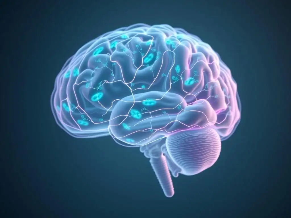
Advanced brain imaging technologies have revolutionized our ability to study the living human brain non-invasively. Techniques like Magnetic Resonance Imaging (MRI), Positron Emission Tomography (PET), and various forms of microscopy allow researchers and clinicians to visualize brain structure, function, and even cellular processes.
While standard structural MRI provides detailed anatomical images and functional MRI (fMRI) measures neural activity by detecting changes in blood flow, these techniques primarily focus on neuronal activity or macroscopic brain regions. However, the increasing recognition of the critical roles of glial cells has spurred the development and refinement of imaging methods specifically capable of probing glial biology.
These advanced imaging modalities offer unique windows into the complex cellular landscape of the brain beyond just neurons. They allow for the detection of specific molecules, the assessment of metabolic activity, the visualization of tissue microstructure, and even the tracking of inflammatory processes mediated by glial cells.
The challenge lies in developing techniques sensitive enough to differentiate glial signals from neuronal signals and to detect subtle changes in glial cell populations or their activity in the context of mental health conditions. The ongoing refinement of these methods is crucial for advancing our understanding of how glial cells contribute to mental well-being and dysfunction.
Specific Imaging Modalities for Glial Cell Research
Magnetic Resonance Spectroscopy (MRS) for Glial Cell Metabolite Analysis
Magnetic Resonance Spectroscopy (MRS) is a non-invasive imaging technique that uses the principles of nuclear magnetic resonance to detect and quantify specific metabolites in a localized brain region. Unlike standard MRI, which provides anatomical images, MRS provides biochemical information. Certain metabolites detected by MRS are predominantly localized within glial cells, making this technique valuable for assessing glial health and function in vivo.
Key glial-specific metabolites include myo-inositol, which is found primarily in astrocytes and can be an indicator of astrocytic health or proliferation. N-acetylaspartate (NAA) is predominantly neuronal, but its ratio relative to glial markers can provide insights into neuronal-glial interactions. Creatine (Cr) and Choline (Cho) are present in both neurons and glia but changes in their ratios or absolute concentrations can reflect alterations in cellular density, membrane turnover, or energy metabolism, processes significantly influenced by glial cells.
For instance, elevated myo-inositol levels have been observed in certain brain regions in conditions like multiple sclerosis and depression, potentially reflecting astrogliosis (an increase in the number or size of astrocytes) or altered astrocytic function. While not a direct count of cells, MRS provides crucial biochemical markers that reflect the metabolic state and potential dysfunction of glial populations in specific brain areas.
Positron Emission Tomography (PET) Imaging of Glial Cell Activity
Positron Emission Tomography (PET) is a nuclear imaging technique that uses small amounts of radioactive tracers (radiotracers) to visualize and measure metabolic processes, blood flow, neurotransmitter activity, or the binding of specific molecules in the brain. PET is particularly powerful for studying glial cells because researchers can design radiotracers that selectively bind to molecules expressed primarily by specific glial subtypes, such as microglia or astrocytes.
One widely used PET tracer for studying neuroinflammation, often mediated by activated microglia, is [11C]PK11195 or its second-generation analogues like [18F]FECT or [18F]DPA-714. These tracers bind to the 18 kDa translocator protein (TSPO), which is upregulated in activated microglia and astrocytes. Increased binding of TSPO tracers in specific brain regions, such as the prefrontal cortex or amygdala, has been reported in studies of individuals with depression, anxiety disorders, and schizophrenia, suggesting a role for neuroinflammation mediated by activated glial cells in these conditions.
For example, some studies have shown a 10-30% increase in TSPO binding potential in certain brain areas of depressed patients compared to healthy controls. PET imaging with TSPO tracers provides a measure of the overall inflammatory state and microglial/astrocyte activation, offering critical insights into the neurobiological underpinnings of mental health challenges and potential targets for mental health support interventions.
Advanced Microscopy Techniques for Glial Cell Morphology and Function
While MRS and PET provide insights into glial biochemistry and activity in vivo (in living individuals), advanced microscopy techniques are essential for visualizing the detailed morphology, distribution, and cellular interactions of glial cells at high resolution, typically in post-mortem tissue or animal models. Techniques like confocal microscopy and two-photon microscopy allow for detailed visualization of individual glial cells, their intricate processes, and their relationship with neurons and synapses.
Electron microscopy offers even higher resolution, enabling the study of ultrastructural details, such as the ensheathment of axons by myelin produced by oligodendrocytes or Schwann cells, or the fine processes of astrocytes wrapping around synapses. Immunofluorescence and immunohistochemistry techniques, often used in conjunction with microscopy, employ antibodies to label specific proteins expressed by different glial cell types (e.g., GFAP for astrocytes, Iba1 for microglia, MBP for myelin) or markers of their activation state or function. These techniques have revealed profound changes in glial morphology and distribution in brain tissue from individuals with mental health conditions, such as reduced astrocytic complexity in depression or altered microglial morphology in schizophrenia. While these are not in vivo human imaging techniques, the detailed cellular insights they provide are crucial for interpreting findings from non-invasive imaging and for understanding the cellular mechanisms underlying mental health disorders, informing potential strategies for glial cells mental health support.
Imaging Other Brain Components Relevant to Mental Health
Vascular Imaging: Assessing Brain Blood Flow and its Impact on Mental Health
Beyond neurons and glial cells, the brain's vascular system plays a critical role in maintaining brain health and function, with direct implications for mental well-being. Brain imaging techniques such as Arterial Spin Labeling (ASL) MRI and dynamic susceptibility contrast (DSC) MRI allow for the non-invasive measurement of regional cerebral blood flow (CBF). Adequate blood flow is essential to supply neurons and glial cells with oxygen and glucose and to remove metabolic waste products.
Alterations in brain blood flow have been observed in various mental health conditions. For instance, reduced blood flow in areas like the prefrontal cortex and hippocampus is a common finding in studies of depression and anxiety disorders. These areas are crucial for mood regulation, executive function, and memory.
Conversely, hyperfrontality (increased blood flow in frontal regions) has sometimes been noted in obsessive-compulsive disorder. Vascular health is intrinsically linked to overall brain resilience, and compromised blood flow can impair neuronal and glial function, contributing to cognitive deficits and mood disturbances. Imaging the brain's vasculature provides an important piece of the puzzle in understanding the complex interplay of factors contributing to mental health challenges.
White Matter Imaging: Exploring the Role of Myelin and Glial Cells in Neural Connectivity
White matter, composed primarily of myelinated axons, forms the communication highways of the brain, connecting different grey matter regions. The integrity of white matter pathways is essential for efficient information processing and coordinated brain function. Diffusion Tensor Imaging (DTI) is an MRI technique that measures the diffusion of water molecules in brain tissue.
Because water diffuses more freely along the direction of myelinated axons than across them, DTI can infer the orientation and integrity of white matter tracts. Metrics derived from DTI, such as fractional anisotropy (FA) and mean diffusivity (MD), are sensitive to changes in myelination, axonal integrity, and tissue microstructure.
Since oligodendrocytes are the glial cells responsible for producing myelin in the central nervous system, DTI findings often reflect the health and function of these crucial glial populations. Abnormalities in white matter integrity, as revealed by DTI (e.g., reduced FA or increased MD), have been consistently reported in studies of schizophrenia, bipolar disorder, depression, and neurodevelopmental disorders like autism spectrum disorder.
These findings suggest that disruptions in the brain's connectivity architecture, influenced by oligodendrocyte health and myelin status, contribute significantly to the symptoms experienced in these conditions. Exploring white matter integrity through imaging is vital for understanding how impaired communication between brain regions impacts mental health.
Neuroinflammation Imaging: Targeting Microglia and Astrocyte Activation
Neuroinflammation, the immune response within the brain, is increasingly recognized as a key player in the development and progression of many mental health disorders. While microglia are the primary mediators of this response, astrocytes also contribute significantly. Imaging techniques specifically targeting markers of neuroinflammation allow researchers to directly visualize and quantify the extent and location of this process in vivo. As mentioned earlier, PET imaging with TSPO tracers is a leading method for detecting activated microglia and reactive astrocytes.
Beyond TSPO PET, other emerging imaging strategies are being developed to provide more specific information about different aspects of neuroinflammation. For example, some PET tracers are being explored to target specific cytokine receptors or enzymes involved in inflammatory pathways. Additionally, advanced MRI techniques sensitive to tissue microstructure changes related to inflammation are under investigation.
Findings from neuroinflammation imaging studies have provided compelling evidence linking elevated levels of glial activation, particularly in limbic and frontal brain regions, to the severity of symptoms in depression, anxiety, and psychosis. This growing body of evidence underscores the importance of addressing neuroinflammation as a potential therapeutic target and highlights the value of imaging in monitoring glial responses and exploring strategies for glial cells mental health support.
Glial Cells and Specific Mental Health Conditions: Imaging Insights
Glial Cells in Depression: Imaging Evidence and Therapeutic Implications
Major depressive disorder (MDD) is a debilitating condition characterized by persistent sadness, loss of interest, and cognitive difficulties. Imaging studies investigating glial cells in depression have provided significant insights. MRS studies have frequently reported altered levels of glial metabolites, such as reduced NAA/Cr ratios (suggesting neuronal or glial dysfunction) and sometimes elevated myo-inositol (potentially indicating astrogliosis) in areas like the prefrontal cortex and hippocampus of depressed individuals. For example, a meta-analysis of MRS studies in MDD found consistent evidence for decreased NAA in the anterior cingulate cortex, a region implicated in emotional regulation.
PET studies using TSPO tracers have often shown increased binding in various brain regions of individuals with active depression, including the anterior cingulate cortex, insula, and prefrontal cortex, suggesting heightened microglial and/or astrocytic activation. Some studies report a 10-25% increase in TSPO binding in specific regions compared to healthy controls.
These imaging findings, corroborated by post-mortem studies showing reduced astrocyte density and altered glial morphology, strongly suggest that glial cell dysfunction and neuroinflammation contribute to the pathophysiology of depression. Understanding these glial alterations through imaging is crucial for developing novel antidepressant strategies, potentially involving targets aimed at modulating glial activity or promoting glial health, thereby improving mental health support for those affected.
Glial Cells in Schizophrenia: Investigating Glial Cell Abnormalities with Brain Imaging
Schizophrenia is a complex psychiatric disorder characterized by symptoms such as hallucinations, delusions, disorganized thinking, and cognitive deficits. Imaging research has pointed towards significant glial involvement in this condition. White matter abnormalities, often assessed using DTI, are consistently reported in schizophrenia, particularly in pathways connecting frontal and temporal brain regions. Reduced fractional anisotropy (FA) in specific white matter tracts, indicative of impaired myelin integrity or axonal packing, is a common finding, potentially reflecting oligodendrocyte dysfunction. Some studies report FA reductions of 5-15% in specific white matter bundles in patients compared to controls.
PET studies investigating neuroinflammation in schizophrenia using TSPO tracers have yielded mixed results, with some studies reporting increased binding in certain brain regions, particularly during acute psychotic episodes, while others find no significant differences or even reduced binding in chronic patients. However, post-mortem studies in schizophrenia have provided more consistent evidence of glial abnormalities, including altered astrocyte morphology, reduced oligodendrocyte numbers or function, and abnormal microglial distribution or activation states.
These converging lines of evidence from imaging and histology suggest that disruptions in glial-neuronal interactions, particularly involving myelination and neuroinflammation, play a role in the complex neural circuit dysfunction underlying schizophrenia, highlighting potential targets for future mental health support interventions.
Glial Cells in Anxiety Disorders: Exploring the Role of Glial Cells in Fear and Stress Responses
Anxiety disorders, such as generalized anxiety disorder, panic disorder, and social anxiety disorder, are characterized by excessive fear and worry. The amygdala, a brain region central to processing fear and emotional responses, is heavily implicated in anxiety. Imaging studies are beginning to explore the role of glial cells in the neural circuits underlying anxiety. PET studies using TSPO tracers have sometimes shown increased binding in the amygdala and other limbic regions in individuals with anxiety disorders or those exposed to stress, suggesting microglial or astrocytic activation in these fear-related circuits.
While direct imaging of glial numbers or specific functions in anxiety in vivo is still an active area of research, preclinical studies have provided strong evidence for glial involvement. For example, studies in rodents have shown that manipulating microglial activity in the amygdala can alter fear responses.
Astrocytes in the amygdala also play a role in regulating synaptic transmission in circuits involved in anxiety. Imaging techniques that can specifically probe glial activity and interactions within these critical emotional processing regions in humans are crucial for understanding the neurobiological basis of anxiety and developing more effective strategies for mental health support.
Glial Cells in Neurodevelopmental Disorders: Imaging Studies in Autism and ADHD
Neurodevelopmental disorders, such as Autism Spectrum Disorder (ASD) and Attention-Deficit/Hyperactivity Disorder (ADHD), emerge early in life and are characterized by altered brain development and connectivity. Glial cells, particularly microglia and oligodendrocytes, play critical roles during brain development, including synaptic pruning, myelination, and shaping neural circuits. Dysfunction in these developmental processes mediated by glia is hypothesized to contribute to the atypical connectivity and cognitive profiles seen in these disorders.
Imaging studies in ASD using techniques like DTI have frequently reported abnormalities in white matter integrity, suggesting altered myelination or connectivity in specific brain pathways, such as those involved in social processing and language. These findings point towards potential oligodendrocyte involvement. PET studies investigating neuroinflammation in ASD have sometimes shown increased TSPO binding, particularly in younger individuals, suggesting altered microglial activity during critical developmental periods.
In ADHD, DTI studies have also revealed altered white matter integrity in pathways connecting areas involved in attention and executive function. While direct in vivo imaging of specific glial functions in these disorders is challenging, these findings, combined with extensive preclinical research, underscore the importance of glial cells in proper brain development and their potential contribution to neurodevelopmental differences, highlighting the need for tailored mental health support approaches.
Future Directions in Glial Cell Imaging and Mental Health Research
Emerging Imaging Technologies for Enhanced Glial Cell Visualization
The field of brain imaging for glial cell research is rapidly advancing, driven by the need for more specific and sensitive methods to visualize different glial subtypes and their dynamic functions in vivo. While TSPO PET has been valuable for assessing inflammation, it lacks specificity for distinguishing between activated microglia and reactive astrocytes. Future developments include the creation of novel PET radiotracers that target molecules more selectively expressed by specific glial populations (e.g., markers for specific astrocyte transporters or microglial receptors) or indicators of specific glial functions (e.g., synaptic pruning activity).
Beyond PET, advancements in MRI technology are also promising. Techniques like diffusion kurtosis imaging (DKI) and neurite orientation dispersion and density imaging (NODDI) can provide more detailed information about tissue microstructure than conventional DTI, potentially offering better sensitivity to subtle changes in glial morphology or density in vivo.
High-field MRI scanners (7T and above) offer increased resolution and signal-to-noise ratio, potentially allowing for the detection of smaller brain structures and more subtle metabolic changes related to glial activity. Combining these advanced MRI techniques with MRS or novel PET tracers holds immense potential for providing a multi-modal view of glial health and function in mental health conditions, opening new avenues for targeted mental health support.
Translational Research: Using Imaging to Develop Glial Cell-Targeted Therapies
The insights gained from imaging glial cells in mental health disorders are crucial for translational research—the process of moving findings from basic science into clinical applications. By identifying specific glial abnormalities or patterns of glial activation associated with particular symptoms or conditions, imaging can help identify potential therapeutic targets. For instance, if PET imaging consistently shows elevated microglial activation in a specific brain region linked to depressive symptoms, it suggests that therapies aimed at reducing neuroinflammation or modulating microglial activity in that region might be beneficial.
Brain imaging can also be used to track the effectiveness of novel glial-targeted therapies in clinical trials. For example, if a new drug is designed to reduce astrocyte reactivity, MRS could potentially be used to monitor changes in astrocytic metabolites, or PET could track changes in TSPO binding, to see if the drug is having its intended effect on glial populations in living patients.
This use of imaging as a biomarker is critical for evaluating treatment response and optimizing dosages. Translational research leveraging glial imaging is a vital step towards developing more precise and effective treatments and improving mental health support by addressing the cellular roots of illness.
The Potential of Glial Cell Imaging for Personalized Mental Health Treatment
One of the major goals in mental health care is to move towards personalized or precision medicine, tailoring treatments to the individual patient's specific biological profile. Glial cell imaging holds significant potential in this regard. Different individuals with the same diagnostic label (e.g., major depressive disorder) may have different underlying neurobiological profiles; some may have prominent neuroinflammation, while others may have more significant white matter abnormalities or astrocytic dysfunction.
By using advanced imaging techniques to assess the specific types and patterns of glial alterations in an individual patient's brain, clinicians in the future might be able to select the most appropriate treatment strategy. For example, a patient showing high levels of microglial activation on a PET scan might benefit more from an anti-inflammatory intervention, while a patient with significant white matter abnormalities revealed by DTI might be a better candidate for therapies aimed at promoting remyelination or improving connectivity.
Glial cell imaging could potentially serve as a diagnostic aid or a predictor of treatment response, helping to match patients with the therapies most likely to be effective for their specific neurobiological profile. This personalized approach, informed by imaging insights into glial cells, represents a promising future direction for delivering more effective and tailored mental health support.
Glial Cell-Targeted Therapies and Mental Health Support
Pharmacological and Non-Pharmacological Strategies Targeting Glial Cells for Mental Health
The growing understanding of glial cells' roles in mental health has spurred interest in developing therapies that directly target these cells. Pharmacological approaches are exploring compounds that can modulate glial activity. For instance, some drugs aim to reduce excessive microglial activation and subsequent neuroinflammation, which might be beneficial in conditions like depression or schizophrenia where inflammation is implicated.
Other strategies focus on promoting the health and function of astrocytes or oligodendrocytes, perhaps by enhancing their ability to clear neurotransmitters, produce neurotrophic factors, or support myelination. Several compounds currently in preclinical or early clinical trials are designed to specifically interact with glial receptors or pathways.
Beyond pharmacological interventions, non-pharmacological strategies are also being investigated for their potential impact on glial cells. Exercise, for example, has been shown in preclinical studies to modulate microglial activity and promote the production of neurotrophic factors by astrocytes, contributing to its known benefits for mood and cognitive function. Dietary interventions and nutritional supplements are also being explored for their anti-inflammatory properties, which could indirectly impact glial activation.
Even psychological therapies might influence glial function through their effects on stress hormones and neural activity patterns, although this area requires more research. The development of therapies that explicitly consider glial biology represents a significant shift, offering new avenues for providing mental health support by addressing fundamental cellular processes in the brain.
Resources and Support Systems for Individuals Affected by Mental Health Conditions and Glial Cell Research
Understanding the complex biological factors, including glial cell dysfunction, that contribute to mental health conditions can be both informative and overwhelming. For individuals affected by these conditions, accessing appropriate resources and support systems is paramount. Mental health support services include therapy (such as cognitive behavioral therapy, dialectical behavior therapy), medication management, support groups, and crisis hotlines. Organizations dedicated to specific conditions like depression, anxiety, schizophrenia, or autism offer valuable information, advocacy, and connection to local resources.
While glial cell research is still largely in the realm of basic science and early clinical translation, staying informed about advancements can offer hope and empower individuals. Reputable mental health organizations and research institutions often publish summaries of new findings in accessible language. For those interested in participating in research, clinical trial databases list studies investigating new treatments, some of which may be exploring glial targets.
Ultimately, effective mental health support involves a multi-faceted approach that includes evidence-based treatments, a strong support network, and staying informed about the evolving scientific understanding of the brain, including the crucial roles played by glial cells. The journey towards better mental health is ongoing, and advancements in understanding glial cell mental health support are contributing vital pieces to the puzzle.
Frequently Asked Questions
How are glial cells relevant to mental health?
Glial cells, often overlooked, play a critical role in brain function by supporting neurons. They are now recognized as significantly impacting mental health disorders, influencing things like neuroinflammation and synaptic plasticity.
What imaging techniques are used to study glial cells in relation to mental health?
Advanced imaging techniques, such as in vivo microscopy and PET scans, allow researchers to visualize glial cell activity and distribution in the brain. These methods help understand how glial dysfunction contributes to mental illness.
Can glial cells be targeted therapeutically for mental health conditions?
Yes, research is exploring therapeutic strategies that target glial cells to reduce neuroinflammation or enhance neuronal support. This represents a promising avenue for developing new treatments for various mental health conditions.
What other brain components, besides neurons and glial cells, are important for mental well-being?
The vascular system is vital for providing oxygen and nutrients. Also, the extracellular matrix plays a role in neuronal communication. Both are important for brain health and mental well-being.
Resources like Mind Care Guide provide comprehensive information on mental well-being. Look for research articles and consult with mental health professionals to learn more about the latest advancements in this field.
Final Reflection: Unveiling the Unsung Heroes of Mental Health
The intricate dance of neurons in the brain has long captivated researchers, but a deeper understanding of mental health requires us to look beyond the neuron—to the glial cells and other supporting components that are equally crucial to its well-being. Ignoring these vital players is akin to studying a symphony by focusing solely on the violins; the richness and complexity of the whole are lost. The implications of this oversight are profound, particularly as we seek more effective treatments for debilitating mental illnesses.
The emerging field of glial cell research is revolutionizing our understanding of mental health conditions. For too long, the focus has been narrowly defined, neglecting the significant contributions of these often-overlooked cells. Their role in modulating neuronal activity, shaping synaptic plasticity, and orchestrating immune responses within the brain is becoming increasingly clear. The brain is not just a collection of neurons; it's a complex ecosystem where glial cells play a critical, often underestimated, role.
This quote encapsulates the shift in perspective that is urgently needed, one that recognizes the interconnectedness of all brain components in maintaining mental well-being.
Consider the potential: What if we could develop targeted therapies that address the dysfunction of glial cells in conditions like depression, anxiety, and schizophrenia? What if we could harness the power of these cells to promote neuroplasticity and resilience? You, the reader, are now equipped with a deeper understanding of this exciting frontier. I urge you to continue exploring this field, to advocate for further research, and to champion a more holistic approach to mental health care that embraces the full complexity of the brain. The future of mental health treatment may well depend on it.
Unveil the Secrets of Your Mind: Explore Brain Imaging Further
Thanks for taking the time to delve into the fascinating world of brain imaging and mental health! We hope you gained a deeper understanding of the crucial role glial cells and other brain components play. Now it's time to take that knowledge further.
- Dive deeper into research: Search for relevant studies on glial cell imaging and its applications in mental health research. This will allow you to stay updated on breakthroughs in the field and further your understanding.
- Share your insights: Discuss this article with friends or family, sparking conversations about the complexities of the brain and mental well-being. Let's break down the stigma together!
- Consider your own brain health: Reflect on how this information might inform your approach to mental wellness. What steps might you take to support your own brain health based on this new knowledge?
Ready to explore the intricate workings of the mind even further? What specific area of brain imaging research piques your interest the most, and why?