
Hippocampus and Mood Disorders: Memory, Stress, and Neurogenesis

Welcome to Mind Care Guide, your compassionate companion on the journey to mental well-being.
Feeling overwhelmed by stress and its impact on your memory and mood? You're not alone. This article delves into the fascinating connection between the hippocampus, stress, and mood disorders, exploring how these intricate pieces of your brain influence your emotional landscape. Discover the vital role of neurogenesis and learn how understanding this relationship can unlock pathways to healing and resilience. Ready to uncover the science and find hope? Let's begin.
Hippocampus and Mood Disorders: Memory, Stress, and Neurogenesis - Mind Care Guide
- Introduction: The Hippocampus - A Key Player in Mood Regulation
- The Hippocampus: Structure, Function, and Memory
- Stress and the Hippocampus: A Detrimental Relationship
- Neurogenesis in the Adult Hippocampus: The Birth of New Neurons
- Hippocampal Volume and Mood Disorders: Evidence from Research
- The Hippocampus, Stress Response, and Mood Dysregulation
- The Role of Neuroinflammation in Hippocampal Dysfunction and Mood Disorders
- Therapeutic Interventions Targeting the Hippocampus to Improve Mood
- Lifestyle Modifications to Protect Hippocampal Health and Improve Mood
- Future Directions in Research: Understanding the Hippocampus and Mood Disorders
- Conclusion: The Hippocampus - A Vital Target for Mood Disorder Treatment and Prevention
-
Frequently Asked Questions
- How does the hippocampus relate to mood disorders?
- What role does stress play in hippocampal function and mood disorders?
- What is neurogenesis and why is it important for mood regulation?
- Can damage or dysfunction of the hippocampus lead to mood disorders?
- What are some potential therapeutic interventions targeting the hippocampus for mood disorders?
- Final Reflection: The Hippocampus: A Bridge Between Memory, Stress, and Mood
- Unlock Your Brain's Potential: Boosting Mood & Memory
Introduction: The Hippocampus - A Key Player in Mood Regulation
Understanding the Role of the Hippocampus in Mental Well-being
The hippocampus, a seahorse-shaped structure nestled deep within the temporal lobe of the brain, is not merely a passive component of our neural architecture. It plays a profoundly active role in shaping our mental landscape, particularly concerning memory formation, spatial navigation, and emotional regulation.
Emerging research consistently highlights the hippocampus's critical involvement in various psychiatric conditions, especially mood disorders like major depressive disorder (MDD) and bipolar disorder. Its intricate connections with other brain regions, including the amygdala (involved in processing emotions) and the prefrontal cortex (involved in decision-making and executive functions), position it as a central hub influencing how we perceive, process, and respond to our internal and external environments.
Understanding the hippocampus's functions provides crucial insights into the biological underpinnings of emotional dysregulation. For instance, its role in contextual memory allows us to differentiate between safe and threatening situations, a function often impaired in anxiety and mood disorders. Furthermore, the hippocampus is one of the few brain regions where neurogenesis – the birth of new neurons – continues throughout adulthood.
This remarkable plasticity is thought to be vital for learning, memory, and potentially, emotional resilience. Disruptions to this process, whether due to chronic stress or other factors, are increasingly linked to the onset and severity of mood-related conditions, underscoring the importance of maintaining hippocampal health for overall mental well-being.
Overview: Connecting Hippocampal Function to Mood Disorders
The intricate relationship between the hippocampus and mood disorders is a significant area of neuropsychiatric research. Studies have repeatedly demonstrated structural and functional abnormalities in the hippocampus of individuals diagnosed with conditions such as depression and bipolar disorder. A prominent finding is the consistent observation of reduced hippocampal volume in many patients, particularly those with a longer duration or greater severity of illness. This atrophy is not merely an incidental finding but is hypothesized to contribute directly to the cognitive and emotional symptoms characteristic of these disorders, including difficulties with memory, concentration, and emotional processing.
Furthermore, the hippocampus is highly susceptible to the detrimental effects of stress hormones, particularly cortisol, which are often elevated in individuals experiencing chronic stress or mood episodes. This vulnerability links the hippocampus, stress, and mood disorders in a complex interplay. Chronic exposure to high levels of cortisol can impair neurogenesis, reduce neuronal survival, and disrupt synaptic plasticity within the hippocampus, potentially leading to the observed volume reductions and functional impairments. Exploring this connection offers promising avenues for developing more effective treatments that not only target symptoms but also address the underlying neurobiological changes associated with these challenging conditions.
The Hippocampus: Structure, Function, and Memory
Anatomy of the Hippocampus: A Detailed Look
The hippocampus is a bilateral structure, meaning there is one in each hemisphere of the brain. Located within the medial temporal lobe, it is part of the limbic system, a network traditionally associated with emotion, motivation, and memory. Its distinctive curved shape, resembling a seahorse (from the Greek "hippokampos"), is formed by tightly packed layers of neurons.
Key subregions include the dentate gyrus (DG), the CA fields (CA1, CA2, CA3), and the subiculum. Information typically flows through the hippocampus via a unidirectional pathway known as the trisynaptic circuit: from the entorhinal cortex to the dentate gyrus, then to CA3, followed by CA1, and finally exiting via the subiculum back to the entorhinal cortex and other brain regions.
Each subregion plays a specialized role. The dentate gyrus is the primary site of adult neurogenesis and is involved in pattern separation, helping to distinguish similar experiences from one another. The CA3 region receives input from the DG and is crucial for pattern completion, allowing the retrieval of an entire memory from a partial cue.
CA1 is a major output region, receiving information from CA3 and projecting to the subiculum and other areas; it is particularly important for encoding new information. The subiculum serves as the main interface between the hippocampus and other cortical and subcortical structures. This intricate anatomical organization underlies the hippocampus's complex functions in memory processing and its susceptibility to various forms of insult, including those related to stress and mood disorders.
The Hippocampus and Memory Formation: Encoding, Consolidation, and Retrieval
The hippocampus is absolutely fundamental to the process of forming new long-term memories, particularly episodic and semantic memories. This process involves several stages: encoding, consolidation, and retrieval. Encoding is the initial learning phase where information is first processed and transformed into a storable format. The hippocampus, especially the CA1 region, is critically involved in rapidly encoding novel experiences. For example, remembering what you ate for breakfast this morning or the details of a recent conversation relies heavily on hippocampal encoding.
Following encoding, consolidation occurs, a process where labile new memories are stabilized and gradually transferred to other cortical regions for long-term storage. The hippocampus plays a crucial, though temporary, role in this phase. It acts as an index, linking together disparate pieces of information stored in different cortical areas that together constitute a complete memory. Over time, the connections between cortical areas strengthen, making the memory less dependent on the hippocampus.
Retrieval is the process of accessing stored information. While retrieval of fully consolidated memories often involves cortical areas, the hippocampus is essential for retrieving detailed, contextual episodic memories, especially newer ones. Damage to the hippocampus, such as from a stroke or chronic stress, can severely impair the ability to form new memories (anterograde amnesia) and recall recent events.
Beyond episodic and semantic memory, the hippocampus is renowned for its vital role in spatial memory and navigation. This function is particularly well-studied, famously illustrated by the discovery of "place cells" in the hippocampus of rodents and later in humans. Place cells are neurons that fire selectively when an animal is in a particular location within an environment, effectively creating a cognitive map of the surroundings. The coordinated activity of many place cells allows the brain to represent spatial locations and navigate efficiently.
The hippocampus's involvement in spatial navigation is critical for tasks ranging from finding your way home to remembering where you parked your car. Studies involving London taxi drivers, who undergo extensive training to navigate the city's complex street network, have shown that they possess larger posterior hippocampi compared to control groups, and the size correlates with the number of years spent as a taxi driver.
This provides compelling evidence of the hippocampus's structural plasticity in response to intense spatial learning demands. Impairments in spatial memory are sometimes observed in neurological conditions affecting the hippocampus, such as Alzheimer's disease, and can also be affected by the same factors that impact other hippocampal functions, including chronic stress and mood disorders.
Stress and the Hippocampus: A Detrimental Relationship
How Chronic Stress Impacts Hippocampal Structure and Function
Chronic stress is a significant adversary to hippocampal health. When we experience stress, the body releases stress hormones, primarily cortisol (in humans) and corticosterone (in rodents), via the hypothalamic-pituitary-adrenal (HPA) axis. While acute, short-term stress can sometimes enhance memory consolidation by activating the hippocampus, prolonged exposure to high levels of these glucocorticoids has profoundly negative effects. Chronic stress leads to a cascade of molecular and cellular changes within the hippocampus, including reduced dendritic branching and spine density in hippocampal neurons, which are essential for synaptic communication.
Furthermore, chronic stress suppresses adult hippocampal neurogenesis, the birth of new neurons, primarily in the dentate gyrus. This reduction in new neurons impairs the hippocampus's ability to adapt and integrate new information, potentially contributing to difficulties in learning and memory observed during periods of prolonged stress. Animal studies have shown that exposure to chronic unpredictable stress can lead to a significant decrease in hippocampal volume, sometimes by as much as 10-15% in specific subregions, mirroring findings seen in humans with chronic stress-related conditions and mood disorders. This structural remodeling directly impairs hippocampal function, affecting memory retrieval and emotional regulation.
Cortisol and Hippocampal Neuronal Damage: The Stress Hormone's Effects
Cortisol, the primary stress hormone, exerts its effects on the hippocampus through glucocorticoid receptors (GRs) and mineralocorticoid receptors (MRs), which are abundantly expressed in this brain region. While MRs are involved in the initial stress response and maintaining baseline activity, GRs mediate the feedback inhibition of the HPA axis, helping to shut down the stress response once the threat has passed. However, under conditions of chronic stress, persistently high levels of cortisol overactivate GRs in the hippocampus.
This prolonged GR activation is neurotoxic. It can lead to decreased glucose uptake by hippocampal neurons, reduced expression of neurotrophic factors like Brain-Derived Neurotrophic Factor (BDNF) which are vital for neuronal survival and plasticity, and increased vulnerability to excitotoxicity (damage caused by excessive stimulation by neurotransmitters like glutamate).
Studies have shown that prolonged exposure to high cortisol can even cause retraction of dendrites and loss of synapses, effectively shrinking the neural network within the hippocampus. This cellular damage contributes directly to the observed reductions in hippocampal volume and the associated cognitive and emotional impairments seen in conditions linked to chronic stress and affecting the hippocampus stress mood disorders axis.
Stress-Induced Memory Impairment: The Hippocampus at Risk
One of the most prominent symptoms of chronic stress is difficulty with memory and concentration. This stress-induced cognitive impairment is largely mediated by the detrimental effects of stress hormones on the hippocampus. The hippocampus's critical role in encoding and retrieving new memories makes it particularly vulnerable. High levels of cortisol can interfere with synaptic plasticity mechanisms, such as long-term potentiation (LTP), which are considered the cellular basis of learning and memory. This interference makes it harder to form new memories and efficiently retrieve existing ones.
Individuals experiencing chronic stress often report subjective memory problems, such as forgetfulness or difficulty focusing. Objective cognitive tests in populations exposed to prolonged stress, such as caregivers of individuals with chronic illness or individuals with PTSD, often show impaired performance on tasks specifically involving the hippocampus, such as verbal and spatial memory tests.
For instance, studies on individuals with Cushing's disease, a condition characterized by excessive cortisol production, frequently reveal significant memory deficits and reduced hippocampal volume, further solidifying the link between elevated cortisol, hippocampal damage, and cognitive decline. This hippocampal vulnerability to stress highlights why managing stress is crucial for preserving cognitive function and overall brain health.
Neurogenesis in the Adult Hippocampus: The Birth of New Neurons
The Importance of Adult Hippocampal Neurogenesis (AHN)
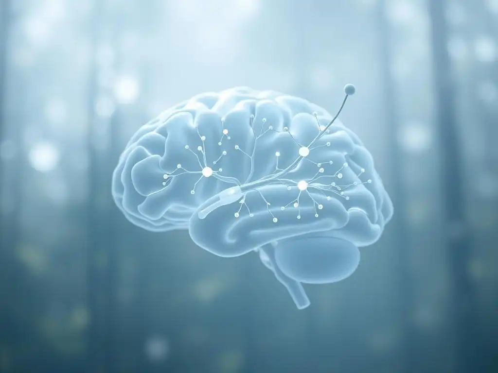
Adult hippocampal neurogenesis (AHN) refers to the fascinating process by which new neurons are generated from neural stem cells in the subgranular zone of the dentate gyrus throughout life. While the rate decreases with age, it persists in humans. These newly born neurons migrate into the granule cell layer, differentiate, integrate into existing neural circuits, and become functional. AHN is not just a biological curiosity; it is believed to play a crucial role in several hippocampal-dependent functions.
AHN is thought to be particularly important for pattern separation, enabling the hippocampus to distinguish between similar experiences and encode them as distinct memories, thereby preventing interference. This function is critical for detailed and precise memory formation. Furthermore, AHN has been strongly implicated in mood regulation and the behavioral effects of antidepressants.
Animal studies consistently show that factors known to improve mood and cognitive function, such as exercise, enriched environments, and antidepressant treatment, increase AHN, while factors that impair mood and cognitive function, such as chronic stress and depression, suppress it. This suggests that AHN may be a key mechanism linking environmental factors and therapeutic interventions to changes in mood and cognitive flexibility, especially in the context of hippocampus stress mood disorders research.
Factors Influencing Neurogenesis: Lifestyle, Environment, and Medications
The rate of adult hippocampal neurogenesis is remarkably sensitive to a wide array of internal and external factors. Positive factors that promote AHN include physical exercise (especially aerobic activity), exposure to enriched environments (environments that are novel, stimulating, and provide opportunities for learning and social interaction), caloric restriction or intermittent fasting, and learning new tasks. For example, running wheels in rodent studies consistently increase the proliferation and survival of new neurons.
Conversely, several factors are known to suppress AHN. These include chronic stress, sleep deprivation, aging, inflammation, high-fat diets, and exposure to certain toxins or drugs of abuse. Chronic stress, as mentioned earlier, is a powerful suppressor of neurogenesis, largely mediated by glucocorticoids. Interestingly, many effective treatments for mood disorders, such as antidepressant medications (SSRIs, SNRIs) and electroconvulsive therapy (ECT), have been shown to increase AHN in animal models, leading to the hypothesis that stimulating neurogenesis might be one way these therapies exert their beneficial effects on mood and cognitive function.
Neurogenesis and Hippocampal Plasticity: Adapting to Change
Adult hippocampal neurogenesis is a prime example of brain plasticity – the brain's ability to change and adapt throughout life. The integration of new neurons into existing hippocampal circuits adds to the structural and functional flexibility of this region. This plasticity is essential for adapting to new experiences, learning, and potentially for maintaining mental resilience in the face of challenges. The ability of the hippocampus to generate and integrate new neurons allows for the continuous updating and refinement of neural circuits involved in memory, spatial navigation, and emotional processing.
Disruptions to this plasticity, particularly the suppression of neurogenesis, are hypothesized to contribute to the pathophysiology of mood disorders. A less plastic hippocampus, less capable of generating new neurons and adapting its circuitry, might be less effective at processing emotional information, discriminating between safe and threatening contexts, or encoding new, positive experiences. This reduced plasticity could contribute to persistent negative mood states and cognitive deficits. Therefore, interventions aimed at promoting neurogenesis and restoring hippocampal plasticity are being explored as potential therapeutic strategies for mood disorders.
Hippocampal Volume and Mood Disorders: Evidence from Research
Reduced Hippocampal Volume in Depression: A Consistent Finding
One of the most replicated structural findings in neuroimaging studies of major depressive disorder (MDD) is a reduction in hippocampal volume. Numerous MRI studies, including large meta-analyses, have consistently reported that individuals with depression tend to have smaller hippocampi compared to healthy control subjects. While the magnitude varies across studies, pooled data from meta-analyses suggest an average volume reduction of approximately 10-15% in patients with recurrent or chronic depression. This reduction is often more pronounced in individuals with a longer history of illness or greater symptom severity.
This observation is significant because it suggests a structural correlate to the functional and cognitive impairments often seen in depression, such as memory problems, difficulty concentrating, and emotional dysregulation. While it's debated whether the reduced volume is a cause or consequence of depression, evidence suggests it is likely a complex interplay. Chronic stress, a known risk factor for depression, is strongly implicated in driving this hippocampal atrophy through mechanisms involving cortisol and suppressed neurogenesis. Understanding this structural change is crucial for comprehending the neurobiological basis of depression and developing targeted interventions.
Hippocampal Atrophy in Bipolar Disorder: Exploring the Link
Similar to depression, studies investigating brain structure in bipolar disorder (BD) have also frequently reported abnormalities in hippocampal volume. While findings in BD can be somewhat more variable than in MDD, meta-analyses and large-scale imaging studies generally indicate that individuals with bipolar disorder, particularly those with a longer illness duration or multiple mood episodes (manic or depressive), also exhibit reduced hippocampal volume compared to healthy individuals. Some studies suggest the reduction might be more pronounced in certain subregions or might differ slightly in pattern compared to MDD, though significant overlap exists.
The link between hippocampal atrophy and bipolar disorder is complex, potentially involving genetic vulnerabilities, stress sensitivity, and recurrent mood episodes. Both depressive and manic episodes can be highly stressful events for the brain. The repeated cycling between extreme mood states, often accompanied by dysregulation of the HPA axis, could contribute to the cumulative impact on hippocampal structure. Research continues to explore whether specific aspects of bipolar disorder, such as the number or type of episodes, or medication effects, differentially influence hippocampal volume, adding layers to our understanding of the hippocampus stress mood disorders connection in this population.
The Severity of Mood Disorder and Hippocampal Size: A Correlation?
Research findings generally suggest a correlation between the severity and duration of mood disorders and the degree of hippocampal volume reduction. Studies have shown that individuals with more severe depressive symptoms, those who have experienced more depressive episodes, or those with a longer duration of illness tend to exhibit greater reductions in hippocampal size compared to those with milder or more recent onset depression. This dose-response relationship supports the idea that the cumulative burden of the illness, including exposure to stress and repeated mood episodes, contributes to structural changes in the hippocampus.
For example, a study might report that for every additional year a person has experienced depression, their hippocampal volume is, on average, slightly smaller. While this correlation does not definitively prove causality, it strongly suggests that the pathological processes underlying chronic or severe mood disorders have a tangible impact on hippocampal structure. This correlation reinforces the importance of early intervention and effective management of mood disorders to potentially mitigate or prevent progressive changes in brain structure and function, thereby protecting the hippocampus stress mood disorders network.
The Hippocampus, Stress Response, and Mood Dysregulation
The HPA Axis and the Hippocampus: A Feedback Loop
The hypothalamic-pituitary-adrenal (HPA) axis is the body's central stress response system. It involves a chain of communication starting in the hypothalamus, which releases corticotropin-releasing hormone (CRH), stimulating the pituitary gland to release adrenocorticotropic hormone (ACTH). ACTH then travels to the adrenal glands, prompting the release of cortisol. The hippocampus plays a crucial role in regulating this axis through a negative feedback loop. It has a high density of glucocorticoid receptors, which bind to circulating cortisol.
When cortisol levels rise, the hippocampus detects this and sends inhibitory signals to the hypothalamus and pituitary gland, effectively dampening the release of CRH and ACTH, and thus reducing cortisol production. This feedback mechanism is essential for returning the body to a state of homeostasis after a stressful event. However, in chronic stress or mood disorders, this feedback loop can become impaired.
If the hippocampus is damaged or its GR function is compromised, it becomes less effective at inhibiting the HPA axis, leading to prolonged elevation of cortisol levels. This dysfunction of the hippocampal-HPA axis feedback contributes significantly to the neuroendocrine abnormalities observed in depression and anxiety disorders.
Hippocampal Dysfunction and Emotional Regulation
Beyond its role in memory, the hippocampus is increasingly recognized for its contribution to emotional regulation. It interacts extensively with the amygdala, which processes the emotional salience of stimuli, and the prefrontal cortex, involved in higher-level cognitive control of emotions. The hippocampus provides contextual information that helps modulate amygdala activity – for instance, differentiating between a safe context where a previously threatening stimulus is no longer dangerous versus a truly threatening context.
In mood disorders, hippocampal dysfunction can impair this contextual processing. This might manifest as difficulty distinguishing between relevant and irrelevant emotional information, leading to exaggerated or prolonged emotional responses. For example, a compromised hippocampus might struggle to provide the context that a past negative event is over and no longer a present threat, contributing to persistent rumination or anxiety. This impaired ability to use contextual cues to regulate emotional responses is a key aspect of emotional dysregulation seen in conditions affecting the hippocampus stress mood disorders circuit.
Impaired Contextual Fear Discrimination in PTSD
A compelling example of hippocampal dysfunction in emotional processing is observed in Post-Traumatic Stress Disorder (PTSD). While PTSD is often characterized by an overactive amygdala, research also highlights significant impairments in hippocampal function. Individuals with PTSD often show reduced hippocampal volume and deficits in tasks requiring contextual processing, particularly contextual fear discrimination.
Contextual fear discrimination is the ability to respond fearfully to a specific threatening context but not to a similar, safe context. Studies have shown that veterans with combat-related PTSD, for instance, exhibit impaired performance on tasks requiring them to differentiate between a safe environment and a threatening one, even if cues are similar. This deficit is strongly linked to hippocampal dysfunction.
The hippocampus's failure to properly process the context of a traumatic event, or to differentiate between the traumatic context and safe, similar contexts, can contribute to the intrusive memories, hypervigilance, and exaggerated fear responses characteristic of PTSD. This specific impairment underscores the hippocampus's vital role in contextualizing emotional memories and regulating fear responses.
The Role of Neuroinflammation in Hippocampal Dysfunction and Mood Disorders
Inflammation and Reduced Neurogenesis in Mood Disorders
Neuroinflammation, the activation of the brain's immune system, is increasingly recognized as a significant factor in the pathophysiology of mood disorders. Studies have shown elevated levels of pro-inflammatory cytokines (signaling molecules like IL-6, TNF-alpha, and CRP) in the blood and cerebrospinal fluid of individuals with depression and bipolar disorder, particularly during acute episodes. This systemic inflammation can translate into inflammation within the brain, affecting vulnerable regions like the hippocampus.
Neuroinflammation has been shown to directly impair adult hippocampal neurogenesis. Pro-inflammatory cytokines can inhibit the proliferation and survival of neural stem cells and interfere with the differentiation and integration of new neurons. For example, studies administering inflammatory stimuli to rodents consistently report a reduction in hippocampal neurogenesis. This inflammation-induced suppression of neurogenesis provides a potential link between systemic inflammation, hippocampal dysfunction, and the development or maintenance of mood symptoms, adding another layer to the complex interplay of hippocampus stress mood disorders.
Inflammatory Markers and Hippocampal Damage
Evidence suggests that elevated inflammatory markers are associated not only with reduced neurogenesis but also with structural changes in the hippocampus. Chronic exposure to inflammatory signals can contribute to oxidative stress and excitotoxicity, processes known to be detrimental to neuronal health and survival. This can lead to synaptic dysfunction, neuronal damage, and ultimately, a reduction in hippocampal volume.
Studies have correlated higher levels of inflammatory markers like C-reactive protein (CRP) with smaller hippocampal volumes in populations at risk for or diagnosed with mood disorders. For instance, a study might find that individuals with elevated CRP levels have, on average, slightly smaller hippocampi compared to those with normal CRP levels, even after accounting for other factors. While the precise mechanisms are still being elucidated, it is clear that neuroinflammation plays a role in damaging the hippocampus, contributing to the structural and functional deficits observed in mood disorders.
Microglial Activation and Synaptic Pruning in Depression
Microglia are the primary immune cells of the brain. In a healthy state, they perform surveillance and support functions, including pruning unnecessary synapses to refine neural circuits. However, in conditions involving neuroinflammation, microglia become activated, shifting to a pro-inflammatory state. Activated microglia release inflammatory cytokines and other potentially neurotoxic molecules.
In the context of depression, post-mortem studies and PET imaging studies have provided evidence of microglial activation in brain regions including the hippocampus. This chronic activation can lead to excessive or inappropriate synaptic pruning, where microglia mistakenly eliminate healthy synapses. Loss of synapses in the hippocampus can impair its function in memory and emotional regulation. Furthermore, activated microglia can impair neurogenesis and contribute to neuronal dysfunction. This aberrant microglial activity and subsequent synaptic changes in the hippocampus are considered key contributors to the neurobiological changes underlying the hippocampus stress mood disorders connection.
Therapeutic Interventions Targeting the Hippocampus to Improve Mood
Antidepressants and Hippocampal Neurogenesis: Restoring Brain Health
Many conventional antidepressant medications, particularly selective serotonin reuptake inhibitors (SSRIs) and serotonin-norepinephrine reuptake inhibitors (SNRIs), have been shown in preclinical studies to promote adult hippocampal neurogenesis. This effect is often observed after several weeks of treatment, which aligns with the typical delay in the onset of clinical antidepressant effects. The hypothesis is that by increasing levels of serotonin and norepinephrine, these medications indirectly enhance the proliferation, survival, and integration of new neurons in the dentate gyrus.
While directly measuring neurogenesis in living humans is challenging, indirect evidence supports this link. Studies using advanced imaging techniques have shown that successful antidepressant treatment can be associated with increases in hippocampal volume or functional changes indicative of improved hippocampal health.
For example, a clinical trial might show that patients who respond well to a specific antidepressant exhibit a modest increase in hippocampal volume over several months of treatment, whereas non-responders do not. This suggests that promoting neurogenesis or restoring hippocampal plasticity might be a key mechanism by which these drugs alleviate symptoms of depression and anxiety, supporting the idea that targeting the hippocampus stress mood disorders link is therapeutically beneficial.
Exercise and Hippocampal Volume: Boosting Brain Power
Physical exercise is a powerful intervention that has consistently demonstrated beneficial effects on hippocampal health and function. Numerous studies, both in animals and humans, have shown that regular aerobic exercise can increase hippocampal volume, particularly in older adults. For instance, a landmark study showed that one year of moderate aerobic exercise resulted in a 2% increase in hippocampal volume in older adults, while a control group showed a decrease.
Exercise promotes hippocampal health through multiple mechanisms, including increasing blood flow, enhancing the production of neurotrophic factors like BDNF (which supports neuronal survival and growth), and stimulating adult hippocampal neurogenesis. These effects contribute to improved memory function, enhanced cognitive performance, and reduced symptoms of depression and anxiety. Incorporating regular physical activity is a widely recommended lifestyle modification for supporting brain health and improving mood, directly impacting the hippocampus stress mood disorders axis.
Cognitive Behavioral Therapy (CBT) and Hippocampal Activity: Re-training the Brain
Cognitive Behavioral Therapy (CBT) is a widely effective psychotherapy for mood and anxiety disorders. While often viewed purely as a psychological intervention, CBT also produces measurable changes in brain function and structure. Studies using fMRI have shown that successful CBT treatment for depression or anxiety is associated with altered activity patterns in brain regions including the prefrontal cortex, amygdala, and hippocampus.
Specifically, CBT is thought to help individuals reframe negative thought patterns and develop more adaptive coping strategies. This process involves learning and memory components that rely on hippocampal function. By teaching individuals to reappraise stressful situations or to engage in behaviors that provide positive reinforcement, CBT may indirectly influence hippocampal activity and plasticity.
For example, a study might show that after a course of successful CBT, patients exhibit normalized activity in the hippocampus during emotional processing tasks compared to before treatment. This suggests that "re-training" the brain through cognitive and behavioral techniques can positively impact the very neural circuits, including the hippocampus, that are disrupted in mood disorders.
Transcranial Magnetic Stimulation (TMS) and Hippocampal Modulation
Transcranial Magnetic Stimulation (TMS) is a non-invasive brain stimulation technique that uses magnetic pulses to stimulate specific areas of the brain. It is an approved treatment for major depressive disorder, typically targeting the prefrontal cortex. However, given the interconnectedness of brain regions, TMS applied to the prefrontal cortex can indirectly influence activity in deeper structures like the hippocampus.
Research suggests that TMS may exert its therapeutic effects, in part, by modulating hippocampal activity and potentially promoting neuroplasticity. Studies using concurrent TMS and fMRI have shown that prefrontal TMS can alter connectivity patterns between the stimulated site and the hippocampus. While the direct mechanisms are still being explored, it is hypothesized that by normalizing activity in prefrontal-limbic circuits, TMS can help restore healthy hippocampal function and reduce the detrimental effects of stress and mood dysregulation on this region. This provides another example of how interventions targeting brain circuitry can impact the hippocampus stress mood disorders pathology.
Lifestyle Modifications to Protect Hippocampal Health and Improve Mood
Diet and Nutrition for a Healthy Hippocampus
The food we eat has a significant impact on brain health, including the hippocampus. A diet rich in omega-3 fatty acids, found in oily fish like salmon and flaxseeds, is particularly beneficial. Omega-3s are essential components of cell membranes and play a role in synaptic function and reducing inflammation, both of which support hippocampal health. Studies have linked higher intake of omega-3s to larger hippocampal volumes and better cognitive function.
Antioxidant-rich foods, such as berries, leafy greens, and nuts, help protect brain cells from damage caused by oxidative stress, which can negatively impact the hippocampus. Following dietary patterns like the Mediterranean diet, which emphasizes fruits, vegetables, whole grains, lean proteins, and healthy fats, has been associated with better cognitive aging and potentially larger hippocampal volumes. Conversely, diets high in saturated fats and refined sugars are linked to inflammation and impaired cognitive function, potentially harming the hippocampus. Prioritizing nutrient-dense foods is a practical step for supporting hippocampal health and resilience against the impacts of stress and mood disorders.
Sleep Hygiene and Hippocampal Function
Adequate and quality sleep is crucial for optimal hippocampal function, particularly for memory consolidation. During sleep, especially slow-wave sleep, the hippocampus is thought to replay recent experiences, transferring information to the cortex for long-term storage. Chronic sleep deprivation disrupts this process, impairing memory formation and retrieval.
Poor sleep hygiene and chronic sleep deprivation have been linked to reduced hippocampal volume and increased vulnerability to the effects of stress. Establishing a regular sleep schedule, creating a relaxing bedtime routine, ensuring a dark and quiet sleep environment, and avoiding stimulants before bed are all components of good sleep hygiene that can help protect hippocampal function and support overall mental well-being, mitigating aspects of the hippocampus stress mood disorders cycle. Aiming for 7-9 hours of quality sleep per night is a key factor in maintaining a healthy hippocampus.
Stress Management Techniques: Protecting the Hippocampus from Damage
Given the detrimental effects of chronic stress on the hippocampus, implementing effective stress management techniques is paramount for protecting this vital brain structure and improving mood. Techniques such as mindfulness meditation, deep breathing exercises, yoga, and progressive muscle relaxation can help activate the body's relaxation response, counteracting the effects of the stress hormone cortisol.
Regular practice of these techniques has been associated with reduced stress levels and positive changes in brain structure and function, including potentially preserving or even increasing hippocampal volume. For example, studies on long-term meditators have shown larger hippocampal volumes compared to non-meditators. Engaging in enjoyable hobbies, spending time in nature, maintaining social connections, and setting realistic boundaries are also effective ways to reduce chronic stress and support hippocampal resilience against the impacts of the hippocampus stress mood disorders axis. Proactive stress management is a powerful tool for preserving cognitive function and emotional health.
Future Directions in Research: Understanding the Hippocampus and Mood Disorders
Novel Treatments Targeting Hippocampal Neurogenesis
Research into the hippocampus stress mood disorders connection is driving the search for novel therapeutic approaches that directly target hippocampal neurogenesis and plasticity. This includes developing drugs that specifically promote the proliferation, survival, or integration of new neurons, potentially bypassing some of the limitations of current antidepressants.
One area of focus is exploring compounds that enhance the expression of neurotrophic factors like BDNF or modulate signaling pathways known to regulate neurogenesis. Another avenue involves investigating the potential of growth factors or stem cell therapies, although these are still largely in preclinical stages for mood disorders. The goal is to develop treatments that not only alleviate symptoms but also promote structural and functional repair within the hippocampus, offering a more fundamental approach to treating mood disorders.
Advanced Imaging Techniques for Studying the Hippocampus
Advances in neuroimaging technology are providing unprecedented insights into the structure and function of the hippocampus in mood disorders. Techniques such as high-resolution structural MRI allow for more precise measurement of hippocampal subregions and their volumes. Diffusion Tensor Imaging (DTI) can assess the integrity of white matter tracts connecting the hippocampus to other brain areas, revealing potential disruptions in neural networks.
Functional MRI (fMRI) enables researchers to study hippocampal activity and connectivity patterns during various cognitive and emotional tasks, highlighting functional abnormalities associated with mood disorders. PET imaging is being used to visualize specific molecular targets, such as inflammatory markers or receptors, within the hippocampus. These advanced tools are crucial for better understanding the subtle changes occurring in the hippocampus in conditions involving hippocampus stress mood disorders and for tracking the effects of interventions.
Investigating the Role of Gut Microbiome in Hippocampal Function and Mood
An exciting and rapidly developing area of research is the investigation of the gut-brain axis, specifically the influence of the gut microbiome on brain health and mood. The trillions of microorganisms residing in our gut communicate with the brain through various pathways, including the vagus nerve, immune system, and production of metabolites.
Emerging research suggests that the composition and diversity of the gut microbiome can impact hippocampal function and neurogenesis. Dysbiosis (an imbalance in gut bacteria) has been linked to increased inflammation and altered stress responses, both of which can negatively affect the hippocampus.
Studies in animals have shown that manipulating the gut microbiome can influence hippocampal neurogenesis and anxiety/depressive-like behaviors. While research in humans is still in early stages, it is hypothesized that targeting the gut microbiome through diet, probiotics, or prebiotics could offer novel strategies for protecting hippocampal health and improving mood, adding a fascinating dimension to the hippocampus stress mood disorders puzzle.
Conclusion: The Hippocampus - A Vital Target for Mood Disorder Treatment and Prevention
Summarizing the Importance of Hippocampal Health for Mental Well-being
The hippocampus stands out as a critical brain structure intricately linked to memory, stress response, and mood regulation. Its vulnerability to chronic stress, coupled with its unique capacity for adult neurogenesis, makes it a key player in the development and persistence of mood disorders like depression and bipolar disorder. Research consistently demonstrates that structural and functional impairments in the hippocampus, including reduced volume and impaired neurogenesis, are associated with the severity and course of these conditions. Understanding the complex interplay between the hippocampus, stress, and mood disorders is fundamental to unraveling the neurobiological basis of these challenging illnesses.
From its role in contextualizing memories that influence emotional responses to its involvement in the HPA axis feedback loop, the hippocampus serves as a central hub in the brain's emotional circuitry. Disruptions here can lead to a cascade of problems, including difficulties with emotional regulation, impaired cognitive function, and heightened vulnerability to stress. Recognizing the hippocampus as a vital component in mental well-being underscores the importance of therapies and lifestyle choices that support its health and plasticity.
Empowering Individuals to Protect Their Hippocampus and Improve Mood.
Given the significant impact of the hippocampus on mood and cognitive function, empowering individuals with knowledge about how to protect and support its health is crucial. While genetic predispositions and life events play a role, lifestyle factors offer tangible ways to build resilience and promote hippocampal plasticity. Embracing strategies known to benefit the hippocampus stress mood disorders axis – such as regular exercise, a nutrient-rich diet, effective stress management techniques, and prioritizing quality sleep – can have a profound positive impact.
Furthermore, seeking evidence-based treatments like psychotherapy (e.g., CBT) and, when necessary, antidepressant medications, can also positively influence hippocampal function and potentially promote recovery. By understanding the vital connection between lifestyle, stress, the hippocampus, and mood, individuals can take proactive steps to enhance their brain health, build resilience against stress, and work towards improved emotional well-being. The science is clear: nurturing your hippocampus is an investment in your mental health.
Frequently Asked Questions
How does the hippocampus relate to mood disorders?
The hippocampus plays a crucial role in mood regulation due to its involvement in memory processing and stress response. Disruptions in its function are often observed in individuals experiencing mood disorders.
What role does stress play in hippocampal function and mood disorders?
Chronic stress can significantly impact the hippocampus, leading to reduced neurogenesis (the creation of new neurons) and potentially contributing to the development or exacerbation of mood disorders.
What is neurogenesis and why is it important for mood regulation?
Neurogenesis is the process of generating new neurons in the brain, including the hippocampus. It's vital for cognitive function and mood, with decreased neurogenesis linked to depression and other disorders.
Can damage or dysfunction of the hippocampus lead to mood disorders?
Yes, damage or dysfunction within the hippocampus can significantly affect mood. This is because the hippocampus is integral to emotional processing and the regulation of the hypothalamic-pituitary-adrenal (HPA) axis, which controls stress responses.
What are some potential therapeutic interventions targeting the hippocampus for mood disorders?
Potential interventions include therapies aimed at enhancing neurogenesis, reducing chronic stress, and promoting hippocampal plasticity. These approaches seek to improve hippocampal function and alleviate symptoms of mood disorders.
Final Reflection: The Hippocampus: A Bridge Between Memory, Stress, and Mood
The intricate relationship between the hippocampus, stress, and mood disorders is not merely a scientific curiosity; it's a crucial key to understanding and treating some of the most prevalent mental health challenges of our time. The vulnerability of this vital brain region to chronic stress, and its subsequent impact on memory and emotional regulation, highlights the urgent need for continued research and innovative therapeutic approaches. This understanding is not just academic; it directly impacts the lives of millions struggling with depression, anxiety, and PTSD.
The ongoing exploration of hippocampal neurogenesis – the brain's remarkable ability to generate new neurons – offers a beacon of hope. This capacity for renewal underscores the brain's plasticity and resilience, suggesting that even after significant damage from chronic stress, recovery and healing are possible. The brain is like a muscle. We must constantly challenge it to keep it strong and healthy.
This quote, while not directly about the hippocampus, perfectly encapsulates the potential for positive change and the importance of nurturing our mental well-being. The more we understand the intricate mechanisms at play within the hippocampus, the better equipped we are to develop targeted interventions that promote its health and function.
Consider the impact of stress in your own life. How does it manifest physically and emotionally? Take a moment to reflect on the ways you can nurture your hippocampal health: Prioritize sleep, engage in regular exercise, cultivate mindfulness practices, and foster strong social connections. These seemingly simple actions can have a profound impact on your brain's resilience and your overall mental well-being. By understanding the delicate balance within the hippocampus, we can empower ourselves and others to navigate the challenges of life with greater strength and resilience.
Unlock Your Brain's Potential: Boosting Mood & Memory
Thanks for taking the time to explore the fascinating connection between the hippocampus, mood, and memory! We hope you gained valuable insights into how stress impacts your brain and the importance of neurogenesis. Now, let's put that knowledge into action.
- Prioritize Daily Mindfulness: Even 10 minutes of mindful meditation or deep breathing can significantly reduce stress hormones and promote hippocampal health. This is your primary step towards a more resilient and positive mindset.
- Challenge Your Brain: Learn something new today! Engage in activities like puzzles, reading, or learning a new language to stimulate neurogenesis and enhance memory.
- Share Your Discoveries: Discuss this article with a friend or family member. Spreading awareness about mental health and brain health is crucial!
What small step will you take today to nurture your hippocampus and improve your mood?