
From Lab to Clinic: Translating Brain Imaging Findings for Mental Health Patients

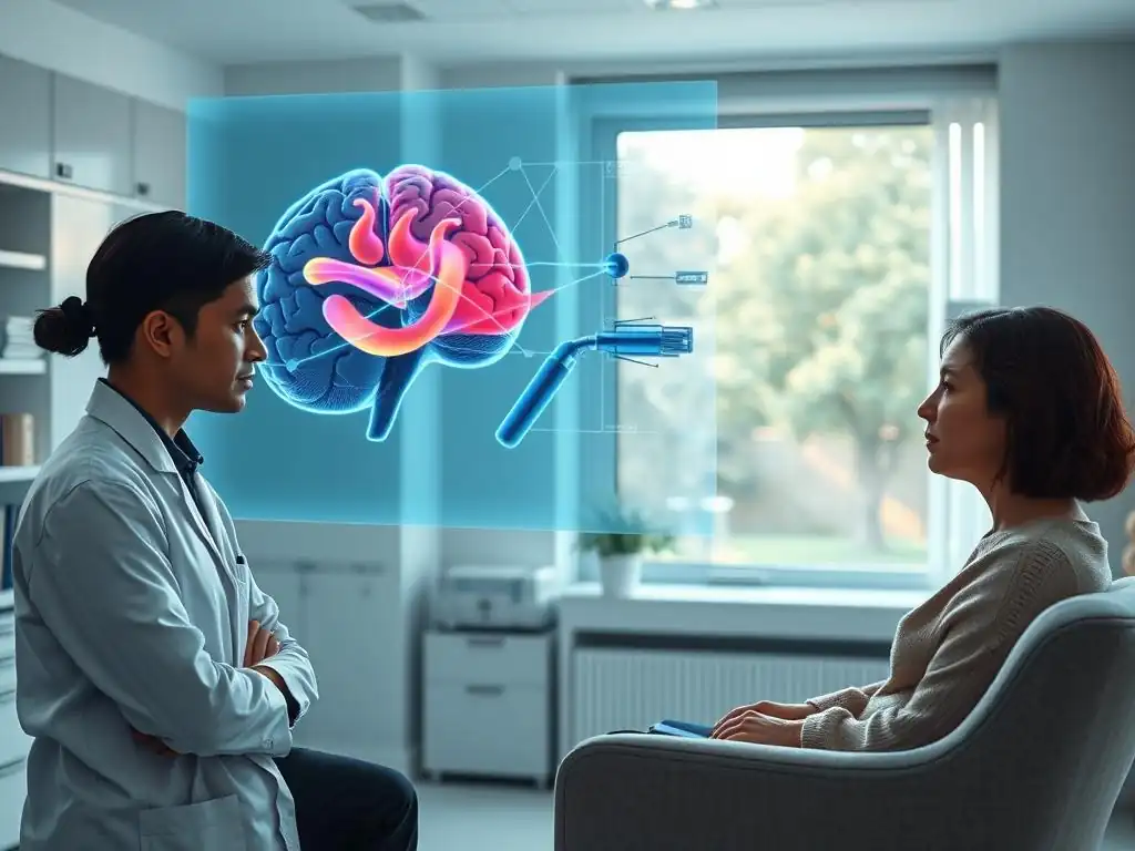
Welcome to Mind Care Guide, a sanctuary for understanding and nurturing your mental well-being.
Are you curious about how science is revolutionizing mental health care? Imagine peering into the very machinery of the mind to unlock the secrets of mental illness. Our latest article, "From Lab to Clinic: Translating Brain Imaging Findings for Mental Health Patients," dives deep into brain imaging mental health insights, revealing how cutting-edge neuroscience is paving the way for more personalized and effective treatments. Discover how these groundbreaking advancements are bringing hope and healing to those facing mental health challenges. Let's explore this fascinating frontier together and empower you on your journey to a healthier mind.
- From Lab to Clinic: Translating Brain Imaging Findings for Mental Health Patients
- Understanding Brain Imaging in Mental Health: Unveiling Insights
- Challenges in Translating Brain Imaging to Clinical Applications
- Overcoming Challenges: Strategies for Effective Translation
- Brain Imaging Mental Health Insights: Examples of Successful Translation
- The Future of Brain Imaging in Mental Healthcare
- Practical Implications for Mental Health Professionals
- Conclusion: Harnessing Brain Imaging for Improved Mental Health Outcomes
-
Frequently Asked Questions
- How can brain imaging techniques contribute to a better understanding of mental health conditions?
- What types of brain imaging methods are used in mental health research and clinical practice?
- How are findings from brain imaging studies translated into practical benefits for mental health patients?
- Are brain imaging results used to diagnose mental health disorders?
- What are the limitations of using brain imaging for mental health purposes?
- Final Reflection: Bridging the Gap Between Neuroscience and Mental Healthcare
- Unlock Your Brain's Potential: Take the Next Step
From Lab to Clinic: Translating Brain Imaging Findings for Mental Health Patients
Mind Care Guide: Bridging the Gap Between Neuroscience and Clinical Practice
Mind Care Guide is dedicated to illuminating the path toward mental well-being by providing accessible and reliable information. A crucial part of understanding mental health involves exploring the intricate workings of the brain. Neuroscience, particularly through advanced brain imaging techniques, is rapidly uncovering fundamental insights into the biological underpinnings of conditions like depression, anxiety, schizophrenia, and bipolar disorder. Our aim is to translate these complex laboratory findings and research insights into practical, understandable knowledge that can benefit individuals, families, and mental health professionals.
Bridging the gap between cutting-edge neuroscience research and clinical practice is essential for improving diagnosis, treatment, and outcomes in mental healthcare. While brain imaging is primarily a research tool today, the insights it provides are beginning to inform clinical understanding and hold immense promise for future personalized approaches. This article explores how brain imaging findings are moving from the research lab into potential clinical applications, offering valuable brain imaging mental health insights and discussing the journey, challenges, and future possibilities of this transformative field.
Understanding Brain Imaging in Mental Health: Unveiling Insights
The Role of Brain Imaging in Mental Health Research: A Primer
Brain imaging plays a pivotal role in mental health research by allowing scientists to non-invasively visualize the structure, function, and connectivity of the living brain. This provides an unprecedented window into the neural circuits and processes that may be altered in various mental health conditions. Researchers use these techniques to compare brain characteristics between individuals with and without specific disorders, investigate the effects of different treatments on brain activity, and explore potential biomarkers that could aid in earlier and more accurate diagnosis.
Through brain imaging, researchers can investigate hypotheses about the neurobiological basis of mental illness. For instance, studies might examine whether specific brain regions show reduced volume or altered activity in individuals experiencing symptoms of depression compared to healthy controls. These investigations contribute significantly to our understanding of why mental health conditions occur, moving beyond purely symptomatic descriptions to explore underlying biological mechanisms. The insights gained from this research are foundational for developing more targeted and effective interventions, offering profound brain imaging mental health insights.
Types of Brain Imaging Techniques Used in Mental Health
A variety of brain imaging techniques are employed in mental health research, each offering unique perspectives on brain structure or function. These tools differ in their resolution, what they measure, and their cost, making certain techniques more suitable for specific research questions. Understanding the basics of these methods is key to appreciating the brain imaging mental health insights they provide.
Generally, brain imaging techniques can be broadly categorized into structural imaging, which reveals the anatomy of the brain, and functional imaging, which measures brain activity. Both types are critical for a comprehensive understanding of the brain in health and disease, and advancements in these areas continue to push the boundaries of what is possible in mental health research.
Structural Imaging: MRI and CT Scans for Mental Health
Structural imaging techniques, such as Magnetic Resonance Imaging (MRI) and Computed Tomography (CT) scans, provide detailed pictures of the brain's physical structure. MRI uses strong magnetic fields and radio waves to create high-resolution images of brain tissue, allowing researchers to measure the volume and integrity of different brain regions.
For example, studies using MRI have shown that certain brain areas, like the hippocampus (involved in memory and emotion) and the prefrontal cortex (involved in decision-making and executive functions), may be smaller in individuals with certain conditions like major depression or PTSD, compared to healthy populations. While these structural differences are not universal biomarkers for diagnosis, they offer important brain imaging mental health insights into potential anatomical correlates of disorders.
CT scans use X-rays to produce cross-sectional images of the brain. While CT provides less detail than MRI for soft tissues like the brain, it is faster and more readily available, often used in emergency settings or when MRI is contraindicated. In mental health contexts, CT might be used to rule out other causes of psychiatric symptoms, such as tumors or strokes. However, for detailed research into the subtle structural differences associated with mental health conditions, MRI is generally the preferred technique due to its superior resolution and ability to differentiate various types of brain tissue.
Functional Imaging: fMRI, PET, and SPECT in Psychiatry
Functional brain imaging techniques measure brain activity, providing dynamic brain imaging mental health insights into how different areas of the brain work together. Functional MRI (fMRI) measures changes in blood flow and oxygenation, which are linked to neural activity. When a brain region is more active, it requires more oxygenated blood.
By detecting these changes, fMRI can map which parts of the brain are engaged during specific tasks or in resting states. For instance, fMRI studies have revealed altered connectivity patterns between brain networks, such as the default mode network or the salience network, in individuals with depression or schizophrenia, suggesting disruptions in how different brain regions communicate.
Positron Emission Tomography (PET) and Single-Photon Emission Computed Tomography (SPECT) use radioactive tracers injected into the bloodstream to measure metabolic activity, blood flow, or the distribution of specific molecules like neurotransmitter receptors. PET, for example, can be used to study dopamine or serotonin systems in the brain, which are implicated in various mental health conditions. Studies using PET have shown differences in dopamine receptor density in individuals with schizophrenia or addiction. While these techniques are more invasive due to the use of radioactive tracers, they offer unique molecular-level brain imaging mental health insights that fMRI cannot provide.
Challenges in Translating Brain Imaging to Clinical Applications
Variability in Brain Imaging Data and Mental Health Diagnoses
One of the primary hurdles in translating brain imaging findings from the lab to the clinic is the inherent variability found in both brain imaging data and mental health diagnoses. Mental health conditions are complex and heterogeneous; individuals diagnosed with the same disorder can present with vastly different symptom profiles, illness trajectories, and underlying biological factors. This clinical heterogeneity is often reflected in the brain imaging data, where findings might vary significantly between individuals with the same diagnosis, making it difficult to identify a single, reliable brain imaging marker that applies to everyone.
Furthermore, brain imaging data itself can be influenced by numerous factors, including individual differences in brain anatomy, age, sex, medication status, comorbidities, and even transient states like stress or sleep deprivation. The acquisition and analysis methods used can also introduce variability.
Consequently, identifying a robust "signature" in brain imaging that accurately and reliably corresponds to a specific mental health diagnosis or predicts treatment response for an individual patient remains a significant challenge. While research groups may report high accuracy in differentiating groups (e.g., patients vs. controls) in specific studies, translating these findings to the individual level in a clinical setting is much harder due to this widespread variability.
Ethical Considerations in Using Brain Imaging for Mental Health Treatment
The potential use of brain imaging in clinical mental healthcare raises important ethical considerations. As our ability to interpret brain imaging mental health insights grows, questions arise about privacy, data security, and potential misuse of sensitive neural information. Brain scans contain highly personal data, and ensuring its confidentiality and protecting against unauthorized access is paramount. There are concerns about discrimination if brain imaging findings were used to make judgments about an individual's mental state or predisposition outside of a clinical context, such as for employment or insurance purposes.
Moreover, there are ethical questions surrounding informed consent, particularly when brain imaging is used in potentially vulnerable populations or for research purposes. Patients need to understand what the imaging technique measures, its limitations, and how the data will be used. There is also the risk of over-interpreting or misinterpreting brain imaging findings in a way that might unduly influence clinical decisions or patient perceptions, potentially leading to unnecessary anxiety or stigma. It's crucial to manage expectations and emphasize that brain imaging is just one piece of the puzzle in understanding a complex individual.
The Cost-Effectiveness of Brain Imaging in Mental Healthcare
The high cost of brain imaging technology presents another significant barrier to its widespread clinical application in mental healthcare. MRI and PET scanners are expensive to purchase, install, and maintain, often costing several million dollars each. Running these machines requires highly trained personnel, including radiologists, technologists, and potentially physicists or specialized analysts, adding to operational expenses. These costs can make advanced brain imaging inaccessible in many clinical settings, particularly in underserved areas or healthcare systems with limited resources.
For brain imaging to become a routine tool in mental healthcare, its clinical utility must clearly justify the substantial financial investment. Currently, brain imaging is not typically used for routine diagnosis of most mental health conditions because clinical assessments based on symptoms and history are generally sufficient and far more cost-effective.
For brain imaging to be adopted clinically, it would need to demonstrate a clear benefit in improving diagnostic accuracy, predicting treatment response, or preventing negative outcomes in a way that significantly outweighs the cost compared to standard care. Research is ongoing to identify specific scenarios where brain imaging offers truly valuable and cost-effective brain imaging mental health insights that change clinical management.
Overcoming Challenges: Strategies for Effective Translation
Standardizing Brain Imaging Protocols for Mental Health Studies
To address the issue of variability and improve the reproducibility of brain imaging findings, significant efforts are being made to standardize acquisition and analysis protocols across different research sites. Variability in scanner hardware, software versions, pulse sequences, and image processing pipelines can all impact the resulting data.
By developing and adopting common standards, researchers can better compare results across studies and build larger, more robust datasets. Initiatives like the ENIGMA Consortium (Enhancing Neuro Imaging Genetics through Meta-Analysis) collect and analyze data from hundreds of research sites worldwide using standardized protocols, enabling large-scale meta-analyses that can identify more reliable patterns and provide deeper brain imaging mental health insights than individual studies alone.
Standardization also involves developing and validating reliable methods for analyzing complex brain imaging data. This includes establishing best practices for denoising images, segmenting brain regions, measuring connectivity, and performing statistical analyses. The goal is to reduce methodological variability as a source of noise, making it easier to identify genuine biological signals associated with mental health conditions. As these protocols become more refined and widely adopted, the consistency and reliability of brain imaging mental health insights derived from research will increase, paving the way for potential clinical applications.
Developing Machine Learning Models for Brain Imaging Data Analysis in Mental Health
Machine learning and artificial intelligence (AI) are playing an increasingly important role in analyzing complex brain imaging datasets. These computational approaches are particularly well-suited for identifying subtle patterns and relationships within high-dimensional data that might not be apparent through traditional statistical methods. Researchers are developing machine learning models trained on large datasets of brain scans from individuals with and without mental health conditions to build classifiers that can potentially distinguish between diagnostic groups or predict outcomes. For example, models are being trained to identify patterns in fMRI connectivity data that might predict an individual's response to antidepressant medication.
While still largely in the research phase, early results show promise. Some studies using machine learning on brain imaging data have reported accuracies in the range of 70-85% for differentiating between groups like individuals with schizophrenia and healthy controls, or predicting treatment response in depression, though these figures are typically achieved in controlled research settings and performance can drop with real-world clinical data. Continued development and validation of these models, along with access to larger, more diverse datasets, are crucial steps towards creating clinically useful tools that can derive actionable brain imaging mental health insights for individual patients.
Integrating Brain Imaging Findings with Clinical Assessments for Personalized Treatment
The most promising path for translating brain imaging into clinical practice likely involves integrating brain imaging mental health insights with other sources of information, rather than relying solely on brain scans for diagnosis or treatment decisions. Clinical assessments, including detailed interviews, symptom questionnaires, and observations, provide rich, nuanced data about an individual's lived experience and current functioning – information that brain imaging alone cannot capture. Genetic testing, cognitive assessments, and physiological measures can also provide valuable context.
The future of personalized mental healthcare may involve combining data from various sources, including brain imaging, using computational approaches to build a more complete picture of an individual's specific needs. For example, a patient presenting with depressive symptoms might undergo a clinical assessment, followed by a brain scan.
Machine learning algorithms could then integrate the imaging data (e.g., connectivity patterns, regional volumes) with clinical factors (e.g., symptom severity, history of trauma, genetic markers) to predict the likelihood of response to different therapies (e.g., specific medications, types of psychotherapy). This integrated approach acknowledges the complexity of mental health and leverages the unique brain imaging mental health insights alongside other critical information to guide tailored treatment plans.
Brain Imaging Mental Health Insights: Examples of Successful Translation
Brain Imaging Biomarkers for Depression: Guiding Treatment Decisions
Major depressive disorder is a highly prevalent condition, yet predicting which treatment will work best for an individual remains challenging. Brain imaging research is actively exploring potential biomarkers that could help guide treatment decisions.
Studies using fMRI have identified patterns of activity and connectivity in specific brain circuits, such as those involving the subcallosal cingulate cortex and prefrontal cortex, that appear to distinguish individuals who are likely to respond to certain interventions like cognitive behavioral therapy or specific antidepressant medications. For instance, research suggests that greater connectivity between certain frontal regions might predict a better response to psychotherapy, while differences in activity in other areas might indicate likely response to medication.
While not yet routine clinical practice, these findings represent significant brain imaging mental health insights. For example, some research centers are exploring the use of imaging-informed approaches in clinical trials to match patients to the treatment most likely to be effective based on their brain activity patterns. Although a single definitive biomarker for depression treatment response has not been established, the consistent findings across multiple studies regarding the involvement of specific circuits offer hope that imaging could one day help clinicians select the most appropriate initial treatment, reducing the trial-and-error process that many patients currently undergo.
Identifying Neural Correlates of Anxiety Disorders Through Brain Imaging
Anxiety disorders encompass a range of conditions, including generalized anxiety disorder, panic disorder, and social anxiety disorder. Brain imaging studies have provided valuable brain imaging mental health insights into the neural underpinnings of these conditions, often pointing to dysfunction in brain circuits involved in fear processing, emotional regulation, and threat detection. For example, fMRI studies of individuals with anxiety disorders often show heightened activity in the amygdala, a brain region crucial for processing fear, when exposed to anxiety-provoking stimuli. Conversely, activity in prefrontal regions, which are involved in regulating emotions, may be reduced or altered.
Structural MRI studies have sometimes reported differences in amygdala volume or connectivity in anxiety disorders, though findings can be inconsistent across studies due to heterogeneity within patient groups. These neural correlates are not diagnostic markers on their own but contribute significantly to our understanding of the biological basis of anxiety. This knowledge informs the development of new therapeutic approaches, such as neurofeedback techniques or targeted neuromodulation therapies (like transcranial magnetic stimulation aimed at specific circuits), that seek to normalize the activity or connectivity of these identified brain regions, leveraging brain imaging mental health insights to design interventions.
Brain Imaging and Schizophrenia: Understanding the Neurobiological Basis
Schizophrenia is a severe mental illness characterized by profound alterations in thought, perception, and behavior. Brain imaging has been instrumental in revealing significant structural and functional abnormalities in the brains of individuals with schizophrenia, offering critical brain imaging mental health insights into its neurobiological basis. Structural MRI studies consistently report reduced gray matter volume in various brain regions in schizophrenia, including the prefrontal cortex, temporal lobes, and hippocampus, compared to healthy controls. While the average volume reduction across multiple studies might be around 5-10% in these areas, there is considerable individual variability.
Functional imaging studies (fMRI, PET) have shown altered activity and connectivity patterns, particularly in networks involved in executive function, attention, and processing sensory information. For instance, disruptions in the connectivity between the prefrontal cortex and other brain regions are frequently observed.
These findings support the view of schizophrenia as a disorder involving widespread disruption of brain circuits, rather than a single localized problem. While brain imaging cannot diagnose schizophrenia on its own (diagnosis relies on clinical symptoms), the insights gained from imaging research have profoundly shaped our understanding of the disorder and are guiding the development of new pharmacological and non-pharmacological treatments aimed at targeting these identified neural abnormalities.
The Future of Brain Imaging in Mental Healthcare
Advancements in Brain Imaging Technology for Mental Health
The field of brain imaging is continuously evolving, with ongoing technological advancements promising even greater brain imaging mental health insights and potential clinical applications. Future developments include higher-field strength MRI scanners, which offer improved resolution and signal-to-noise ratio, allowing for visualization of finer brain structures and more subtle functional changes. Development of more sensitive and specific PET tracers will enable better targeting and measurement of specific neurotransmitter systems or molecular targets implicated in mental illness.
Furthermore, advancements in portable and lower-cost imaging technologies, such as more sophisticated forms of electroencephalography (EEG) or near-infrared spectroscopy (NIRS), could potentially bring some level of brain activity monitoring outside of specialized centers, making it more accessible. The integration of different imaging modalities (e.g., simultaneously acquiring fMRI and EEG data) could provide a more comprehensive picture of brain activity. These technological leaps are expected to yield richer datasets and more precise brain imaging mental health insights, potentially leading to the identification of more reliable biomarkers and the development of novel, imaging-guided therapies.
Personalized Mental Healthcare: Tailoring Treatment Based on Brain Imaging Profiles
The ultimate goal of translating brain imaging research into clinical practice is to enable personalized mental healthcare. This vision involves using brain imaging data, in conjunction with other clinical and biological information, to tailor treatment strategies to the individual patient.
Instead of a one-size-fits-all approach or lengthy trial-and-error, clinicians could potentially use an individual's unique brain imaging profile to predict their likelihood of responding to specific medications, psychotherapies, or neuromodulation techniques. For example, imaging might help determine if a patient with depression is more likely to benefit from an antidepressant targeting serotonin versus norepinephrine, or if transcranial magnetic stimulation applied to a specific brain region is indicated.
Achieving personalized mental healthcare based on brain imaging mental health insights requires robust, validated biomarkers that are reliable at the individual level. This is a complex challenge given the variability discussed earlier. However, ongoing research, fueled by larger datasets and advanced machine learning, is steadily moving towards identifying these personalized profiles. The ability to match patients to the most effective treatment from the outset could significantly improve outcomes, reduce healthcare costs, and minimize the burden of ineffective therapies for individuals struggling with mental health conditions.
The Impact of Brain Imaging on Mental Health Stigma
Brain imaging has the potential to significantly impact the stigma surrounding mental illness. By demonstrating that mental health conditions are associated with measurable differences or changes in brain structure and function, imaging helps to frame these disorders as legitimate biological illnesses, similar to neurological conditions or heart disease. This can challenge the misconception that mental illnesses are simply a matter of poor character, personal weakness, or something that individuals should just "snap out of."
Presenting brain imaging mental health insights in an accessible and empathetic way can help patients and their families understand that their struggles have a biological basis, potentially reducing self-blame and shame. While it is crucial to avoid biological determinism (mental health is complex and influenced by many factors beyond biology), highlighting the brain's involvement can foster greater public understanding and compassion. As brain imaging becomes more integrated into discussions about mental health, it can contribute to a more informed and less stigmatizing societal view of these conditions, encouraging individuals to seek help without fear of judgment.
Practical Implications for Mental Health Professionals
Interpreting Brain Imaging Reports for Mental Health Patients
For brain imaging to be useful in clinical mental healthcare, mental health professionals need to understand how to interpret imaging reports and integrate these findings into their overall assessment of a patient. Currently, routine clinical brain scans (like those performed in neurology or radiology departments) are primarily used to rule out structural abnormalities such as tumors, cysts, or stroke, which might mimic psychiatric symptoms.
Reports from these scans typically focus on gross anatomical features. However, as research imaging findings move closer to clinical utility, mental health professionals may encounter reports that include information on subtle structural differences or functional connectivity patterns potentially relevant to psychiatric conditions.
Interpreting these reports requires a basic understanding of neuroanatomy and the specific imaging technique used. It's crucial for clinicians to understand the limitations of current brain imaging in diagnosing mental illness and to avoid over-interpreting findings that might be within the range of normal variation or not specific to a particular condition. Collaboration with neuroradiologists and researchers specializing in psychiatric neuroimaging is essential to accurately interpret complex brain imaging mental health insights and understand their potential relevance, or lack thereof, to an individual case.
Communicating Brain Imaging Findings to Patients and Families
Communicating brain imaging findings to patients and their families is a sensitive and crucial task that requires empathy and clarity. It's important to explain why a scan was performed (e.g., to rule out other causes, as part of a research study, or potentially in the future, to help guide treatment) and what the findings mean in the context of the individual's overall clinical picture. Avoid presenting complex imaging data without clear explanation. Use analogies or simple diagrams if helpful, but ensure accuracy.
When discussing findings potentially related to a mental health condition, emphasize that these are often subtle differences observed across groups and that brain imaging is just one piece of the puzzle. Avoid language that is overly technical, deterministic, or potentially stigmatizing.
For example, instead of saying "your scan shows brain damage," one might explain, "studies show that people with similar symptoms sometimes have differences in how certain brain areas are connected, and your scan shows a pattern that is sometimes seen in this condition, which helps us understand the biological factors at play." The focus should remain on how the findings contribute to understanding the individual's experience and informing their care plan, using the brain imaging mental health insights to empower rather than alarm.
Integrating Brain Imaging into Mental Health Treatment Planning: A Step-by-Step Guide
Integrating brain imaging into mental health treatment planning is currently more common in research settings or specialized clinics, but here is a potential framework for how it might be incorporated in the future, or how research findings are considered today:
- Clinical Assessment: Begin with a comprehensive clinical evaluation, including history, symptoms, and functional impact.
- Identify Potential Utility: Determine if brain imaging might offer valuable brain imaging mental health insights for this specific patient, based on current research and clinical questions (e.g., differentiating between conditions, predicting treatment response, ruling out neurological causes).
- Select Appropriate Imaging Technique: Based on the clinical question, choose the most relevant imaging modality (e.g., structural MRI to check for gross abnormalities, fMRI or PET if investigating functional or molecular aspects relevant to research protocols or future clinical tests).
- Acquire and Process Data: Perform the brain scan using standardized protocols. Process the raw data using validated analysis pipelines.
- Interpret Findings: A clinician, potentially in consultation with a neuroradiologist or neuroimaging specialist, interprets the imaging report in the context of the clinical assessment. Consider if the findings align with known patterns for the suspected condition or offer predictive information based on current research.
- Integrate with Other Data: Combine brain imaging insights with clinical information, genetic data, cognitive test results, etc.
- Inform Treatment Plan: Use the integrated information to refine the diagnosis (if applicable), select potential treatment options (e.g., type of therapy, medication class, neuromodulation target), or monitor treatment response. This step heavily relies on future validation of imaging biomarkers.
- Communicate with Patient: Discuss the findings and how they informed the treatment plan with the patient and family in an understandable and empathetic manner.
- Monitor and Re-evaluate: Treatment plans should be dynamic. Future imaging or other assessments might be used to monitor progress and adjust the plan as needed.
This step-by-step process highlights that brain imaging is not a standalone diagnostic tool for most mental illnesses but rather a potentially powerful source of complementary brain imaging mental health insights that, when integrated thoughtfully, can contribute to more precise and personalized care.
Conclusion: Harnessing Brain Imaging for Improved Mental Health Outcomes
Brain Imaging: A Powerful Tool for Understanding and Treating Mental Illness
Brain imaging has revolutionized our understanding of the brain's involvement in mental health conditions. Techniques like MRI, fMRI, and PET have provided unprecedented brain imaging mental health insights into the structural, functional, and molecular alterations associated with disorders such as depression, anxiety, and schizophrenia. While currently primarily a research tool, the findings from brain imaging are fundamentally changing how we conceptualize mental illness, moving towards a model that integrates biological understanding with psychological and social factors.
The journey from laboratory discovery to routine clinical application is complex, facing challenges related to data variability, cost, and ethical considerations. However, ongoing advancements in technology, data analysis methods (including machine learning), and research standardization are steadily paving the way for brain imaging to play a more direct role in clinical decision-making. The potential to use brain imaging mental health insights to improve diagnostic accuracy, predict treatment response, and enable truly personalized mental healthcare is immense, holding the promise of better outcomes for individuals struggling with mental health challenges.
The Importance of Continued Research and Collaboration in Brain Imaging and Mental Health
The translation of brain imaging findings into clinical practice requires continued, robust research and enhanced collaboration across disciplines. Researchers need to continue refining imaging techniques, developing more reliable biomarkers, and conducting large-scale studies to validate findings across diverse populations. Collaboration between neuroscientists, psychiatrists, psychologists, radiologists, computer scientists, and ethicists is essential to navigate the technical, clinical, and ethical complexities involved.
Investing in research infrastructure, fostering data sharing initiatives, and supporting interdisciplinary training programs are critical steps. As our understanding grows and technology advances, brain imaging has the potential to transform mental healthcare, offering more precise diagnostics, targeted treatments, and ultimately, improved well-being for millions. The ongoing pursuit of brain imaging mental health insights is a vital endeavor in the effort to alleviate the burden of mental illness.
Frequently Asked Questions
How can brain imaging techniques contribute to a better understanding of mental health conditions?
Brain imaging allows researchers and clinicians to visualize brain structure and function, revealing neurological differences associated with various mental health disorders. This helps in understanding the biological basis of these conditions.
What types of brain imaging methods are used in mental health research and clinical practice?
Common methods include MRI, fMRI, PET scans, and EEG. These techniques provide different types of information, such as brain structure, activity, and neurochemical processes.
How are findings from brain imaging studies translated into practical benefits for mental health patients?
Researchers are working to identify biomarkers that can predict treatment response or diagnose specific conditions. This can lead to personalized treatment plans based on individual brain characteristics.
Are brain imaging results used to diagnose mental health disorders?
While brain imaging can provide valuable insights, it's generally used as part of a comprehensive evaluation, alongside clinical interviews and psychological assessments. It's not usually a standalone diagnostic tool.
What are the limitations of using brain imaging for mental health purposes?
Brain imaging can be expensive and time-consuming, and interpreting the results can be complex. Furthermore, group-level findings may not always apply to individual patients.
Final Reflection: Bridging the Gap Between Neuroscience and Mental Healthcare
The convergence of advanced brain imaging techniques and our understanding of mental health disorders is no longer a futuristic dream; it's a rapidly evolving reality with profound implications for diagnosis, treatment, and patient care. The insights gleaned from fMRI, EEG, and other neuroimaging modalities are transforming how we perceive and approach conditions like depression, anxiety, and schizophrenia, moving beyond solely symptom-based diagnoses towards a more precise, biologically informed approach. This shift promises a future where personalized treatment plans, tailored to individual brain patterns, become the norm.
The journey from lab to clinic, however, is not without its challenges. The ethical considerations surrounding data privacy, the need for standardized protocols across research and clinical settings, and the ongoing need for more robust research to validate findings are all crucial aspects that must be addressed. The greatest advances in medicine have come from understanding the underlying biology of disease. This is equally true for mental illness.
This quote underscores the importance of continued investment in brain imaging research and its translation into practical clinical applications, ensuring that the benefits of this technology reach those who need it most.
Consider the potential impact on your own life, or the lives of those you know, who struggle with mental health challenges. Imagine a future where early detection and personalized interventions, guided by brain imaging data, could significantly improve outcomes and reduce the suffering associated with these conditions. This is not merely a scientific endeavor; it is a humanitarian one. Let us embrace the potential of brain imaging, advocate for responsible research and ethical implementation, and work together to bridge the gap between the lab and the clinic, ultimately improving the lives of countless individuals affected by mental illness.
Unlock Your Brain's Potential: Take the Next Step
Thanks for diving into the world of brain imaging and its impact on mental health! We hope you gained a clearer understanding of how these incredible advancements are shaping the future of treatment. Now, let's put that knowledge into action.
- Schedule a consultation with a mental health professional: Discuss your mental health and explore whether brain imaging could benefit your journey to wellness. This is the most impactful action you can take based on today's article.
- Share this article with someone you care about: Help spread awareness about the potential of brain imaging in mental health treatment. A simple share can make a significant difference.
- Explore reputable resources on brain imaging and mental health: Delve deeper into this exciting field by researching reputable organizations and studies online.
What steps will you take to better understand and advocate for advancements in mental health care?