
The Brain on the Mend: What Scans Show After Years of Therapy


Welcome to Mind Care Guide, a space dedicated to nurturing your mental well-being.
Ever wondered what truly happens within your brain during years of dedicated therapy? We delve into the fascinating neuroscience behind healing. In our latest article, "The Brain on the Mend: What Scans Show After Years of Therapy," we explore the groundbreaking insights revealed by brain scans regarding therapy effectiveness. Prepare to be amazed by what science is uncovering about the resilience and adaptability of the human mind. Ready to see the impact for yourself?
Rewiring the Anxious Brain: Neuroimaging After Years of CBT
- Introduction: Mapping Brain Changes Through Long-Term Cognitive Behavioral Therapy
- Neuroimaging Techniques for Evaluating CBT Outcomes
- Long-Term CBT Outcomes and Observable Brain Changes
- Interpreting Neuroimaging Data: Correlating Brain Changes with Clinical Improvement
- Case Studies: Illustrating Neuroimaging Results After Long-Term CBT
- Limitations and Future Directions in Neuroimaging Research for CBT
- Conclusion: Neuroimaging as a Tool for Understanding CBT's Impact on the Anxious Brain
-
Frequently Asked Questions
- What is the main purpose of using brain scans to assess therapy effectiveness?
- What types of brain scans are typically used to evaluate the impact of therapy?
- How can brain scans help personalize therapy approaches?
- What are some limitations of relying solely on brain scans to determine therapy effectiveness?
- Are brain scans a standard practice in monitoring the progress of all types of mental health therapy?
- Final Reflection: The Enduring Power of Healing
- Unlock Your Brain's Potential: Take the Next Step
Introduction: Mapping Brain Changes Through Long-Term Cognitive Behavioral Therapy
Anxiety disorders affect millions globally, significantly impacting quality of life. While various treatments exist, Cognitive Behavioral Therapy (CBT) stands out as a highly effective, evidence-based psychotherapy. For many individuals, engaging in long-term CBT leads to profound and lasting reductions in anxiety symptoms.
But beyond the subjective experience of feeling better, what changes are actually happening in the brain? This article delves into how neuroimaging techniques are providing remarkable insights into the neural mechanisms underlying long-term CBT success, offering a window into the brain's capacity for change, or neuroplasticity, in the context of therapeutic intervention. Understanding the brain on the mend after years of dedicated therapeutic work highlights the physical reality of mental health recovery.
Exploring the effectiveness of long-term therapy through objective measures like brain scans therapy effectiveness offers a powerful testament to the impact of psychological treatment. By comparing brain activity and structure before and after extensive CBT, researchers can pinpoint specific neural circuits that are altered, providing concrete evidence of the therapy's ability to "rewire" the anxious brain. This not only validates the experiences of those who benefit from CBT but also opens avenues for understanding how and why it works at a fundamental biological level. It's a fascinating intersection of psychology and neuroscience, revealing the tangible results of therapeutic effort.
Understanding the Neurobiology of Anxiety Disorders and CBT's Impact
Anxiety disorders are associated with dysregulation in specific brain circuits, particularly those involved in processing threat and regulating emotion. Key players include the amygdala, often hyperactive in anxiety and responsible for detecting potential danger, and the prefrontal cortex (PFC), crucial for executive functions like decision-making, impulse control, and emotion regulation. In anxious individuals, the PFC may show reduced activity or connectivity with the amygdala, impairing the brain's ability to dampen excessive fear responses.
Other regions, such as the insula (involved in sensing internal states) and the anterior cingulate cortex (ACC, involved in conflict monitoring), also play roles in the complex neural architecture of anxiety. Understanding these baseline differences is crucial for appreciating how therapy facilitates change.
Cognitive Behavioral Therapy targets maladaptive thought patterns and behaviors that perpetuate anxiety. Through techniques like cognitive restructuring (challenging distorted thoughts) and exposure therapy (gradually confronting feared situations), CBT aims to modify how the brain processes threats and regulates emotional responses. The hypothesis is that by repeatedly practicing new ways of thinking and reacting, individuals can strengthen the regulatory pathways from the PFC to the amygdala, effectively increasing the brain's capacity to manage fear and worry.
This process leverages the brain's inherent neuroplasticity – its ability to reorganize itself by forming new neural connections throughout life. Long-term engagement with these therapeutic strategies is believed to solidify these changes, leading to more stable and enduring symptom reduction.
The Role of Neuroimaging in Assessing CBT Effectiveness
Neuroimaging provides non-invasive methods to visualize the brain's structure and activity, offering objective data to complement subjective self-reports and behavioral observations in assessing therapy outcomes. Before the advent of modern scanning techniques, the effectiveness of psychological therapies was primarily gauged through symptom questionnaires and clinical interviews. While invaluable, these methods don't directly reveal the underlying biological changes. Brain scans therapy effectiveness research allows scientists to investigate whether clinical improvements correlate with measurable alterations in brain function or structure, providing a deeper understanding of the therapeutic process.
Utilizing neuroimaging before, during, and after a course of long-term CBT enables researchers to track changes over time. This longitudinal approach is particularly powerful for studying neuroplasticity, as it can demonstrate how specific brain regions or networks adapt in response to therapeutic interventions.
For instance, scans might reveal a decrease in amygdala reactivity to threat cues alongside a reduction in anxiety symptoms, or an increase in connectivity between the PFC and the amygdala as cognitive control over emotional responses improves. These findings not only validate CBT's mechanisms of action but also contribute to building a more comprehensive, biologically-informed model of mental health recovery.
Neuroimaging Techniques for Evaluating CBT Outcomes
Several advanced neuroimaging techniques are employed to investigate the neural correlates of CBT effectiveness, each offering unique insights into different aspects of brain function and structure. The choice of technique depends on the specific research question, whether it focuses on the size of brain regions, the level of activity within them, or the communication between different parts of the brain. Combining insights from multiple modalities often provides a more complete picture of the brain's response to long-term therapy.
Types of Neuroimaging Used to Monitor Treatment Progress
MRI (Magnetic Resonance Imaging) and Identifying Structural Adaptations
Structural MRI provides detailed images of the brain's anatomy, allowing researchers to measure the size and volume of different brain regions. Studies using structural MRI have explored whether long-term CBT leads to observable changes in the gray matter density or volume of areas implicated in anxiety, such as the amygdala, hippocampus (involved in memory and context processing, often affected by chronic stress), and various prefrontal cortex subregions.
For example, some research suggests that successful treatment of PTSD with trauma-focused CBT may be associated with increases in hippocampal volume, potentially counteracting stress-induced atrophy. While findings can vary across studies and anxiety types, structural MRI offers clues about the enduring physical changes in brain tissue architecture that might support sustained recovery.
These structural studies contribute to understanding the long-term impact of therapy by showing that therapeutic interventions can influence the physical makeup of the brain. Although changes in volume or density might be subtle and require sophisticated analysis techniques, their detection provides compelling evidence that psychological treatment isn't just about changing thoughts; it can be associated with tangible alterations in brain structure. This reinforces the concept of neuroplasticity at a macroscopic level, demonstrating the brain's remarkable capacity to adapt and reorganize over time in response to therapeutic input.
fMRI (Functional Magnetic Resonance Imaging) and Mapping Neural Circuit Changes
Functional MRI (fMRI) measures brain activity by detecting changes in blood flow. When a brain region is more active, it requires more oxygenated blood, which fMRI can detect.
This technique is particularly valuable for studying how brain circuits respond to specific tasks (like viewing fearful faces or imagining anxious scenarios) or during resting states (measuring spontaneous activity and connectivity between regions). In the context of brain scans therapy effectiveness research, fMRI is frequently used to assess changes in the reactivity of the amygdala to threat stimuli and alterations in the connectivity between the amygdala and regulatory regions in the prefrontal cortex before and after long-term CBT.
Studies using fMRI have consistently shown shifts in neural activity patterns following successful CBT. For instance, many individuals with social anxiety disorder show excessive amygdala activation when anticipating or experiencing social evaluation. After long-term CBT, fMRI scans often reveal a significant reduction in this amygdala hyperresponsiveness. Simultaneously, connectivity between the amygdala and parts of the PFC, such as the ventromedial prefrontal cortex (vmPFC), which is involved in emotion regulation, may increase, indicating a stronger top-down control over fear responses. These functional changes observed via fMRI provide dynamic evidence of how therapy alters the brain's active processing of emotional information.
PET (Positron Emission Tomography) Scans: Observing Neurotransmitter Modulation
Positron Emission Tomography (PET) is another powerful neuroimaging technique that uses radioactive tracers to visualize and measure metabolic processes, blood flow, or the distribution of specific molecules like neurotransmitter receptors or transporters in the brain. While perhaps less common than fMRI in standard CBT outcome research due to its cost and invasiveness, PET scans offer unique insights into the neurochemical changes that might underpin therapeutic success. For example, PET can be used to study the dopamine or serotonin systems, neurotransmitters often implicated in mood and anxiety disorders and targeted by pharmacological treatments, but potentially also modulated by psychological interventions.
Although direct evidence linking long-term CBT specifically to widespread changes in neurotransmitter receptor availability via PET is still an active area of research, some studies have explored related mechanisms. For instance, research using tracers for the benzodiazepine receptor site on the GABA-A receptor (a key inhibitory system often dysregulated in anxiety) has shown altered binding potential in certain brain regions in anxiety disorders.
While not solely focused on CBT, PET's capacity to map these molecular targets provides a complementary perspective to structural and functional imaging, suggesting potential avenues for future research into how long-term psychotherapy might influence neurochemical balance alongside circuit-level changes, further illuminating brain scans therapy effectiveness.
Establishing a Baseline: Initial Neuroimaging Before CBT
A critical step in using neuroimaging to assess the effectiveness of long-term CBT is establishing a reliable baseline scan before treatment begins. This initial scan serves as a crucial reference point against which all subsequent scans are compared. Without a pre-treatment measure, it is impossible to definitively attribute any observed neural changes to the therapy itself.
The baseline scan captures the individual's brain structure, function, or neurochemistry in their anxious state, providing a neural signature of their condition prior to intervention. This allows researchers to track changes over the course of therapy, often involving follow-up scans after several months or years of treatment, depending on the study design.
Establishing a robust baseline also helps account for individual variability in brain structure and function. No two brains are exactly alike, and pre-existing differences can influence how individuals respond to therapy. By comparing a patient's post-treatment scan to their own baseline, researchers can isolate the changes that are most likely attributable to the therapeutic process, rather than being inherent individual traits. This personalized approach to neuroimaging analysis is essential for accurately assessing brain scans therapy effectiveness on an individual level and across groups in research studies.
Long-Term CBT Outcomes and Observable Brain Changes
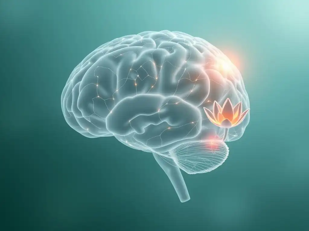
Years of research utilizing neuroimaging have provided compelling evidence that long-term CBT is associated with significant and observable changes in brain function, particularly within the networks implicated in anxiety and emotion regulation. These changes are not merely transient shifts but appear to represent more enduring adaptations in neural processing, aligning with the sustained symptom relief experienced by many individuals who undergo comprehensive therapy. The consistent findings across numerous studies bolster the understanding of CBT as a treatment that facilitates tangible neural transformation.
The focus of much of this research has been on how CBT influences the interplay between subcortical regions like the amygdala, which are involved in automatic emotional responses, and cortical regions, especially the prefrontal cortex, which exert cognitive control. Successful therapy seems to shift the balance within this system, strengthening the capacity of the prefrontal cortex to regulate and modulate activity in the amygdala, leading to a reduction in excessive fear and worry responses. This observed neural shift provides a biological correlate for the clinical improvements seen in patients.
CBT and its Impact on Amygdala and Prefrontal Cortex Function
Amygdala Hypoactivation: Evidence of Reduced Threat Response in Anxiety
One of the most consistent findings in neuroimaging research on CBT for anxiety disorders is a reduction in amygdala activity in response to threat-related stimuli after successful treatment. Before therapy, individuals with anxiety often show exaggerated amygdala responses when presented with cues related to their fears, such as fearful faces for social anxiety or panic-related images for panic disorder.
Following a course of long-term CBT, studies using fMRI have frequently reported a significant decrease in this hyperactivation. While the precise percentage varies between studies and anxiety types, research often indicates reductions in amygdala reactivity ranging approximately from 15% to 30% when patients are re-exposed to these stimuli after therapy.
This observed hypoactivation of the amygdala post-CBT is interpreted as evidence that the brain's initial, automatic alarm system is less reactive to perceived threats. Through repeated exposure and cognitive restructuring, the individual learns that feared situations are not as dangerous as initially appraised, and this learning is reflected in a dampened neural response in the amygdala. This reduced "threat signal" likely contributes significantly to the decrease in subjective feelings of fear and anxiety experienced by patients, providing a neural basis for the clinical benefits of brain scans therapy effectiveness.
Prefrontal Cortex Hyperactivation: Enhanced Cognitive Control and Emotion Regulation
Complementary to the reduced amygdala activity, studies often reveal increased activation or altered connectivity within regions of the prefrontal cortex (PFC) after successful long-term CBT. The PFC, particularly areas like the ventromedial prefrontal cortex (vmPFC) and dorsolateral prefrontal cortex (dlPFC), plays a crucial role in evaluating the emotional significance of stimuli, regulating emotional responses, and exerting cognitive control. In anxious individuals, the functional connectivity between the PFC and the amygdala is often weaker.
Following long-term therapy, fMRI studies have demonstrated increased activity in these prefrontal regions during tasks requiring emotion regulation or exposure to anxiety triggers. For example, some research suggests that successful treatment can lead to increased activation in the dlPFC by perhaps 10-25% during reappraisal tasks, indicating enhanced cognitive control abilities. Furthermore, connectivity analyses often show strengthened pathways between the vmPFC and the amygdala. This suggests that CBT facilitates a more effective "top-down" regulation of the amygdala by the prefrontal cortex, allowing individuals to better manage their fear responses and apply learned coping strategies.
Neuroplasticity and Long-Term CBT Success
How the Brain Adapts: A Focus on Synaptic Plasticity
The observable changes in brain structure and function following long-term CBT are tangible evidence of neuroplasticity at work. At a fundamental level, neuroplasticity involves changes in the strength and number of connections between neurons, a process known as synaptic plasticity. When individuals repeatedly engage in new thought patterns or behaviors during therapy – like challenging a catastrophic thought or staying in a feared situation without performing avoidance rituals – they are essentially activating new neural pathways and weakening old ones. This repeated activation strengthens the relevant synaptic connections, making those pathways more efficient over time.
For instance, practicing cognitive reappraisal strengthens the connections within the prefrontal circuits involved in evaluating and reinterpreting threatening information. Similarly, successful exposure therapy helps build new inhibitory pathways from the PFC to the amygdala, reducing the automatic fear response. Over months and years of therapy, these microscopic changes accumulate, leading to the macroscopic alterations in regional activity and connectivity observed with neuroimaging. This ongoing process of synaptic remodeling is the biological engine driving the brain's adaptation and recovery facilitated by long-term CBT.
Neuroimaging Confirmation of Structural and Functional Changes After CBT
Neuroimaging techniques provide the empirical confirmation that these neuroplastic processes indeed occur in response to long-term CBT. Structural MRI findings, though sometimes subtle, can suggest changes in gray matter volume or density that might reflect alterations in neuronal size, glial cell numbers, or synaptic density in relevant brain regions after extended therapy. Functional MRI data, showing altered activity patterns and strengthened or weakened connectivity between brain areas, directly illustrates the reorganization of neural circuits.
For example, a study might show reduced gray matter volume in the amygdala (perhaps reflecting reduced neuronal activity or size) alongside decreased fMRI reactivity to fearful faces after long-term CBT for panic disorder. Another study might demonstrate increased connectivity between the vmPFC and hippocampus after trauma-focused CBT for PTSD, correlating with improved emotional regulation and memory processing. These diverse findings, observed across different imaging modalities and anxiety subtypes, collectively reinforce the understanding that brain scans therapy effectiveness is grounded in the brain's fundamental capacity for neuroplastic change initiated and guided by therapeutic intervention.
Interpreting Neuroimaging Data: Correlating Brain Changes with Clinical Improvement
While neuroimaging provides powerful objective data on brain changes, interpreting these findings and correlating them with clinical improvement is a complex process. It's not simply a matter of pointing to a brain scan and saying "this person is better." Researchers must carefully analyze the imaging data, consider potential confounds, and integrate the findings with traditional measures of symptom severity and functional improvement. Understanding the nuances of this interpretation is key to accurately assessing brain scans therapy effectiveness.
The goal is to establish a reliable link between specific neural alterations observed in the scans and the reported reduction in anxiety symptoms, improved coping skills, and enhanced quality of life documented in clinical assessments. This requires sophisticated statistical analysis and careful consideration of study design. The journey from brain scan to clinical insight involves multiple steps and challenges, but the potential rewards in terms of understanding treatment mechanisms are significant.
Challenges in Interpreting Neuroimaging Findings
Differentiating Treatment Effects from Individual Variability
One significant challenge in interpreting neuroimaging data is accounting for the vast inherent variability in brain structure and function across individuals. Brains differ in size, shape, and activity patterns even among healthy individuals. Furthermore, factors unrelated to therapy, such as medication use (if applicable), lifestyle changes, or even the passage of time, could potentially influence brain measures. Researchers must employ rigorous study designs, such as using control groups (who may receive a different treatment or be on a waiting list) and longitudinal assessments, to help isolate the effects specifically attributable to the long-term CBT intervention.
Statistical methods must also be carefully applied to determine whether observed group-level changes are statistically significant and clinically meaningful, rather than simply reflecting random noise or individual differences. Interpreting brain scans therapy effectiveness requires acknowledging that while group trends may show consistent patterns of change, the precise nature and magnitude of these changes can vary considerably from one person to the next, reflecting the unique journey of recovery.
The Importance of Longitudinal Neuroimaging Studies
Cross-sectional studies, which compare groups of people who have received therapy to those who haven't at a single time point, can provide initial insights, but they cannot definitively show that therapy caused the brain changes. Longitudinal studies, which scan the same individuals before and after a course of long-term CBT, are far more powerful for demonstrating treatment-induced neuroplasticity. Tracking changes within the same brain over time allows researchers to directly observe the trajectory of neural adaptation as symptoms improve.
These studies, however, are resource-intensive, requiring multiple scanning sessions and long-term follow-up. They also face challenges such as participant dropout and the potential for practice effects on tasks performed during fMRI scans. Despite these difficulties, longitudinal neuroimaging studies are essential for building a robust evidence base for brain scans therapy effectiveness and for understanding the dynamic nature of recovery at the neural level. They provide the strongest evidence that observed brain changes are a consequence of the therapeutic process.
Integrating Neuroimaging Data with Clinical Assessments
Combining Objective Measures with Subjective Patient Reports and Behavioral Data
For neuroimaging data to be truly informative about the effectiveness of long-term CBT, it must be integrated with traditional clinical assessment methods. Brain scans provide objective, biological measures, but they do not capture the entirety of the patient's experience. Subjective reports from validated questionnaires (e.g., Beck Anxiety Inventory, Generalized Anxiety Disorder 7-item scale) detail the severity and frequency of symptoms from the patient's perspective. Behavioral assessments, such as performance in laboratory tasks designed to elicit anxiety or observations of real-world functioning, provide objective measures of how anxiety impacts behavior.
By correlating changes observed in brain scans (e.g., reduced amygdala reactivity) with improvements in self-reported symptoms and behavioral performance (e.g., decreased avoidance of feared situations), researchers can build a more comprehensive picture of treatment response. This integration strengthens the evidence for brain scans therapy effectiveness, demonstrating that the neural changes are not isolated phenomena but are functionally linked to meaningful improvements in how individuals feel and behave. It underscores that recovery is a multi-faceted process involving both the brain and the person's lived experience.
Creating a Comprehensive Assessment of Treatment Response
A comprehensive assessment of treatment response ideally combines neuroimaging data, subjective symptom reports, and objective behavioral measures. This multi-modal approach provides a richer and more nuanced understanding of how long-term CBT impacts an individual. For example, a patient might show a significant reduction in amygdala activity (neuroimaging), report a substantial decrease in worry (subjective), and demonstrate increased engagement in social activities they previously avoided (behavioral). When all these data points align, they provide compelling evidence of successful treatment and illustrate the interconnectedness of biological, psychological, and behavioral aspects of recovery.
Furthermore, integrating these data types can help identify individuals who might be "responders" at a neural level even if their subjective symptoms haven't fully resolved, or vice versa. This can inform treatment planning and provide valuable feedback to both the therapist and the patient. Ultimately, using brain scans therapy effectiveness alongside other measures contributes to a more holistic and scientifically grounded approach to evaluating the impact of long-term therapy for anxiety.
Case Studies: Illustrating Neuroimaging Results After Long-Term CBT
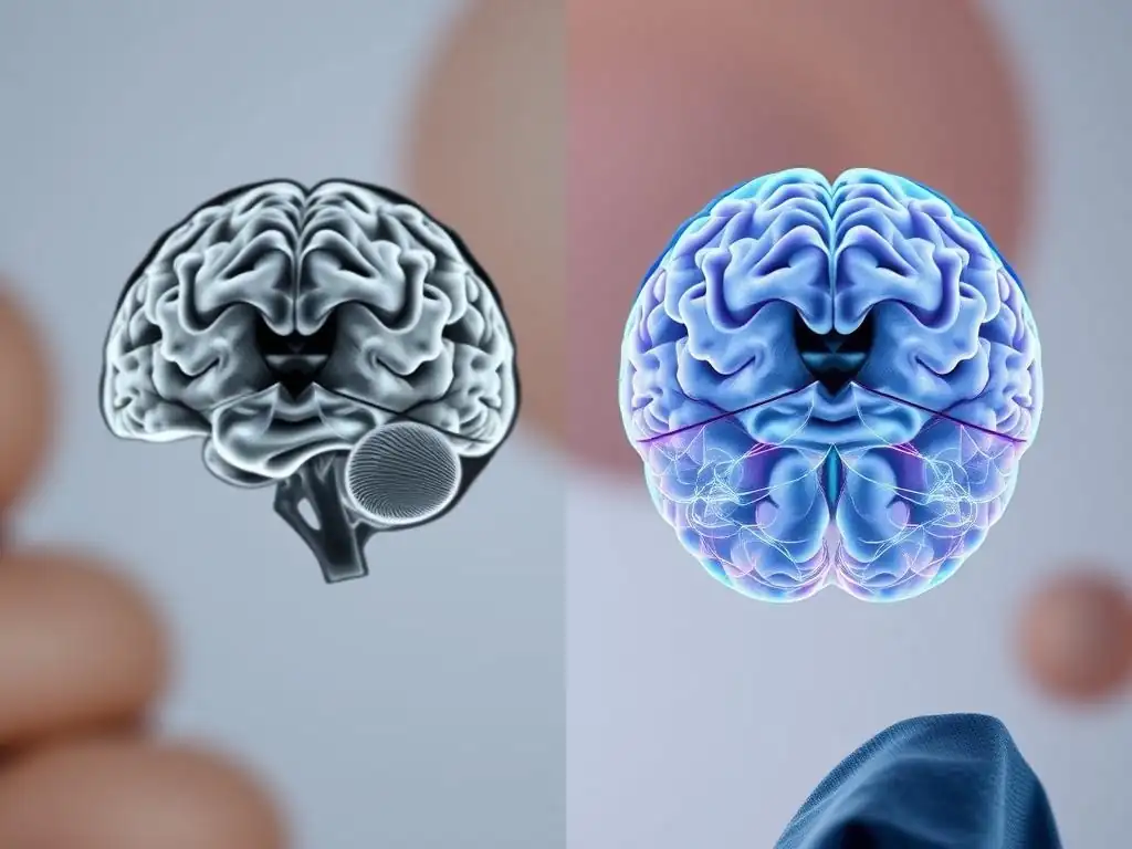
To better understand how neuroimaging findings manifest in practice, let's consider illustrative examples based on common patterns observed in research studies. These hypothetical case studies demonstrate the potential for brain scans to reveal the neural underpinnings of recovery after years of dedicated therapy.
Case Study 1: Generalized Anxiety Disorder – Amygdala Activity Before and After CBT
Consider "Sarah," a 45-year-old woman with a long history of Generalized Anxiety Disorder (GAD), characterized by persistent and excessive worry about everyday matters. Before starting long-term CBT (say, 2 years of weekly sessions followed by monthly maintenance), Sarah participated in an fMRI scan while viewing images designed to evoke worry-related thoughts (e.g., an overflowing inbox, a weather report showing a storm). Her scan showed high levels of activity in the amygdala and reduced connectivity between the amygdala and her prefrontal cortex, consistent with an anxious brain state.
After two years of intensive CBT, during which she learned to challenge her catastrophic thinking and practice acceptance of uncertainty, Sarah reported a significant reduction in daily worry and physical tension. A follow-up fMRI scan, using the same stimuli, showed a noticeable decrease in amygdala activation – perhaps a 20% reduction compared to her baseline scan – and increased functional connectivity between her vmPFC and amygdala.
This neural pattern aligned with her clinical improvement, suggesting that her brain had become less reactive to potential threats and better equipped to regulate emotional responses, providing tangible evidence of brain scans therapy effectiveness in GAD.
Meet "David," a 28-year-old man who struggled with severe social anxiety disorder, leading him to avoid social gatherings and public speaking. Before his long-term CBT (e.g., 18 months), an fMRI scan revealed hyperactivation in brain regions associated with social threat processing, including the amygdala and parts of the dorsal medial prefrontal cortex (dmPFC) involved in self-referential thinking and anticipating negative social evaluation. His scans also showed weaker functional connectivity within networks supporting social cognition and emotion regulation.
After completing his therapy, which included extensive exposure exercises (like giving speeches to small groups) and cognitive restructuring of fears of judgment, David felt significantly more comfortable in social situations and actively sought them out. A follow-up fMRI scan showed reduced activity in the amygdala and dmPFC when anticipating social interaction.
Crucially, analyses revealed strengthened functional connectivity between his vmPFC (involved in safety signaling) and the amygdala, as well as improved connectivity within broader social cognition networks. These changes suggested that his brain was now processing social information less defensively and with better regulatory control, illustrating brain scans therapy effectiveness in rewiring social fear circuits.
Case Study 3: Panic Disorder – Prefrontal Cortex Modulation of Fear Circuitry
Consider "Maria," a 35-year-old woman with a history of panic disorder, experiencing recurrent, unexpected panic attacks and persistent worry about future attacks. Before her long-term CBT (e.g., 2 years of weekly sessions), an fMRI scan during exposure to physical sensations mimicking panic (like rapid breathing) showed heightened activity in her insula (involved in sensing bodily states) and amygdala, with relatively weak activation in prefrontal areas responsible for interpreting these sensations non-catastrophically.
After mastering techniques like interoceptive exposure (intentionally triggering physical sensations in a controlled environment) and cognitive restructuring (relabeling sensations as benign), Maria's panic attacks became infrequent and less intense, and her anticipatory worry significantly decreased. A post-CBT fMRI scan showed reduced activity in the insula and amygdala during similar interoceptive challenges.
More notably, there was increased activation in her dorsolateral prefrontal cortex (dlPFC), a region involved in cognitive control and reappraisal, when experiencing these sensations. This suggested that her brain had developed stronger top-down control, allowing her to apply learned coping strategies and reinterpret bodily signals, demonstrating how long-term CBT can enhance prefrontal modulation of fear circuitry, a key aspect of brain scans therapy effectiveness.
Limitations and Future Directions in Neuroimaging Research for CBT
While neuroimaging has provided invaluable insights into the neural mechanisms of long-term CBT, the field still faces limitations and presents exciting avenues for future exploration. The technology is complex, expensive, and requires specialized expertise for both data acquisition and interpretation. Furthermore, current neuroimaging techniques offer correlational data – they show associations between brain changes and clinical outcomes, but establishing definitive causality can be challenging. The resolution and speed of scanning technologies continue to improve, but there are still limits to how precisely we can observe neural activity at the level of individual neurons or rapid synaptic events.
Despite these challenges, ongoing advancements in neuroimaging technology and analytical methods promise to deepen our understanding. Future research aims to refine the ability to predict treatment response based on baseline brain characteristics, identify specific neural markers that indicate readiness for or success in therapy, and potentially use imaging data to personalize therapeutic approaches. The ethical considerations surrounding the use of neuroimaging in clinical practice also warrant careful attention as the technology becomes more sophisticated and potentially more accessible.
Ethical Considerations in Clinical Neuroimaging
As neuroimaging moves from primarily a research tool towards potential clinical application in mental health, ethical considerations become paramount. Issues include the privacy and security of sensitive brain data, the potential for misinterpretation or over-simplification of complex findings when communicating with patients, and ensuring equitable access to potentially costly scanning technologies. There is also the ethical challenge of interpreting individual scans; while group-level findings in research are robust, applying these findings to predict outcomes for a single person requires caution and further validation.
Clinicians and researchers must work together to develop guidelines for the responsible use of neuroimaging in mental healthcare. This includes transparent communication with patients about what scans can and cannot reveal, avoiding deterministic language about brain findings, and ensuring that imaging data is used to complement, not replace, clinical judgment and the patient's subjective experience. As brain scans therapy effectiveness research progresses, maintaining a strong ethical framework will be crucial.
The Future of Personalized CBT: Tailoring Treatment Based on Neuroimaging Biomarkers
One of the most promising future directions for brain scans therapy effectiveness research is the potential for personalized treatment. If specific patterns of brain activity or structure predict who is most likely to benefit from long-term CBT versus another type of therapy (or a combination treatment), neuroimaging could potentially serve as a biomarker to help tailor treatment selection. For example, perhaps individuals with a certain pattern of prefrontal-amygdala connectivity might respond exceptionally well to exposure-based CBT, while those with different patterns might benefit more from therapies focusing on cognitive restructuring or mindfulness.
Research is beginning to explore these possibilities, using machine learning algorithms to analyze neuroimaging data and identify predictive patterns. While this is still in its early stages and not yet ready for routine clinical use, the vision is that one day, a baseline brain scan, combined with other clinical information, could help guide therapists and patients towards the most effective therapeutic path, maximizing the chances of successful long-term outcomes. The potential for using brain scans therapy effectiveness insights to create more precise and effective interventions is a significant driving force in the field.
Conclusion: Neuroimaging as a Tool for Understanding CBT's Impact on the Anxious Brain
Neuroimaging has revolutionized our understanding of how the brain changes in response to psychological therapy. Through techniques like fMRI and structural MRI, researchers have gathered compelling evidence that long-term Cognitive Behavioral Therapy for anxiety disorders is associated with measurable alterations in brain structure and function, particularly in regions involved in threat processing and emotion regulation, such as the amygdala and prefrontal cortex. These findings provide objective, biological support for the subjective and behavioral improvements observed in individuals who successfully complete comprehensive CBT.
The consistent observation of reduced amygdala reactivity and enhanced prefrontal control after long-term therapy underscores the profound neuroplastic capacity of the brain. CBT appears to facilitate a "rewiring" process, strengthening adaptive neural pathways and weakening maladaptive ones, leading to more resilient and less anxious patterns of brain activity. This growing body of evidence on brain scans therapy effectiveness not only validates CBT as a powerful intervention but also deepens our understanding of the biological basis of mental health recovery.
Reinforcing the Link Between Neural Changes and Symptom Reduction
The core message from decades of neuroimaging research on CBT is the strong link between observed neural changes and clinically significant symptom reduction. When individuals report feeling less anxious, worrying less, and engaging more freely in life, brain scans often reveal corresponding shifts towards more balanced and regulated neural activity. This correlation reinforces the understanding that mental health challenges are not merely "in the mind" in an abstract sense, but are deeply rooted in brain function, and that effective psychological therapies directly influence these neural processes.
By demonstrating that long-term CBT can lead to observable and relatively enduring changes in brain circuits, neuroimaging strengthens the scientific foundation of psychotherapy. It provides a powerful visual and quantitative illustration of the brain on the mend, offering hope and validation to individuals undergoing therapy and encouraging further investment in psychological treatments. The evidence for brain scans therapy effectiveness continues to grow, highlighting the biological reality of therapeutic change.
The Ongoing Evolution of Neuroimaging Technology in Anxiety Research
The field of neuroimaging is constantly evolving, with new techniques offering higher resolution, faster acquisition times, and more sophisticated ways to analyze complex brain networks. Future advancements, such as improvements in diffusion tensor imaging (DTI) to study white matter connectivity, or advancements in simultaneous EEG-fMRI recordings to capture both fast and slow neural dynamics, promise to provide even more detailed insights into the neural impact of long-term CBT.
As technology progresses, the potential for neuroimaging to inform clinical practice, perhaps through identifying biomarkers for treatment response or monitoring progress at a neural level, becomes increasingly tangible. While challenges remain, the ongoing evolution of neuroimaging technology ensures that research into brain scans therapy effectiveness will continue to push the boundaries of our understanding of how psychological interventions change the brain and promote lasting mental well-being.
Frequently Asked Questions
What is the main purpose of using brain scans to assess therapy effectiveness?
Brain scans offer a visual representation of brain activity and structure to determine if therapy has induced positive changes in the brain related to mental health conditions. This can help objectively measure progress.
What types of brain scans are typically used to evaluate the impact of therapy?
Common scans include fMRI (functional Magnetic Resonance Imaging) to measure brain activity during tasks, and structural MRI to assess brain structure. Other options like PET scans can also provide valuable data on brain function.
How can brain scans help personalize therapy approaches?
By identifying specific brain regions affected by a condition and monitoring changes during therapy, scans can help tailor treatment plans. This allows for a more targeted and effective therapeutic intervention.
What are some limitations of relying solely on brain scans to determine therapy effectiveness?
Brain scans are just one piece of the puzzle. Factors like individual variability, the complexity of mental health conditions, and the need for subjective assessments alongside objective data must be considered.
Are brain scans a standard practice in monitoring the progress of all types of mental health therapy?
No, brain scans are not typically used for all types of therapy. They are more commonly employed in research settings or for complex cases where objective measures are needed to complement clinical assessments.
Final Reflection: The Enduring Power of Healing
The images captured by brain scans, once solely diagnostic tools, are now revealing the profound and lasting impact of therapy on the brain's architecture. This isn't just about symptom reduction; it's about witnessing tangible, measurable changes in neural pathways, demonstrating the brain's remarkable plasticity and our capacity for healing. This understanding offers a powerful new perspective on the efficacy of therapeutic interventions, moving beyond anecdotal evidence to concrete neurological proof.
The journey of healing, as depicted through these evolving brain scans, underscores the importance of perseverance and hope. It's a testament to the human spirit's resilience, a reminder that even after years of struggle, the brain can rewire itself, forging new connections and finding pathways to well-being. The brain is the most complex organ in the body, but it is also the most adaptable.
This adaptability, revealed through these scans, offers a beacon of hope for those navigating mental health challenges. It is a powerful counter-narrative to the often-pervasive feelings of hopelessness and despair that can accompany mental illness.
Consider the implications of these findings. What does this new understanding of therapeutic efficacy mean for you? Perhaps it inspires you to seek help, to embrace the potential for change, or to advocate for increased access to effective therapies. Let the images of healing brains, captured through advanced neuroimaging, serve as a powerful reminder of your own inherent capacity for growth, resilience, and transformation. The journey may be long, but the destination – a healthier, more integrated self – is within reach.
Unlock Your Brain's Potential: Take the Next Step
Thanks for taking the time to explore the fascinating world of brain recovery with us! You've gained valuable insights into the long-term effects of therapy – now it's time to put that knowledge into action.
Here's what you can do next:
- Schedule a consultation with your therapist to discuss your progress and explore new strategies based on the latest research. This is the most impactful step you can take towards continued healing.
- Share this article with someone who might find it inspiring – their journey to recovery could be enriched by this information. Let's build a supportive community!
- Try a simple mindfulness exercise today – even five minutes can make a difference in fostering the brain's plasticity and resilience.
So, what small step will you take today to nurture your brain's incredible ability to heal and grow?