
Decoding the Brain: What Scans Reveal About Therapy for OCD

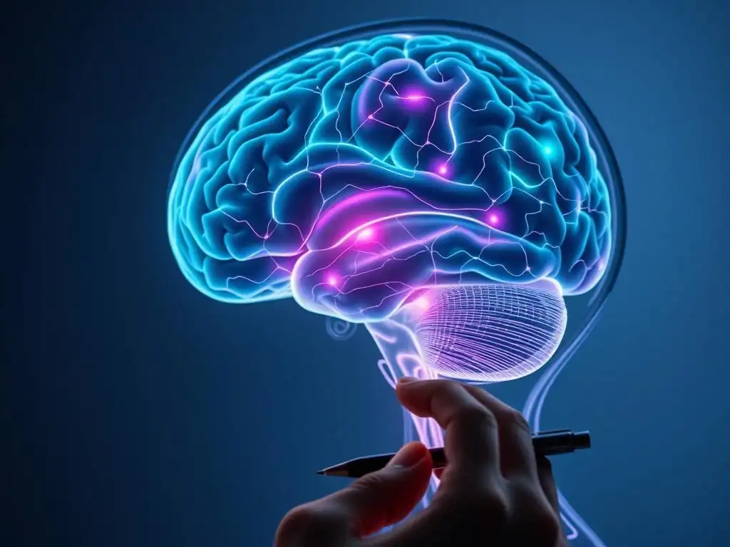
Welcome to Mind Care Guide, your trusted companion on the journey to mental well-being. Ever wondered what actually happens inside your brain during therapy, especially when tackling something as complex as OCD? This isn't just about talking; it's about tangible changes. Dive into "Decoding the Brain: What Scans Reveal About Therapy for OCD" and discover how OCD brain scan therapy offers a powerful window into understanding and overcoming this challenging condition. Could this be the key to unlocking a calmer, clearer mind? Let's explore the fascinating neuroscience behind it.
Decoding the Brain: What Scans Reveal About Therapy for OCD
- Introduction: Understanding OCD Through Brain Imaging
- How OCD Affects the Brain: Identifying Key Neural Correlates
- Brain Scans Used in OCD Research and Clinical Practice
- OCD Brain Scan Therapy: Exploring the Impact of Treatment
- Long-Term Therapy Outcomes and Brain Changes in OCD
- Case Studies: Real-World Examples of OCD Brain Scan Therapy
- The Future of OCD Treatment: Personalized Approaches Based on Brain Scans
- Limitations of Brain Scans in OCD Treatment
- Conclusion: Harnessing Neuroscience for Better OCD Care
- Frequently Asked Questions
- Final Reflection: Unveiling the Mind's Landscape in OCD Treatment
- Unlock Your Brain's Potential: Start Your OCD Journey Today
Introduction: Understanding OCD Through Brain Imaging
Obsessive-Compulsive Disorder (OCD) is a complex condition characterized by intrusive thoughts (obsessions) and repetitive behaviors (compulsions). For many years, our understanding of OCD was primarily based on clinical observation and patient reports. However, advancements in neuroscience, particularly brain imaging techniques, have provided invaluable insights into the neural underpinnings of this disorder. These technologies allow researchers and clinicians to look inside the living brain, observing its structure and activity, offering a new dimension to understanding the challenges faced by individuals with OCD.
The advent of sophisticated brain scanning technologies has revolutionized the study of mental health conditions like OCD. By visualizing the brain, we can identify specific areas or circuits that may function differently in individuals with OCD compared to those without the disorder. This objective, biological perspective complements traditional psychological and behavioral assessments, painting a more complete picture of OCD. This deeper understanding is crucial not only for diagnosis but also for developing and refining treatments, including the emerging concept of OCD brain scan therapy, where insights from imaging inform treatment strategies.
The Role of Brain Scans in Understanding and Monitoring OCD Treatment
Brain scans play a pivotal role in both understanding the neural basis of OCD and monitoring the effects of therapeutic interventions. Before treatment begins, scans can help researchers identify neural markers associated with the disorder, such as altered activity in specific brain regions known to be involved in fear, decision-making, and habit formation. This baseline information is crucial for research aimed at understanding the root causes of OCD and identifying potential targets for new therapies.
Furthermore, brain imaging allows clinicians and researchers to observe changes in brain structure and function after a patient undergoes therapy, whether it's psychotherapy like Cognitive Behavioral Therapy (CBT) or medication. By comparing scans taken before and after treatment, studies can demonstrate how effective therapies literally reshape or re-pattern brain activity. This provides objective evidence of treatment effectiveness at a biological level and helps explain how therapies work, moving beyond just observing symptom reduction to understanding the underlying neural recovery process in OCD. This evidence strengthens the rationale for various forms of OCD brain scan therapy approaches.
The Importance of Integrating Neuroscience with Clinical Practice for OCD
Integrating neuroscience findings with clinical practice is becoming increasingly important in the field of OCD treatment. While clinical assessment based on symptoms remains the cornerstone of diagnosis and treatment planning, insights from brain imaging offer a powerful complementary lens. Understanding which brain circuits are implicated in OCD symptoms can help clinicians tailor interventions to target those specific neural pathways, potentially leading to more effective and personalized treatment strategies.
Moreover, the integration of neuroscience can help destigmatize OCD by framing it, in part, as a disorder with identifiable biological correlates, similar to other medical conditions. This can be empowering for patients and their families.
As our understanding grows, the insights gained from studying the brain through scans are informing the development of more targeted interventions, including novel brain stimulation techniques or neurofeedback, which are forms of OCD brain scan therapy that directly engage with neural activity. Bridging the gap between brain research and clinical care is essential for advancing the field and improving outcomes for those affected by OCD.
How OCD Affects the Brain: Identifying Key Neural Correlates
Research using brain imaging has consistently pointed to specific brain regions and circuits that function differently in individuals with OCD. These findings have helped shape our understanding of the disorder not just as a psychological struggle but also as a condition rooted in specific neural dysfunctions. Identifying these key neural correlates is fundamental to developing targeted interventions and understanding how treatments impact the brain.
The primary circuit implicated in OCD is often referred to as the cortico-striato-thalamo-cortical (CSTC) circuit. This complex network involves areas responsible for processing information, making decisions, and controlling behavior. In individuals with OCD, this circuit appears to be overactive or dysregulated, leading to the characteristic symptoms of intrusive thoughts and compulsive behaviors. Understanding this circuit is central to exploring the potential of OCD brain scan therapy.
Overactive Brain Regions in OCD: The Orbitofrontal Cortex and Anterior Cingulate Cortex
Two key players in the dysregulated CSTC circuit in OCD are the orbitofrontal cortex (OFC) and the anterior cingulate cortex (ACC). The OFC is involved in processing information related to reward, punishment, and decision-making, particularly in evaluating the emotional significance of thoughts and actions. In many studies, individuals with OCD show heightened activity in the OFC, especially when provoked by stimuli related to their obsessions. This overactivity is thought to contribute to the feeling of 'not right' or excessive concern associated with obsessions.
Similarly, the anterior cingulate cortex (ACC) is involved in error detection, conflict monitoring, and motivation. Research using fMRI has frequently found increased activity in the ACC in individuals with OCD, particularly when they are resisting compulsions or processing anxiety-provoking information. This heightened ACC activity may reflect a constant state of alarm or an inability to disengage from intrusive thoughts, driving the compulsive need for resolution. These findings are crucial when considering how OCD brain scan therapy aims to normalize activity in these areas.
Neurotransmitters and OCD: The Role of Serotonin, Dopamine, and Glutamate
Beyond specific brain regions, imbalances in certain neurotransmitters—chemical messengers in the brain—are also strongly implicated in OCD. Serotonin is perhaps the most well-known, given that selective serotonin reuptake inhibitors (SSRIs) are a first-line medication treatment for OCD. Studies using PET scans have investigated serotonin transporter and receptor binding in individuals with OCD, often finding alterations in serotonin system function, although findings can be complex and vary across studies.
Dopamine, another key neurotransmitter involved in reward, motivation, and habit formation, is also thought to play a role. Some research suggests that dopamine dysfunction, particularly in the striatum (part of the CSTC circuit), may contribute to the compulsive behaviors seen in OCD. Glutamate, the brain's primary excitatory neurotransmitter, is also gaining attention, with studies suggesting altered glutamate levels or signaling in certain brain areas in OCD. Understanding these neurochemical imbalances is vital for developing pharmacological interventions and provides targets for researchers using brain scans to understand how treatments affect the brain chemistry related to OCD.
Structural Brain Differences in Individuals with OCD: Gray Matter Volume
Structural MRI studies have investigated whether there are observable differences in the physical structure of the brain in individuals with OCD. While findings are not always consistent across all studies, some research points towards subtle differences in gray matter volume (GMV) in certain brain regions. Gray matter consists primarily of neuronal cell bodies and dendrites, and its volume can reflect the number of neurons and synapses in an area.
Meta-analyses of structural MRI studies have sometimes reported reduced gray matter volume in areas like the orbitofrontal cortex, anterior cingulate cortex, and striatum in individuals with OCD compared to healthy controls. Conversely, some studies have reported increased gray matter volume in other regions or within sub-regions of the implicated circuit. These structural differences are thought to either predispose individuals to OCD or result from the chronic patterns of activity associated with the disorder. Investigating how these structural differences change with treatment is an area of interest for research into the long-term effects of OCD brain scan therapy.
Brain Scans Used in OCD Research and Clinical Practice
A variety of brain imaging techniques are employed in OCD research and, to a lesser extent currently, in clinical practice. Each technique provides a different window into brain function or structure, offering unique insights into the neural basis of the disorder and how it responds to treatment. Understanding these methods is key to interpreting findings related to OCD brain scan therapy.
These technologies range from those that measure metabolic activity and blood flow to those that map the physical structure of the brain or the distribution of specific molecules. The choice of scanning method depends on the specific question being asked, whether it's about real-time brain activity during a task, the concentration of a neurotransmitter, or the volume of gray matter in a particular region.
Functional Magnetic Resonance Imaging (fMRI) for OCD Assessment: Measuring Brain Activity
Functional Magnetic Resonance Imaging (fMRI) is one of the most commonly used brain imaging techniques in OCD research. fMRI measures brain activity by detecting changes in blood flow. When a brain region is more active, it requires more oxygenated blood, and fMRI can detect the magnetic properties of oxygenated versus deoxygenated hemoglobin. By measuring the blood-oxygen-level-dependent (BOLD) signal, fMRI can show which parts of the brain are more or less active during specific tasks, or even in a resting state.
In OCD research, fMRI is frequently used to observe brain activity while individuals are exposed to stimuli related to their obsessions or performing tasks that challenge the affected brain circuits, such as decision-making or conflict resolution tasks. Studies have consistently shown heightened activity in the OFC and ACC during symptom provocation tasks in individuals with OCD. Critically, fMRI is also used after treatment to see if this heightened activity has decreased or normalized, providing direct evidence of therapy's impact on brain function, a core aspect of OCD brain scan therapy research.
Positron Emission Tomography (PET) Scans and OCD Neurochemistry: Receptor Binding Studies
Positron Emission Tomography (PET) scans offer a different type of insight by measuring metabolic activity or the distribution of specific molecules in the brain. This is achieved by injecting a small amount of a radioactive tracer substance that binds to or is taken up by specific molecules or cells. For instance, tracers can be designed to bind to neurotransmitter receptors or transporters.
PET scans have been instrumental in studying the neurochemical basis of OCD. Researchers have used PET to investigate the serotonin system, observing the density of serotonin transporters or receptors in different brain regions. Studies have also explored the dopamine and glutamate systems using PET. While more complex and less widely available than fMRI, PET provides unique information about the biochemical environment of the brain, helping researchers understand how medications like SSRIs affect neurotransmitter systems and how these changes relate to symptom improvement in the context of OCD brain scan therapy.
Structural MRI: Identifying Brain Volume Differences and White Matter Integrity in OCD
Structural Magnetic Resonance Imaging (MRI) provides detailed images of the brain's anatomy. Unlike fMRI, structural MRI focuses on the physical structure, shape, and volume of brain tissues. It can differentiate between gray matter, white matter (composed of nerve fibers connecting different brain regions), and cerebrospinal fluid. This technique is used to identify potential structural abnormalities or differences in brain regions implicated in OCD.
As mentioned earlier, structural MRI studies have investigated differences in gray matter volume in areas like the OFC, ACC, and striatum. Additionally, techniques like Diffusion Tensor Imaging (DTI), a type of structural MRI, can assess the integrity and organization of white matter tracts, which are the communication pathways between different brain regions. Alterations in white matter connectivity within the CSTC circuit have also been reported in OCD. Tracking structural changes over time, potentially after treatment, is another way brain scans contribute to understanding OCD brain scan therapy.
OCD Brain Scan Therapy: Exploring the Impact of Treatment
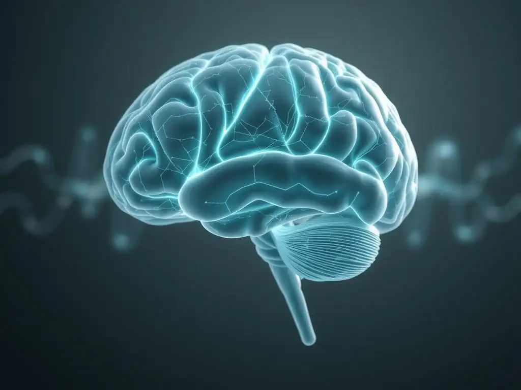
The concept of OCD brain scan therapy isn't about using the scan itself as a treatment, but rather using the information gained from brain scans to understand how existing therapies work, predict who might respond best to which treatment, and potentially develop new, more targeted interventions. Research using brain imaging has provided compelling evidence that effective treatments for OCD, both psychotherapy and medication, lead to measurable changes in brain function and structure.
These studies demonstrate the brain's plasticity – its ability to change and adapt in response to experience and intervention. Observing these changes through brain scans provides a biological correlate to the psychological and behavioral improvements seen in patients, reinforcing the power of evidence-based treatments for OCD.
Cognitive Behavioral Therapy (CBT) and Brain Activity Changes: Modulation of Frontostriatal Circuits
Cognitive Behavioral Therapy (CBT), particularly its exposure and response prevention (ERP) component, is a highly effective psychotherapy for OCD. Brain imaging studies have shown that successful CBT treatment is associated with significant changes in activity within the very same brain circuits implicated in OCD, particularly the CSTC loop involving the frontal cortex and striatum.
For instance, fMRI studies comparing scans before and after successful CBT often report reduced activity in the orbitofrontal cortex and anterior cingulate cortex when patients are confronted with anxiety-provoking stimuli. This suggests that CBT helps to "turn down" the overactive alarm signals in these regions.
Furthermore, some studies indicate increased activity in areas involved in executive control and cognitive reappraisal, such as parts of the prefrontal cortex, suggesting that therapy strengthens the brain's ability to regulate thoughts and behaviors. These observed neural changes provide biological backing for the effectiveness of CBT as a form of OCD brain scan therapy research.
Exposure and Response Prevention (ERP) and Neural Plasticity: Strengthening Adaptive Neural Pathways
Exposure and Response Prevention (ERP), a core technique within CBT, directly targets the cycle of obsessions and compulsions. By gradually exposing individuals to their feared situations or thoughts without allowing them to perform compulsions, ERP helps them learn that their fears are often unfounded and that they can tolerate the anxiety without engaging in rituals. Brain imaging studies suggest that ERP facilitates neural plasticity, helping to "rewire" the brain.
Studies using fMRI and other techniques after successful ERP have shown not only decreased activity in previously overactive areas like the OFC and ACC but sometimes increased activity in areas associated with cognitive control and habit inhibition, such as the dorsal striatum or specific prefrontal regions. This suggests that ERP helps build new, more adaptive neural pathways that bypass the old, compulsive ones. Effectively, ERP helps train the brain to respond differently to triggers, and brain scans provide evidence of this neural retraining, supporting the idea that insights from imaging enhance our understanding of OCD brain scan therapy.
Medication's Influence on OCD Brain Scans: SSRIs and Beyond - Effects on Serotonin Transporters
Medication, primarily selective serotonin reuptake inhibitors (SSRIs), is another mainstay of OCD treatment. Brain imaging, particularly PET scans, has been crucial in understanding how these medications work at a neurochemical level and how these chemical changes relate to symptom improvement. SSRIs are believed to increase serotonin levels in the brain by blocking its reuptake.
PET studies using tracers that bind to serotonin transporters (SERTs) have shown that SSRIs effectively occupy these transporters. Furthermore, studies have investigated how SSRI treatment affects activity in brain regions implicated in OCD. Similar to CBT, successful medication treatment is often associated with reductions in activity in the OFC and ACC, as observed with fMRI.
Some research also suggests changes in receptor binding or metabolic activity in other parts of the CSTC circuit. While the exact mechanisms are still being fully elucidated, brain scans provide objective evidence that medications induce physiological changes in the brain that correlate with symptom reduction, contributing to our understanding of OCD brain scan therapy from a pharmacological perspective.
Long-Term Therapy Outcomes and Brain Changes in OCD
Understanding the long-term effects of therapy on the brain is crucial for evaluating treatment durability and potential for recovery. Brain imaging studies that follow patients over extended periods after successful treatment provide valuable insights into whether the neural changes observed immediately post-treatment are sustained and whether they correlate with long-term symptom remission.
These studies help address important questions about the trajectory of brain recovery in OCD and whether certain neural markers are associated with a lower risk of relapse. Investigating these long-term outcomes using brain scans adds a vital dimension to understanding the enduring impact of effective OCD brain scan therapy.
Sustained Remission and Normalization of Brain Function: Long-Term Follow-Up Studies
Long-term follow-up studies using brain imaging aim to determine if the brain changes observed after initial successful treatment persist over months or even years. Research in patients who achieve sustained remission from OCD after CBT or medication often shows that the normalization of activity in key brain regions, such as the OFC and ACC, is maintained. For example, a study following patients for two years after successful treatment might find that the reduced activity in the OFC seen immediately post-treatment is still present, mirroring their continued lack of significant OCD symptoms.
These findings suggest that effective therapy doesn't just offer temporary relief but can lead to lasting changes in brain function, potentially reflecting a recalibration of the dysregulated circuits. While complete "normalization" to the level of healthy controls isn't always observed in every region, the pattern of activity often shifts significantly towards a less pathological state. This provides a biological basis for the hope of long-term recovery in OCD and highlights the potential for sustained benefits from OCD brain scan therapy approaches.
The Relationship Between Symptom Reduction and Brain Scan Results: Correlation Analyses
A key area of research involves correlating the degree of symptom reduction reported by patients with the magnitude of changes observed in their brain scans. For instance, studies might analyze whether a greater decrease in OFC activity post-treatment is associated with a larger drop in Yale-Brown Obsessive Compulsive Scale (Y-BOCS) scores, the standard measure of OCD symptom severity.
Numerous studies have found significant correlations between symptom improvement and changes in brain activity or structure. For example, research might show that patients with the most significant reduction in compulsions after ERP exhibit the largest increases in gray matter volume in specific prefrontal areas or the most pronounced decreases in ACC activity during conflict tasks. These correlations strengthen the link between the observed neural changes and the clinical improvements, suggesting that the brain imaging findings are not merely incidental but are direct indicators of the therapeutic effect and are central to the concept of OCD brain scan therapy research.
Predicting Treatment Response with Pre-Treatment Brain Scans: Identifying Neural Predictors
An exciting area of research involves using brain scans taken before treatment to predict how well a patient might respond to a specific therapy (e.g., CBT vs. medication). If researchers could identify neural markers that predict treatment success or failure, it could pave the way for more personalized and effective treatment selection.
Preliminary studies have explored this possibility. For example, some research has suggested that the level of activity in certain brain regions, or the connectivity between them, at baseline might predict response to SSRIs or CBT.
One study, for instance, might find that patients with a particular pattern of connectivity within the CSTC circuit are more likely to respond well to ERP than those with a different pattern. While this field is still developing and not yet used routinely in clinical practice, identifying such neural predictors through brain scans holds significant promise for optimizing OCD brain scan therapy by matching patients to the interventions most likely to benefit them.
Case Studies: Real-World Examples of OCD Brain Scan Therapy
While the term "OCD brain scan therapy" primarily refers to research using scans to understand and refine treatment, hypothetical or generalized case studies based on research findings can illustrate the insights gained. These examples demonstrate how brain imaging has provided concrete evidence of the biological impact of effective OCD treatments on the brain.
These case studies, drawn from the patterns observed across numerous research participants, help to make the often-abstract concepts of neural circuits and activity changes more tangible. They highlight the potential for brain scans to objectively show that therapy is not just "in the mind" but is fundamentally changing how the brain functions.
Case Study 1: fMRI Before and After CBT - Reduced Activity in the Orbitofrontal Cortex
Consider a hypothetical patient, "Sarah," who struggled severely with contamination obsessions and washing compulsions. Before starting a course of intensive CBT with ERP, Sarah underwent an fMRI scan during which she was asked to handle objects she perceived as contaminated. Her pre-treatment scan showed significantly heightened activity in her orbitofrontal cortex (OFC) and anterior cingulate cortex (ACC) when exposed to these triggers, compared to neutral objects. This heightened activity correlated with her intense anxiety and urge to wash.
After 15 sessions of successful CBT, which included extensive ERP, Sarah's symptoms dramatically improved, and her Y-BOCS score dropped significantly. A follow-up fMRI scan, performed under the same conditions, showed a marked reduction in activity in her OFC and ACC when exposed to the previously feared objects. The activity levels in these regions were much closer to those observed in healthy controls. This case illustrates how fMRI can visually demonstrate the neural changes underlying successful psychotherapeutic OCD brain scan therapy.
Case Study 2: PET Scan Results and Medication Management - Changes in Serotonin Receptor Binding
Imagine a patient, "David," diagnosed with OCD, for whom medication was recommended. Before starting an SSRI, David participated in a research study involving a PET scan using a tracer that binds to a specific type of serotonin receptor known to be implicated in OCD. His baseline scan revealed a particular pattern of receptor binding in areas like the striatum.
After several months of consistent SSRI treatment, David experienced a significant reduction in his obsessive thoughts and compulsive behaviors. A follow-up PET scan showed a change in the pattern of serotonin receptor binding in the striatum and other brain regions. While complex, these changes in neurochemistry observed via PET scans are thought to be part of the mechanism by which SSRIs exert their therapeutic effect on the brain circuits involved in OCD. This kind of study showcases how PET can provide insights into the neurochemical impact of medication in the context of OCD brain scan therapy research.
Case Study 3: Structural MRI Changes After ERP - Increased Gray Matter Volume in Specific Regions
Consider "Michael," who underwent a course of ERP therapy for checking compulsions. While structural changes are often more subtle and slower than functional changes, research has explored how intensive therapy might impact brain structure over time. Michael's structural MRI scan before therapy might have shown certain characteristics, perhaps related to gray matter volume in areas like the prefrontal cortex.
After several months of successful ERP and sustained symptom reduction, a follow-up structural MRI scan might, based on some research findings, reveal an increase in gray matter volume in specific prefrontal regions associated with cognitive control and decision-making. This hypothesized change could reflect increased neural density or synaptic connections in areas that are strengthened by the process of learning to resist compulsions and tolerate uncertainty during ERP. This type of structural change, while still an active area of research, suggests that effective OCD brain scan therapy might lead to lasting physical alterations in the brain's structure.
The Future of OCD Treatment: Personalized Approaches Based on Brain Scans
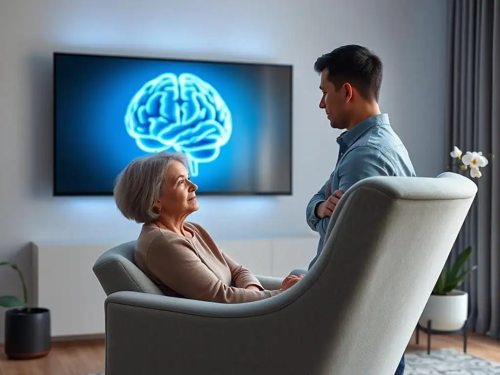
The insights gained from brain imaging are pointing towards a future where OCD treatment could become more personalized and targeted. Moving beyond a one-size-fits-all approach, researchers hope to use brain scan data to identify biomarkers that predict treatment response, integrate this information into clinical decision-making, and develop novel brain-based interventions.
This vision of personalized OCD brain scan therapy holds the promise of improving treatment outcomes, reducing the time it takes to find an effective therapy for an individual, and potentially offering new options for those who don't respond to current standard treatments.
Identifying Biomarkers for OCD Treatment Response: Neural Signatures of Treatment Success
One of the most promising future applications of brain imaging in OCD is the identification of biomarkers – measurable biological indicators – that can predict how a patient will respond to a specific treatment. Imagine a scenario where a pre-treatment brain scan could tell a clinician whether a patient is more likely to benefit from CBT or medication.
Research is actively exploring various potential neural biomarkers, such as patterns of activity in certain brain circuits during specific tasks (measured by fMRI), levels of certain neurotransmitters or receptor binding (measured by PET), or structural characteristics like white matter integrity (measured by DTI). Identifying reliable neural signatures of treatment success could revolutionize how we approach treatment selection, making OCD brain scan therapy a more predictive and personalized process.
Integrating Brain Scan Data into Treatment Decisions: Stratified Treatment Approaches
Once reliable neural biomarkers are identified, the next step is integrating this brain scan data into clinical decision-making. This could lead to stratified treatment approaches, where patients are grouped based on their brain characteristics and then directed towards the therapy most likely to be effective for their specific neural profile.
For example, a patient whose scan shows a particular pattern of serotonin transporter availability might be prioritized for SSRI medication, while a patient with a specific pattern of connectivity in the CSTC circuit might be identified as a strong candidate for ERP. This data-driven approach to treatment selection, informed by brain scans, has the potential to significantly improve response rates and reduce the trial-and-error process that many patients with OCD currently face in finding effective help. This represents a significant step towards truly personalized OCD brain scan therapy.
The Potential of Neurofeedback and Brain Stimulation for OCD: Targeting Specific Brain Circuits
Insights from brain imaging are also directly informing the development of novel therapeutic techniques that directly target the dysregulated brain circuits in OCD. Neurofeedback, for instance, uses real-time fMRI or EEG (electroencephalography) to allow patients to see their own brain activity and learn to regulate it. If a scan shows overactivity in the ACC, neurofeedback could potentially train the patient to decrease activity in that specific region.
Brain stimulation techniques, such as Transcranial Magnetic Stimulation (TMS) or Deep Brain Stimulation (DBS) (though DBS is typically for severe, treatment-resistant cases), also rely on understanding which brain areas are implicated in OCD. TMS can non-invasively stimulate or inhibit activity in specific cortical regions, while DBS involves surgically implanting electrodes to stimulate deeper brain structures like the striatum. These emerging therapies are direct examples of interventions rooted in neuroscience findings from brain scans and represent a potential future direction for OCD brain scan therapy methods.
Limitations of Brain Scans in OCD Treatment
Despite the exciting potential of brain imaging in understanding and potentially guiding OCD treatment, it's important to acknowledge the current limitations. Brain scans are powerful research tools, but their direct application in routine clinical practice for individual patient care is still evolving and faces significant hurdles.
These limitations include practical considerations like cost and accessibility, ethical questions surrounding the use of brain data, and the inherent complexity of interpreting brain imaging results. Recognizing these challenges is crucial for setting realistic expectations about the current role of brain scans in the treatment of OCD.
Cost and Accessibility of Brain Imaging Techniques: Practical Considerations
One of the most significant limitations is the cost and accessibility of advanced brain imaging techniques like fMRI and PET. These scans require expensive equipment and specialized personnel to operate and interpret. As a result, they are typically available only at major research institutions or large medical centers, making them inaccessible for routine use in most standard clinical settings.
The high cost means that using brain scans for every patient to inform treatment decisions is currently not feasible within most healthcare systems. While research continues to explore the value of these scans, the practical constraints of cost and limited availability mean that, for now, OCD brain scan therapy is primarily a research concept and not a widely implemented clinical practice.
Ethical Considerations in Using Brain Scans for Mental Health: Privacy and Stigma
The use of brain scans in the context of mental health also raises important ethical considerations. Brain imaging data is highly personal and potentially sensitive. Issues surrounding data privacy, who has access to this information, and how it is stored and used are paramount. There are concerns about potential discrimination based on brain scan findings, for example, in insurance or employment.
Furthermore, while neuroscience can help destigmatize mental illness by highlighting biological factors, there's also a risk of over-medicalization or reducing a complex disorder like OCD solely to a brain circuit problem, potentially overlooking the psychological and environmental factors that are also critical. Careful ethical guidelines and open discussions are needed as OCD brain scan therapy evolves to ensure patient privacy and avoid exacerbating stigma.
Interpretation Challenges and the Need for Expert Analysis: Complexity of Brain Data
Interpreting brain imaging data is complex and requires significant expertise. Raw scan data needs to be processed and analyzed using sophisticated software and statistical methods. Findings are often based on group averages in research studies, and applying these findings to an individual patient can be challenging. A single scan might show variations that are within the range of normal variability or influenced by factors unrelated to OCD.
Identifying reliable biomarkers that are robust enough for individual clinical prediction is difficult. The brain is highly dynamic, and activity patterns can change depending on the person's state, the specific task being performed, and even minor variations in the scanning environment. Therefore, integrating brain scan data into clinical decisions requires not only technological advancements but also highly trained experts who can accurately interpret the complex findings, which is a current bottleneck for widespread OCD brain scan therapy.
Conclusion: Harnessing Neuroscience for Better OCD Care
The journey into decoding the brain's role in OCD through imaging has profoundly advanced our understanding of this challenging disorder. Brain scans have moved OCD from being viewed solely as a psychological affliction to one with identifiable neural underpinnings, providing objective evidence for the biological reality of the condition and the physical impact of effective treatments.
While significant limitations exist in applying these techniques to routine clinical practice today, the ongoing research into OCD brain scan therapy holds immense promise. It represents a future where neuroscience is not just an academic pursuit but a direct contributor to improving patient care, offering hope for more precise, effective, and personalized interventions for OCD.
The Importance of Brain Scans in Understanding OCD: Complementing Clinical Assessment
Brain scans are invaluable tools that complement traditional clinical assessment in understanding OCD. While patient interviews and behavioral observations remain essential for diagnosis and understanding the individual's lived experience, imaging provides an objective window into the brain's structure and function. It allows researchers to identify the specific neural circuits and neurotransmitter systems that are dysregulated in OCD, providing a biological framework for the symptoms observed clinically.
This deeper understanding is critical for advancing the field, informing the development of targeted therapies, and validating the impact of existing treatments at a biological level. Brain scans reinforce that OCD is a real, biologically influenced disorder, helping to reduce stigma and encouraging a comprehensive view that integrates mind and brain.
The Value of Evidence-Based OCD Treatment: Combining Therapy and Neuroscience
The findings from brain imaging studies strongly underscore the value of evidence-based treatments for OCD, such as CBT (especially ERP) and medication. By demonstrating that these therapies lead to measurable, positive changes in brain activity and structure, neuroscience provides compelling biological evidence for their effectiveness. This reinforces the importance of accessing and adhering to these proven treatments.
Furthermore, the research highlights how therapy and medication both impact the brain, albeit through different mechanisms. This supports the clinical practice of combining treatments when necessary or tailoring approaches based on individual patient needs. The insights gained from OCD brain scan therapy research provide a scientific foundation for these clinical decisions, emphasizing that effective treatment leads to tangible brain changes correlated with symptom relief.
Hope for the Future: Advancements in OCD Brain Scan Therapy and Precision Medicine
The field of OCD brain scan therapy is still in its nascent stages clinically, but the rapid advancements in imaging technology and data analysis offer significant hope for the future of OCD treatment. Research is moving towards identifying specific neural biomarkers that could predict treatment response, allowing for a future where patients are matched to the most effective therapy from the outset, reducing suffering and improving outcomes.
The potential integration of brain scan data into personalized treatment plans, along with the development of novel brain-targeted interventions like neurofeedback and stimulation techniques informed by imaging, points towards a future of precision medicine for OCD. While challenges remain, the ability to decode the brain through scans is paving the way for more effective, individualized, and biologically informed care for individuals living with Obsessive-Compulsive Disorder.
Frequently Asked Questions
What is an OCD brain scan and how does it relate to therapy?
An OCD brain scan, like fMRI or PET scans, can reveal neurological activity associated with OCD symptoms. This information can help understand the effectiveness of therapy by showing changes in brain activity pre- and post-treatment.
Can a brain scan diagnose OCD?
While brain scans cannot definitively diagnose OCD on their own, they can support a clinical diagnosis. Scans reveal patterns of brain activity often seen in individuals with OCD, such as hyperactivity in certain brain regions.
What types of brain scans are used in OCD research and therapy?
Common scans include fMRI (functional Magnetic Resonance Imaging), which measures brain activity through blood flow, and PET (Positron Emission Tomography) scans, which use radioactive tracers to visualize brain function. These scans help researchers understand brain changes linked to OCD and therapy.
How can brain scans help personalize OCD therapy?
By identifying specific brain regions affected in an individual with OCD, scans can potentially guide the selection of more targeted therapy approaches. This could lead to more personalized and effective treatment plans.
Are brain scans a necessary part of OCD treatment?
Brain scans are not typically a routine part of OCD treatment. They are primarily used in research settings to better understand OCD and evaluate the effectiveness of different therapies, but may play a role in the future of personalized treatment.
Final Reflection: Unveiling the Mind's Landscape in OCD Treatment
The intricate workings of the OCD brain, once shrouded in mystery, are increasingly illuminated by advanced neuroimaging techniques. This offers not just a deeper understanding of the disorder, but a powerful roadmap for developing more effective and targeted therapies. The ability to visualize the neural pathways involved in obsessive-compulsive behaviors provides a level of precision previously unimaginable, paving the way for personalized treatment approaches tailored to individual brain responses.
The journey of understanding OCD through brain scans has been profoundly impactful, shifting the conversation from solely behavioral interventions to a more holistic approach that considers the underlying neurological mechanisms. This shift has fostered a sense of hope and empowerment for those struggling with OCD, demonstrating that their experiences are rooted in tangible biological processes, not simply a matter of willpower or personal failing. The brain is not a machine to be fixed, but a garden to be tended.
This quote resonates deeply, as brain imaging allows us to understand the specific "weeds" that need tending in the case of OCD, guiding the development of more effective "gardening" techniques.
Consider the implications of this research. What if we could predict treatment response based on pre-therapy brain scans? What if we could monitor treatment progress in real-time, adjusting interventions as needed?
This is no longer science fiction, but a rapidly approaching reality. You, the reader, are now equipped with a greater understanding of the neurological underpinnings of OCD and the potential of brain imaging to revolutionize its treatment. Take a moment to reflect on the power of this knowledge, and consider how it might inform your own approach to mental health, whether personally or through supporting others on their journey to recovery.
Unlock Your Brain's Potential: Start Your OCD Journey Today
Thanks for diving deep into the world of OCD and brain scans with us! We hope you gained valuable insights into the powerful connection between therapy and brain activity. Ready to take the next step?
- Schedule a consultation with a therapist specializing in OCD. This is the most crucial step toward understanding and managing your symptoms.
- Explore mindfulness techniques discussed in the article. Even small daily practices can make a big difference.
- Share this article with someone who might benefit from understanding OCD better – you never know who you might help.
What specific mindfulness technique resonates most with you, and how do you plan to incorporate it into your daily routine?