
Rewiring Anxiety: Brain Scan Proof of Therapy's Lasting Impact

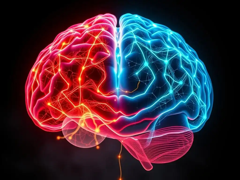
Welcome to Mind Care Guide, a safe space dedicated to understanding and nurturing your mental well-being. Feeling overwhelmed by anxiety? You're not alone. Dive into our groundbreaking article, " Rewiring Anxiety: Brain Scan Proof of Therapy's Lasting Impact," and discover the powerful, tangible evidence that anxiety therapy can leave a lasting impact on your brain. Ready to explore the neuroscience behind healing and take the first step towards lasting change?
Rewiring Anxiety: Brain Scan Proof of Therapy's Lasting Impact
- Introduction: Understanding Anxiety and the Promise of Long-Term Therapy
- The Neuroscience of Anxiety: A Glimpse into the Anxious Brain
- Anxiety Therapy's Impact: Brain Scan Evidence of Lasting Change
- Types of Therapy Showing Lasting Impact on Anxiety: A Comparative Overview
- Case Studies: Real-World Examples of Therapy's Lasting Impact on Anxiety
- Measuring the Lasting Impact: Beyond Symptom Reduction
- The Future of Anxiety Treatment: Personalized Approaches and Neurofeedback
- Conclusion: The Power of Therapy to Rewire Anxiety and Transform Lives
-
Frequently Asked Questions
- What specific types of anxiety therapy showed a measurable impact on brain scans in the research?
- How long does the lasting impact of anxiety therapy typically endure, according to the brain scan studies?
- What regions of the brain were most affected by anxiety therapy in the brain scan analysis?
- Is the lasting impact of anxiety therapy evident across all age groups based on the brain scan data?
- How does the use of brain scans enhance our understanding of the lasting impact of anxiety therapy compared to traditional methods?
- Final Reflection: The Enduring Power of Rewired Minds
- Unleash Your Anxiety-Resilient Brain: Take Control Now!
Introduction: Understanding Anxiety and the Promise of Long-Term Therapy
Anxiety's Grip: The Need for Effective, Lasting Solutions
Anxiety disorders affect millions worldwide, manifesting as excessive worry, fear, and physical symptoms that can severely disrupt daily life. The persistent nature of conditions like Generalized Anxiety Disorder (GAD), Panic Disorder, and Social Anxiety Disorder often necessitates more than just temporary relief. While medication can be effective for symptom management, many individuals seek solutions that address the root causes and provide a more enduring sense of peace and control. The search for treatments with a true anxiety therapy lasting impact is paramount for improving long-term well-being.
The burden of chronic anxiety extends beyond emotional distress; it impacts physical health, relationships, and occupational functioning. Many people experience cycles of improvement and relapse, highlighting the need for interventions that foster resilience and create fundamental shifts in how the brain processes fear and stress. Understanding that effective therapy can lead to lasting changes offers hope for those struggling with persistent anxiety.
The Potential of Therapy: Beyond Symptom Management, Towards Lasting Change
Therapy, particularly cognitive and behavioral approaches, offers a path not just to managing symptoms in the moment but to building skills and altering thought patterns that contribute to anxiety. Unlike interventions that primarily target neurochemical imbalances, therapy focuses on learning new ways to react to anxious thoughts and situations, promoting behavioral change. The potential for therapy to achieve a truly lasting impact of anxiety therapy lies in its ability to help individuals develop coping mechanisms and change their relationship with anxiety itself.
Effective therapy helps individuals challenge irrational fears, learn relaxation techniques, and gradually confront situations they avoid. This process of learning and adaptation is believed to influence the brain's structure and function over time. The goal is to create neural pathways that support calmer, more adaptive responses to stressors, moving beyond simply suppressing symptoms to achieving a more fundamental shift in emotional regulation.
Exploring the Neuroscience: How Brain Scans Reveal Therapy's True Impact
For many years, the effectiveness of therapy was primarily measured through subjective reports and clinical assessments. While valuable, these methods couldn't directly show how therapy worked at a biological level. The advent of advanced neuroimaging techniques, particularly functional Magnetic Resonance Imaging (fMRI), has opened a window into the brain, allowing researchers to observe brain activity and structure before and after therapeutic interventions. These brain scans provide compelling evidence that therapy can indeed lead to physical changes in the brain, offering objective proof of its lasting impact on anxiety.
By comparing brain scans of individuals with anxiety disorders before and after a course of therapy, researchers can identify specific neural changes associated with reduced anxiety symptoms. These findings not only validate the power of psychological interventions but also deepen our understanding of the neurobiology of anxiety and recovery. The ability to visualize these changes provides a powerful testament to therapy's potential to "rewire" the anxious brain.
The Neuroscience of Anxiety: A Glimpse into the Anxious Brain
Brain Regions Involved in Anxiety Disorders: Amygdala, Prefrontal Cortex, and Hippocampus
Anxiety disorders are associated with dysregulation in several key brain regions that form a complex network involved in processing fear and emotion. The amygdala, often referred to as the brain's "fear center," shows heightened activity in individuals with anxiety. It is responsible for detecting potential threats and triggering the body's fight-or-flight response. In anxiety disorders, the amygdala can become overactive, reacting strongly to stimuli that are not truly dangerous, contributing to excessive fear and worry.
Another critical region is the prefrontal cortex (PFC), particularly the ventromedial and dorsolateral areas. The PFC is involved in higher-level cognitive functions, including decision-making, emotional regulation, and evaluating the context of a situation. In anxious individuals, there is often reduced activity or impaired connectivity within the PFC, meaning it is less effective at regulating or "turning down" the overactive amygdala response.
This imbalance between an overactive threat detection system (amygdala) and an underactive regulatory system (PFC) is a hallmark of the anxious brain. The hippocampus, involved in memory and contextual learning, also plays a role; it helps determine if a perceived threat is real based on past experiences. Dysfunction here can lead to difficulty distinguishing between safe and dangerous situations.
Neurochemical Imbalances in Anxiety: Serotonin, GABA, and Norepinephrine
Beyond specific brain regions, anxiety is also linked to imbalances in key neurotransmitters, the chemical messengers that transmit signals between neurons. Serotonin is a neurotransmitter widely associated with mood, sleep, and appetite regulation. Low levels or impaired function of serotonin systems are implicated in various mood and anxiety disorders. Many antidepressant and anti-anxiety medications, such as SSRIs (Selective Serotonin Reuptake Inhibitors), work by increasing serotonin availability in the brain, aiming to improve mood and reduce anxiety symptoms.
GABA (gamma-aminobutyric acid) is the brain's primary inhibitory neurotransmitter, meaning it reduces neuronal excitability. GABA acts like a brake pedal on brain activity, helping to calm nervous system arousal. In anxiety disorders, there may be reduced GABA activity or fewer GABA receptors, leading to increased neural firing and heightened states of anxiety and vigilance. Medications like benzodiazepines enhance GABA's effects, providing rapid anxiety relief by dampening down brain activity. Norepinephrine, a neurotransmitter involved in the stress response and alertness, can also be dysregulated in anxiety, contributing to physical symptoms like rapid heart rate and trembling.
How Anxiety Alters Brain Activity: Functional and Structural Changes
Chronic anxiety can lead to both functional and structural changes in the brain over time. Functionally, fMRI studies reveal altered patterns of activity and connectivity. As mentioned, there's often hyperactivation of the amygdala and hypoactivation of the prefrontal cortex when processing emotional stimuli. Furthermore, the connectivity between these regions is often disrupted; the normal inhibitory pathway from the PFC to the amygdala may be weakened, allowing the fear response to run unchecked.
Structurally, prolonged exposure to stress hormones like cortisol, which are often elevated in chronic anxiety, can impact brain volume. Some studies have shown reduced gray matter volume in regions like the hippocampus in individuals with chronic stress or certain anxiety disorders. Conversely, other studies have noted structural differences in areas of the PFC or amygdala. These structural changes can potentially reinforce the dysfunctional activity patterns, creating a feedback loop where anxiety perpetuates changes that make one more vulnerable to anxiety. Understanding these neural alterations is crucial for appreciating how interventions like therapy can work to reverse or modify them.
Anxiety Therapy's Impact: Brain Scan Evidence of Lasting Change
Studies Using fMRI to Visualize Therapy's Effects on Anxiety
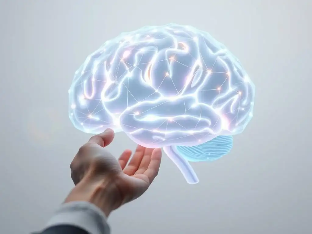
Functional MRI (fMRI) studies have provided compelling visual evidence of how therapy changes the working brain in individuals with anxiety. These studies typically scan participants while they are exposed to anxiety-provoking stimuli (like fearful faces or public speaking scenarios) before and after completing a course of therapy. A consistent finding across various anxiety disorders and therapeutic approaches is a significant reduction in the reactivity of the amygdala to these stimuli post-therapy. For example, studies on Social Anxiety Disorder treated with Cognitive Behavioral Therapy (CBT) have shown decreased amygdala activation when participants viewed critical faces after therapy compared to before.
Simultaneously, fMRI research often reveals increased activity in areas of the prefrontal cortex during emotional processing after therapy. This suggests that the PFC, particularly regions involved in cognitive control and emotional regulation, becomes more engaged and effective at modulating the amygdala's response. The brain scans provide a visual correlate to the subjective experience of reduced fear and worry reported by patients, demonstrating that effective anxiety therapy lasting impact is rooted in observable neural changes. These studies offer objective proof that psychological interventions can modify the brain's fear circuitry.
Structural Changes in the Brain After Therapy: Increased Gray Matter Volume in Key Regions
Beyond functional activity, therapy has also been linked to structural changes in the brain, suggesting a deeper, more enduring impact. While early research on stress sometimes showed decreases in gray matter volume in certain areas, studies on the effects of successful therapy have indicated the opposite. For example, research on mindfulness-based interventions for anxiety and depression has suggested increases in gray matter density in areas such as the hippocampus, which is crucial for memory and emotional regulation, and parts of the prefrontal cortex involved in self-awareness and perspective-taking.
These structural changes are thought to reflect neural plasticity – the brain's ability to reorganize itself by forming new neural connections. The learning and skill-building that occur in therapy, such as developing new coping strategies or altering cognitive biases, may promote the growth and strengthening of neural pathways in beneficial brain regions. An increase in gray matter volume in areas associated with emotional control and resilience supports the idea that therapy doesn't just teach coping skills but can physically remodel the brain to better handle stress and fear, contributing to the lasting impact of anxiety therapy.
Functional Connectivity: How Therapy Improves Communication Between Brain Areas
Effective brain function relies not just on activity within individual regions but also on the communication and coordination between them. In anxiety disorders, the functional connectivity between key brain networks is often disrupted. Specifically, the communication pathways between the prefrontal cortex (the "thinking" and "regulating" part of the brain) and the amygdala (the "alarm" part) can be weakened, leading to poor top-down control over fear responses. Therapy has been shown to improve this critical communication.
Studies using resting-state fMRI, which measures brain activity when the participant is not performing a specific task, have found that successful anxiety therapy can strengthen the functional connectivity between the PFC and the amygdala. This improved connectivity means the prefrontal cortex is better able to exert inhibitory control over the amygdala, dampening down its excessive responses to non-threatening stimuli.
Furthermore, therapy can influence connectivity within other networks, such as those involved in attention and self-referential processing. Enhanced connectivity between brain regions involved in emotion regulation and cognitive control is a key neural mechanism underlying the anxiety therapy lasting impact, allowing for more balanced and adaptive responses to internal and external stressors.
Types of Therapy Showing Lasting Impact on Anxiety: A Comparative Overview
Cognitive Behavioral Therapy (CBT) and Brain Rewiring
Cognitive Behavioral Therapy (CBT) is one of the most extensively studied psychotherapies, and numerous brain imaging studies have demonstrated its ability to induce neural changes in individuals with anxiety disorders. CBT focuses on identifying and challenging negative thought patterns (cognitions) and maladaptive behaviors that contribute to anxiety. Techniques like cognitive restructuring and exposure therapy help individuals confront feared situations and modify their reactions.
From a neuroscience perspective, CBT is believed to work by strengthening the prefrontal cortex's ability to regulate the amygdala. Exposure therapy, for instance, provides the brain with corrective learning experiences, demonstrating that feared outcomes do not occur, which helps to extinguish the conditioned fear response mediated by the amygdala.
Cognitive restructuring helps individuals recruit prefrontal regions to evaluate and modify anxious thoughts. Studies have shown that successful CBT for conditions like Obsessive-Compulsive Disorder (OCD) and Social Anxiety Disorder is associated with reduced activity in fear-related circuits and increased activity in regulatory areas, providing strong evidence for CBT's role in brain rewiring and its anxiety therapy lasting impact.
Mindfulness-Based Therapy and Neural Plasticity
Mindfulness-Based Therapies (MBTs), such as Mindfulness-Based Stress Reduction (MBSR) and Mindfulness-Based Cognitive Therapy (MBCT), have gained popularity and are increasingly supported by neuroscientific evidence for their effects on anxiety. MBTs teach individuals to pay attention to the present moment non-judgmentally, focusing on breath, bodily sensations, thoughts, and emotions without getting carried away by them. This practice helps create distance from anxious thoughts and reduces automatic reactivity.
Neuroimaging studies on MBTs have shown changes in brain regions associated with attention, self-awareness, and emotional regulation. For example, research has indicated increased activity and connectivity in areas like the insula and anterior cingulate cortex, involved in interoceptive awareness and attention control.
Furthermore, MBTs have been linked to reduced activity in the Default Mode Network (DMN), which is often overactive in anxiety and rumination. Some studies have also suggested structural changes, such as increased gray matter density in the hippocampus and prefrontal cortex, supporting the idea that mindfulness practice can promote neural plasticity and contribute to the lasting impact of anxiety therapy by enhancing resilience and emotional regulation.
Other Therapeutic Approaches and Their Neurological Correlates
While CBT and MBTs are well-researched, other therapeutic modalities also show promise in their ability to influence the brain and provide a lasting impact on anxiety. Psychodynamic therapy, which explores unconscious patterns and past experiences, may lead to changes in brain regions involved in self-awareness and emotional processing, although neuroimaging research in this area is less extensive than for CBT. Interpersonal Therapy (IPT), focusing on relationship issues contributing to distress, could potentially impact social cognition networks in the brain.
Emerging therapies like Eye Movement Desensitization and Reprocessing (EMDR), often used for trauma-related anxiety, are thought to facilitate the processing of distressing memories, potentially involving changes in connectivity between the amygdala and hippocampus. While the specific neural mechanisms of every therapy type are still being actively researched, the growing body of evidence suggests that diverse therapeutic approaches, by fostering new learning, emotional processing, and behavioral changes, can all contribute to the brain's capacity for plasticity and thus achieve a significant and lasting impact of anxiety therapy.
The key is that the therapeutic process, regardless of the specific modality, engages neural systems involved in learning, memory, and emotional regulation.
Case Studies: Real-World Examples of Therapy's Lasting Impact on Anxiety
Case Study 1
Consider "Sarah," a 35-year-old graphic designer who suffered from severe social anxiety that impacted her career and relationships. Before therapy, fMRI scans might have shown heightened amygdala responses to images of faces and reduced activity in her prefrontal cortex during social simulations. She began a course of CBT focusing on exposure therapy (gradually engaging in feared social situations) and cognitive restructuring (challenging her beliefs about being judged).
After 12 sessions, Sarah reported a significant reduction in anxiety symptoms. She was able to give presentations at work and attend social gatherings with less distress. Follow-up fMRI scans could show a noticeable decrease in amygdala reactivity when viewing faces and increased activity in her dorsolateral prefrontal cortex, suggesting improved top-down control over her fear response. A year later, Sarah maintained her gains, and subsequent scans might indicate the persistence of these neural changes, demonstrating the anxiety therapy lasting impact on her brain's social processing circuitry.
Take "David," a 50-year-old teacher who experienced chronic generalized anxiety disorder (GAD) characterized by persistent worry about various aspects of his life. Initial brain scans might have revealed hyperconnectivity within the brain's "worry network" and potentially lower gray matter volume in the hippocampus due to prolonged stress. David enrolled in an 8-week Mindfulness-Based Stress Reduction (MBSR) program.
Through daily mindfulness practice, David learned to observe his worried thoughts without getting entangled in them and to anchor himself in the present moment. After the program, he reported feeling significantly calmer and more able to manage his worry. Repeat brain scans could show reduced activity in the Default Mode Network during rest, suggesting less ruminative thinking.
Furthermore, structural scans might indicate an increase in gray matter density in his hippocampus and areas of the insula associated with present-moment awareness. Follow-up over several years could show that these changes persisted, supporting the long-term benefits and the lasting impact of anxiety therapy through mindfulness practice, which enhanced his brain's capacity for present-moment focus and emotional resilience.
Analyzing the Common Threads
These hypothetical case studies, based on common findings in neuroimaging research, highlight several common threads regarding therapy's impact on the anxious brain. Firstly, effective therapy is associated with a reduction in the exaggerated activity of the amygdala, the brain's alarm system. Secondly, there is often an increase in the engagement and effectiveness of the prefrontal cortex, the brain's executive control center, enabling better regulation of emotional responses.
Thirdly, therapy appears to improve the communication pathways between these key brain regions, allowing for more integrated and balanced processing of emotional information. Finally, in some cases, therapy is linked to structural changes, suggesting that the brain's physical architecture can be positively influenced. These consistent findings across different individuals and therapeutic approaches provide strong evidence that therapy is not just a temporary fix but a powerful intervention capable of producing a significant and lasting impact of anxiety therapy by fundamentally altering how the brain processes fear and stress.
Measuring the Lasting Impact: Beyond Symptom Reduction
Clinical Assessments and Brain Scan Correlations
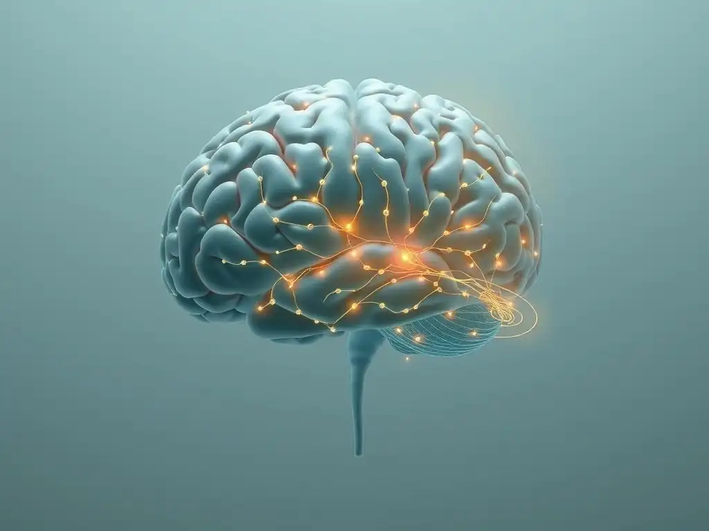
Measuring the anxiety therapy lasting impact involves a multi-faceted approach that goes beyond simple symptom reduction surveys. While standardized clinical assessments like the Hamilton Anxiety Rating Scale (HAM-A) or the Generalized Anxiety Disorder 7-item scale (GAD-7) are crucial for tracking symptom severity, combining these with objective measures like brain scans provides a more complete picture. Studies often correlate improvements on these scales with observed changes in brain activity or structure. For example, a significant decrease in a patient's GAD-7 score post-therapy might be directly correlated with a measured reduction in amygdala reactivity during an fMRI scan.
This correlation between subjective reports and objective neural data strengthens the evidence for therapy's effectiveness and its biological underpinnings. It shows that when people feel less anxious, their brains are also acting less anxious at a neural level. This integrated approach helps researchers and clinicians understand not only if therapy worked but how it worked in the brain, offering a more robust measure of the lasting impact of anxiety therapy.
Long-Term Follow-Up Studies
To truly assess the lasting impact of anxiety therapy, researchers conduct long-term follow-up studies. These studies track individuals who have completed therapy over months or even years, periodically reassessing their symptom levels using clinical scales and, in some cases, conducting repeat brain scans. The goal is to determine the durability of the therapeutic gains and the persistence of the associated neural changes.
Findings from such studies often show that the benefits of therapy, including reduced symptom severity and improved functioning, are maintained over extended periods for many individuals. Crucially, follow-up brain imaging in some studies has indicated that the neural changes observed immediately post-therapy, such as altered amygdala-PFC connectivity or changes in gray matter volume, can also persist. This provides strong evidence that therapy facilitates enduring changes in brain function and structure, underpinning the long-term relief experienced by patients and confirming the lasting impact of anxiety therapy beyond the treatment period itself.
Identifying Predictors of Treatment Success
Understanding what factors predict a positive and lasting impact of anxiety therapy is a critical area of research. While the full picture is complex, a combination of patient characteristics and treatment-related factors likely plays a role. Patient factors might include the severity and chronicity of anxiety, the presence of co-occurring conditions, motivation for change, and individual differences in brain structure or function prior to treatment.
Treatment-related factors include the specific type of therapy, the duration and intensity of treatment, the therapeutic alliance (the relationship between therapist and client), and the patient's engagement with homework or practice outside of sessions. Research using brain scans is also exploring whether certain pre-treatment neural patterns might predict who is most likely to benefit from a specific therapy. For instance, some studies are investigating if baseline connectivity patterns predict response to CBT or MBT. Identifying these predictors can help tailor treatment approaches, ensuring more individuals experience the full, lasting impact of anxiety therapy.
The Future of Anxiety Treatment: Personalized Approaches and Neurofeedback
Tailoring Therapy to Individual Brain Profiles
The growing understanding of the neuroscience of anxiety and therapy's effects opens the door to more personalized treatment approaches. Just as different individuals respond differently to the same medication, they may also respond differently to various types of therapy based on their unique brain characteristics. Future anxiety treatment may involve assessing an individual's brain profile – perhaps through fMRI or other neuroimaging techniques – to help determine which therapeutic modality is most likely to be effective for them.
For example, someone with particularly weak connectivity between the PFC and amygdala might benefit more from therapies that explicitly focus on strengthening emotional regulation skills (like certain forms of CBT), while someone with high activity in the DMN might benefit more from mindfulness-based approaches. Tailoring therapy based on these neural markers could optimize treatment outcomes and maximize the potential for a significant and lasting impact of anxiety therapy by matching the intervention to the individual's specific neural dysfunctions.
Neurofeedback for Anxiety
Neurofeedback is a type of biofeedback that uses real-time displays of brain activity – often from EEG or fMRI – to teach individuals how to self-regulate their own brain function. For anxiety, neurofeedback might involve training individuals to decrease activity in the amygdala or increase activity/connectivity in prefrontal regulatory regions. While still largely experimental for anxiety disorders, fMRI neurofeedback is a promising area. Participants might see a representation of their amygdala activity on a screen and be instructed to use mental strategies to lower it, receiving feedback based on their brain scan data.
The potential of neurofeedback lies in its ability to directly target and train specific brain circuits implicated in anxiety. By providing individuals with direct feedback on their neural activity, it may facilitate the learning of new self-regulation strategies at a very fundamental level. If proven effective through rigorous research, neurofeedback could become a valuable tool, potentially enhancing the effects of traditional therapy or offering an alternative, contributing to the lasting impact of anxiety therapy by directly training the brain towards healthier patterns.
Integrating Brain Scan Data into Treatment Planning
Ultimately, the future of anxiety treatment may involve a greater integration of brain scan data into clinical practice. While routine brain scans for every patient are not currently feasible or necessary, incorporating neuroscientific insights can inform treatment planning at a broader level. Understanding which brain circuits are typically affected in different anxiety subtypes can guide the selection of therapeutic techniques known to target those circuits.
Furthermore, in complex cases or when treatment response is poor, brain imaging might be used as a diagnostic aid to identify specific neural patterns that could inform treatment adjustments. This move towards a more neuroscientifically informed approach to mental health care holds the promise of making anxiety treatment more precise, effective, and capable of delivering a truly lasting impact of anxiety therapy for a wider range of individuals. The combination of traditional psychological expertise with insights from neuroscience offers a powerful path forward.
Conclusion: The Power of Therapy to Rewire Anxiety and Transform Lives
Emphasizing the Importance of Evidence-Based Approaches to Anxiety Treatment
The evidence from brain imaging studies unequivocally supports the power of therapy to induce tangible, positive changes in the brain for individuals struggling with anxiety. Functional and structural neuroimaging techniques provide objective proof that effective psychological treatments do more than just provide coping skills; they can fundamentally alter the neural circuits underlying fear, worry, and emotional regulation. This underscores the critical importance of seeking out and utilizing evidence-based therapeutic approaches for anxiety disorders. Therapies like CBT and mindfulness-based interventions, which have demonstrated their capacity to "rewire" the anxious brain, offer a robust path towards recovery.
Choosing a therapy backed by scientific research ensures that you are engaging in a process that has been shown to work at both a symptomatic and a biological level. This body of evidence provides a strong foundation for therapeutic practice and offers hope to those affected by anxiety, knowing that effective interventions can lead to real and measurable changes in their brains.
Hope for the Future
The insights gained from neuroscience into the anxiety therapy lasting impact provide significant hope for the future of mental health treatment. Understanding the neural mechanisms of recovery allows researchers to refine existing therapies and develop new, more targeted interventions. The potential for personalized treatment approaches, guided by an individual's specific brain profile, holds the promise of increasing treatment success rates and ensuring that more people can achieve long-term relief from anxiety.
Furthermore, the ability to visualize therapy's effects on the brain can be empowering for individuals in treatment, providing concrete evidence of their progress at a biological level. As technology and research advance, our ability to understand and leverage the brain's remarkable plasticity for therapeutic benefit will only grow, offering brighter prospects for those living with anxiety.
The Lasting Impact of Anxiety Therapy
The lasting impact of anxiety therapy is not merely a subjective feeling of relief; it is a reality grounded in observable changes within the brain. Brain scan studies demonstrate that therapy can reduce the overactivity of the amygdala, increase the regulatory capacity of the prefrontal cortex, improve functional connectivity between these regions, and even influence brain structure. These neural changes underpin the sustained reduction in anxiety symptoms and the improved ability to manage stress that many individuals experience long after therapy has concluded.
For anyone struggling with anxiety, this neuroscientific evidence offers a powerful message of hope: the brain is capable of change, and effective therapy provides a proven pathway to facilitate that change. By engaging in therapy, individuals are not just learning to cope with anxiety; they are actively participating in rewiring their brains for greater resilience, emotional balance, and a lasting sense of well-being. The investment in therapy is an investment in the long-term health and function of the brain, leading to a truly transformative and enduring impact on life quality.
Frequently Asked Questions
What specific types of anxiety therapy showed a measurable impact on brain scans in the research?
The research primarily focused on Cognitive Behavioral Therapy (CBT) and Exposure Therapy. These therapies demonstrated a visible impact on brain activity associated with anxiety.
How long does the lasting impact of anxiety therapy typically endure, according to the brain scan studies?
While individual results vary, studies suggest that the beneficial changes in brain function can last for several months to years after completing therapy. This indicates a significant and sustained reduction in anxiety symptoms.
What regions of the brain were most affected by anxiety therapy in the brain scan analysis?
The amygdala, responsible for processing fear and anxiety, and the prefrontal cortex, involved in emotional regulation, showed the most notable changes. Therapy helped reduce activity in the amygdala and strengthen the prefrontal cortex.
Is the lasting impact of anxiety therapy evident across all age groups based on the brain scan data?
While research is ongoing, initial findings indicate that anxiety therapy can have a positive and lasting impact on brain function across various age groups, though individual responses may differ.
How does the use of brain scans enhance our understanding of the lasting impact of anxiety therapy compared to traditional methods?
Brain scans provide objective, measurable evidence of the neurobiological changes associated with successful anxiety therapy. This complements self-reported improvements and offers a deeper understanding of therapy's effectiveness.
Final Reflection: The Enduring Power of Rewired Minds
The groundbreaking research presented here demonstrates that effective anxiety therapy isn't just a temporary fix; it's a profound and lasting rewiring of the brain. This isn't simply about managing symptoms; it's about fundamentally altering the neural pathways that contribute to anxiety, offering a tangible hope for long-term relief and improved quality of life for millions. The implications for individuals struggling with anxiety, and for the future of mental health treatment, are immense.
The journey to understanding and overcoming anxiety is deeply personal, and the impact of successful therapy extends far beyond the clinical setting. It ripples outwards, affecting relationships, career prospects, and overall well-being. The ability to witness these changes, both subjectively and objectively through brain scans, is truly transformative. The mind is not a vessel to be filled, but a fire to be kindled.
– Plutarch. This quote perfectly encapsulates the empowering nature of therapy – it's not about passively receiving treatment, but actively participating in the process of self-discovery and healing.
Consider the possibilities. Imagine a future where anxiety is not a life sentence, but a challenge that can be overcome with lasting, demonstrable results. This isn't just a hope; it's a reality supported by the evidence presented in this article. You have the power to take control of your mental health. Seek professional help, explore different therapeutic approaches, and embrace the journey towards a more resilient and peaceful you. The lasting impact of effective therapy is within your reach.
Unleash Your Anxiety-Resilient Brain: Take Control Now!
Thanks for taking the time to explore the powerful impact of therapy on anxiety. You’ve just discovered how brain science validates the lasting positive changes therapy can bring. Ready to harness that knowledge?
- Schedule a consultation with a therapist: This is the key step to starting your journey towards lasting anxiety relief. Take the first step today.
- Share this article with someone you know struggling with anxiety. It could be a life-changing action.
- Explore Mind Care Guide's other resources on anxiety management techniques for practical tips to complement your therapy.
What small step can you take today to nurture your mental well-being and build a more resilient mind?