
Beyond Talk Therapy: What Scans Show About Brain Changes


Welcome to Mind Care Guide, a sanctuary for understanding your mental well-being.
Ever wondered what's really happening inside your brain when you're struggling with a mental health challenge? We delve deeper than conventional wisdom, exploring the fascinating world of neuroscience. In our latest article, "Beyond Talk Therapy: What Scans Show About Brain Changes," we uncover how brain scans are revealing unprecedented insights into mental health changes. Prepare to have your understanding challenged and your hope ignited as we explore the science behind healing. Intrigued? Read on to discover how this groundbreaking research could reshape the future of mental healthcare.
The Promise of Neuroimaging: Brain Changes in Mental Health Treatment
- Introduction: Integrating Neuroscience and Mental Health
- Understanding Brain Plasticity Through Therapy
-
Types of Brain Scans Used in Mental Health Research
- MRI (Magnetic Resonance Imaging): Visualizing Brain Structure After Therapy
- fMRI (Functional Magnetic Resonance Imaging): Tracking Brain Activity During Therapeutic Processes
- PET (Positron Emission Tomography): Assessing Metabolic Changes
- EEG (Electroencephalography): Analyzing Brainwave Patterns and Therapy Outcomes
-
Brain Scan Evidence: Specific Mental Health Conditions and Therapy-Induced Changes
- Depression: Brain Scan Findings After Cognitive Behavioral Therapy (CBT) and Medication
- Anxiety Disorders: Examining Amygdala Activity Changes with Exposure Therapy and Mindfulness
- PTSD (Post-Traumatic Stress Disorder): Brain Imaging and EMDR Therapy Effectiveness
- Schizophrenia: Evaluating Brain Volume and Activity Changes with Antipsychotics and Therapy
- Advantages of Using Brain Scans to Assess Therapy Effectiveness
- Limitations and Ethical Considerations of Brain Scans in Therapy
- Future Directions: The Integration of Brain Scans into Mental Health Practice
- Case Studies: Real-World Examples of Brain Scan-Informed Therapy
- Conclusion: The Future of Mental Health is Neurological
-
Frequently Asked Questions
- What role do brain scans play in understanding mental health challenges?
- How can brain scans help determine the effectiveness of mental health treatments?
- What types of mental health changes can be observed through brain scans?
- Are brain scans a replacement for traditional talk therapy in mental health treatment?
- What are the limitations of using brain scans for diagnosing and treating mental health conditions?
- Final Reflection: Rewiring the Mind: The Promise and Potential of Brain Scans in Mental Health
- Unlock Your Brain's Potential: Discover Lasting Change
Introduction: Integrating Neuroscience and Mental Health
The Intersection of Therapy and Brain Imaging
For decades, mental health treatment primarily relied on subjective reports from patients and clinical observations from therapists. While invaluable, this approach offered limited insight into the biological underpinnings of conditions and how interventions physically impact the brain. The advent of sophisticated neuroimaging techniques has begun to bridge this gap, allowing researchers and clinicians to peek inside the living brain and observe its structure and function in real-time or across periods. This convergence of traditional therapeutic practices with neuroscience, particularly through brain scanning technologies, is transforming our understanding of mental well-being and the mechanisms by which therapy facilitates healing.
This integration is crucial because it moves mental healthcare beyond symptom management towards understanding and potentially modifying the neural circuits involved in distress. By observing how the brain changes during or after therapeutic interventions, we gain objective evidence of therapy's effectiveness. This provides a deeper, more nuanced understanding than purely behavioral or emotional reports can offer, confirming that therapy is not just "talking" but a process that leads to tangible, physical changes within the nervous system, directly impacting how individuals feel, think, and behave.
The Role of Brain Scans in Understanding Mental Well-being
Brain scans provide unparalleled insights into the complex network of neurons, pathways, and regions that underpin our thoughts, emotions, and behaviors. In the context of mental health, these technologies help researchers identify neural correlates of various conditions, such as altered activity patterns, structural differences, or connectivity issues. More importantly, they allow us to study how these neural signatures respond to treatment, offering objective evidence of the impact of interventions like psychotherapy or medication. Understanding how brain scans relate to mental health changes is becoming a cornerstone of modern psychiatric research.
For example, studies using functional magnetic resonance imaging ( fMRI) can show which brain areas are more or less active when someone experiences anxiety or processes emotions. By comparing scans taken before and after a course of therapy, researchers can observe shifts in activity levels or connectivity patterns that correlate with symptom improvement. This provides concrete, scientific backing for the efficacy of therapeutic approaches, demonstrating that therapy induces plastic changes in the brain, essentially helping to "rewire" maladaptive circuits associated with mental health challenges.
Understanding Brain Plasticity Through Therapy
How Therapy Influences Brain Structure and Function
The human brain is remarkably adaptable, a property known as neuroplasticity. This means the brain can reorganize itself by forming new neural connections throughout life. Therapy, particularly evidence-based psychotherapies like Cognitive Behavioral Therapy (CBT) or Dialectical Behavior Therapy (DBT), capitalizes on this plasticity. By learning new coping mechanisms, challenging negative thought patterns, and developing healthier emotional regulation strategies, individuals are essentially engaging in a process that strengthens some neural pathways and weakens others. Brain scans offer a window into these dynamic changes.
Research using structural MRI, for instance, has shown changes in gray matter volume in specific brain regions after successful therapy. For example, some studies on depression have noted changes in the hippocampus and prefrontal cortex following treatment. Functional imaging like fMRI reveals shifts in how different brain regions communicate; therapy can strengthen connectivity in circuits related to emotional regulation or executive control while potentially reducing activity or connectivity in areas associated with fear or rumination. These observed brain scans mental health changes underscore the profound biological impact of psychological interventions.
Measuring Impact: Neuroimaging in Therapy Research
Neuroimaging techniques are invaluable tools for quantifying the impact of therapy on the brain. By using standardized protocols and comparing brain scans taken at different points in the treatment process (e.g., baseline, mid-treatment, post-treatment), researchers can measure specific changes in brain structure, activity, or connectivity. This allows for objective assessment of therapeutic outcomes at a neural level, complementing traditional measures like symptom severity scores or patient questionnaires.
For instance, studies on the treatment of phobias using exposure therapy have utilized fMRI to track reductions in amygdala (the brain's fear center) activity when patients are presented with phobic stimuli after successful treatment. Similarly, research into mindfulness-based therapies has employed EEG to observe changes in brainwave patterns, noting shifts towards alpha and theta frequencies associated with relaxed alertness and meditation states. These quantitative measures derived from brain scans provide compelling evidence of therapy's effectiveness and help refine our understanding of how different therapeutic modalities work at a biological level, furthering the study of brain scans mental health changes.
Types of Brain Scans Used in Mental Health Research
MRI (Magnetic Resonance Imaging): Visualizing Brain Structure After Therapy
Magnetic Resonance Imaging ( MRI) is a non-invasive imaging technique that uses strong magnetic fields and radio waves to create detailed images of brain structure. In mental health research, structural MRI is used to measure the size and integrity of different brain regions. By taking MRI scans before and after a course of therapy, researchers can look for changes in gray matter volume (which contains neuron cell bodies) or white matter integrity (which consists of nerve fibers connecting different brain areas).
For example, studies have shown that successful treatment for Post-Traumatic Stress Disorder (PTSD) can be associated with an increase in hippocampal volume, a region often found to be smaller in individuals with chronic PTSD. Similarly, research into the effects of long-term meditation practices, which share some goals with therapy, has suggested increases in gray matter density in areas related to attention and emotional regulation. These structural brain scans reveal mental health changes that reflect neural adaptation and potentially recovery processes.
fMRI (Functional Magnetic Resonance Imaging): Tracking Brain Activity During Therapeutic Processes
Functional Magnetic Resonance Imaging ( fMRI) measures brain activity by detecting changes in blood flow. When a brain region is more active, it requires more oxygenated blood. fMRI captures these changes, providing insights into which parts of the brain are engaged during specific tasks, emotional states, or even during therapeutic interventions themselves. This allows researchers to observe real-time brain function and how it is altered by therapy.
Researchers use fMRI in several ways to study therapy's impact. They might scan patients while they perform tasks related to their condition (e.g., viewing emotional faces for anxiety, recalling traumatic memories for PTSD) before and after therapy to see how brain responses change. Studies using fMRI on individuals with depression undergoing CBT have demonstrated decreased activity in regions associated with negative self-referential thought (like the medial prefrontal cortex) and increased activity in areas involved in cognitive control. These functional brain scans provide dynamic evidence of mental health changes induced by therapy.
PET (Positron Emission Tomography): Assessing Metabolic Changes
Positron Emission Tomography ( PET) is an imaging technique that uses small amounts of radioactive tracers to measure metabolic activity, blood flow, or the distribution of neurotransmitter receptors in the brain. PET scans can provide information about the biochemical processes underlying mental health conditions and how these processes are affected by treatment, including therapy.
For instance, PET scans have been used to study glucose metabolism in the brain, which is an indicator of neural activity. Studies have shown altered metabolic rates in specific brain regions in conditions like depression or obsessive-compulsive disorder ( OCD). Following successful therapy or medication, PET scans can reveal normalization or changes in these metabolic patterns. PET can also use tracers that bind to specific neurotransmitter receptors (like serotonin or dopamine receptors), allowing researchers to investigate how therapy might influence neurotransmitter systems, offering another perspective on brain scans mental health changes.
EEG (Electroencephalography): Analyzing Brainwave Patterns and Therapy Outcomes
Electroencephalography ( EEG) is a non-invasive technique that measures electrical activity in the brain using electrodes placed on the scalp. EEG is particularly good at capturing rapid changes in brain activity and is used to analyze different brainwave frequencies (e.g., alpha, beta, theta, delta), which are associated with various states of consciousness and cognitive processes. While not providing detailed spatial resolution like MRI or PET, EEG offers excellent temporal resolution.
In mental health research, EEG can be used to identify abnormal brainwave patterns associated with conditions like anxiety, depression, or attention deficits. Studies have used EEG to observe shifts in these patterns after therapy. For example, some research on mindfulness and meditation, often integrated into therapeutic approaches, has noted increases in alpha waves (associated with relaxation) and theta waves (associated with deep relaxation and meditation). EEG can also track event-related potentials ( ERPs), which are brain responses to specific stimuli, revealing how information processing changes after therapy, contributing to our understanding of brain scans mental health changes.
Brain Scan Evidence: Specific Mental Health Conditions and Therapy-Induced Changes
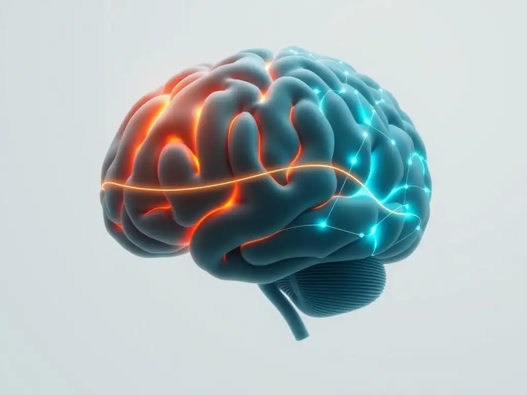
Depression: Brain Scan Findings After Cognitive Behavioral Therapy (CBT) and Medication
Major depressive disorder is associated with observable differences in brain structure and function. Common findings include reduced volume in areas like the hippocampus and prefrontal cortex, as well as altered activity and connectivity in circuits involved in mood regulation, reward processing, and cognitive control. Both psychotherapy, particularly CBT, and antidepressant medications have been shown to induce changes in these areas, as evidenced by brain scans.
Studies using fMRI have found that successful CBT for depression is associated with increased activity in the dorsal prefrontal cortex, a region involved in cognitive control and regulation of emotions, and decreased activity in the amygdala, which processes fear and emotional responses. Similarly, MRI studies have sometimes reported increased hippocampal volume in depressed individuals after effective treatment. While findings can vary across studies, the consistent observation is that interventions for depression lead to measurable brain scans mental health changes, supporting the idea that therapy is a biological treatment.
Anxiety Disorders: Examining Amygdala Activity Changes with Exposure Therapy and Mindfulness
Anxiety disorders, such as phobias, panic disorder, and generalized anxiety disorder, are often characterized by hyperactive responses in brain regions associated with fear and threat processing, most notably the amygdala. Therapeutic approaches like exposure therapy, which involves gradually confronting feared situations, and mindfulness-based therapies, which cultivate present-moment awareness, aim to reduce this heightened reactivity. Brain imaging studies have provided evidence for their effectiveness at the neural level.
Using fMRI, researchers have shown that exposure therapy for phobias or PTSD leads to a significant reduction in amygdala activity when individuals are exposed to fear-inducing stimuli after treatment. For example, one study on spider phobia showed reduced amygdala response to spider images post-exposure therapy. Mindfulness practices have also been linked to decreased amygdala reactivity and increased connectivity between the amygdala and prefrontal cortex, suggesting improved top-down regulation of emotional responses. These brain scans highlight the mental health changes resulting from therapy targeting fear circuits.
PTSD (Post-Traumatic Stress Disorder): Brain Imaging and EMDR Therapy Effectiveness
Post-Traumatic Stress Disorder (PTSD) is associated with distinct changes in brain structure and function, including reduced hippocampal volume, altered amygdala reactivity, and dysfunctional connectivity between the amygdala and the medial prefrontal cortex (involved in extinction of fear responses). Therapies like Trauma-Focused Cognitive Behavioral Therapy (TF-CBT) and Eye Movement Desensitization and Reprocessing (EMDR) are effective treatments. Brain imaging helps us understand how they work.
Studies using fMRI and structural MRI have investigated the neural effects of EMDR and TF-CBT for PTSD. Research suggests that successful treatment is associated with a normalization of amygdala activity during the processing of trauma-related stimuli and potentially an increase in hippocampal volume. Furthermore, changes in connectivity between the prefrontal cortex and the amygdala have been observed, indicating improved regulatory control over fear responses. These brain scans underscore the significant mental health changes achievable through trauma-focused therapies.
Schizophrenia: Evaluating Brain Volume and Activity Changes with Antipsychotics and Therapy
Schizophrenia is a complex disorder associated with widespread changes in brain structure and function, including reductions in gray matter volume in various cortical regions, enlarged ventricles, and altered activity and connectivity in networks involved in perception, cognition, and emotion. While antipsychotic medications are the primary treatment for positive symptoms (hallucinations, delusions), psychosocial therapies like CBT for psychosis and social skills training play a crucial role in managing symptoms, improving functioning, and enhancing quality of life. Brain imaging helps evaluate the impact of these combined interventions.
Brain scans in individuals with schizophrenia often show progressive gray matter loss, although the trajectory varies. Antipsychotic medications can influence brain volume, sometimes associated with changes in basal ganglia.
Studies investigating the effects of CBT for psychosis using fMRI have shown changes in activity in areas like the prefrontal cortex and temporal lobe, which are involved in reality testing and auditory processing, correlating with a reduction in symptom severity, particularly negative symptoms and delusions. These findings demonstrate that even in a complex disorder like schizophrenia, interventions can lead to detectable brain scans mental health changes, highlighting the importance of integrated care.
Advantages of Using Brain Scans to Assess Therapy Effectiveness
Objective Measures: Complementing Subjective Reports in Mental Health
One of the significant advantages of using brain scans in mental health assessment is their ability to provide objective data. Traditional clinical assessments rely heavily on self-report questionnaires, interviews, and behavioral observations, all of which are inherently subjective and can be influenced by factors like mood, memory, or desire to please. Brain scans offer a biological perspective, providing quantifiable metrics of neural structure, activity, or connectivity that are less susceptible to subjective bias.
This objective data from brain scans serves as a powerful complement to subjective reports. For example, a patient might report feeling less anxious after therapy, and an fMRI scan could provide neural evidence for this improvement by showing reduced amygdala reactivity to stressful stimuli. This can help validate patient experiences, provide therapists with additional information about a patient's progress at a biological level, and offer compelling evidence for the effectiveness of different therapeutic approaches. Observing these brain scans mental health changes provides a more complete picture of recovery.
Personalized Treatment: Tailoring Therapy Based on Brain Scan Results
As our understanding of brain-behavior relationships in mental health grows, brain scans hold the potential to contribute to more personalized treatment approaches. Different individuals with the same diagnosis might have variations in the underlying neural circuits affected. By using brain imaging to identify these individual neural profiles, clinicians could potentially tailor therapeutic interventions to target the specific brain networks most relevant to a patient's difficulties.
For instance, fMRI scans could reveal that a patient's depression is characterized more by dysfunction in reward circuits than in cognitive control circuits. This information could potentially guide the selection of specific therapeutic techniques or the combination of therapy with medication or other interventions like Transcranial Magnetic Stimulation (TMS), which targets specific brain areas. While still largely a research area, the prospect of using brain scans to predict treatment response or identify optimal therapy types represents a significant step towards truly personalized mental healthcare, leveraging brain scans to predict mental health changes.
Identifying Potential Therapy Resistance Through Brain Imaging
Brain imaging may also offer insights into why some individuals do not respond well to standard therapeutic approaches. By comparing the baseline brain scans of responders versus non-responders, researchers can look for neural signatures that predict treatment outcome. Identifying such biomarkers could help clinicians determine early on whether a particular therapy is likely to be effective for a specific patient, potentially saving time and reducing frustration.
For example, research is ongoing to identify fMRI patterns that might predict response to CBT in depression or anxiety. Early findings suggest that certain connectivity patterns in the prefrontal cortex might be associated with a lower likelihood of responding to standard talk therapy alone. While not yet a standard clinical tool, the ability to use brain scans to identify potential therapy resistance could allow for quicker adjustments to treatment plans, such as trying a different therapy modality, adding medication, or considering alternative interventions, based on observed brain scans and anticipated mental health changes.
Limitations and Ethical Considerations of Brain Scans in Therapy
Cost and Accessibility: Ensuring Equitable Access to Neuroimaging
Despite the potential of brain scans in mental health, significant limitations exist. One major barrier is the high cost of neuroimaging equipment and the expertise required to operate it and interpret the results. An MRI scan can cost several hundred to thousands of dollars, making routine scanning inaccessible for many patients and mental health practices, particularly in underserved areas. This raises concerns about equitable access to potentially beneficial diagnostic or monitoring tools.
Currently, brain scans are primarily used in research settings or in specialized clinical contexts, such as ruling out organic causes for symptoms (e.g., tumors, stroke) rather than routinely guiding therapy for common conditions like depression or anxiety. For brain imaging to become a more integrated part of mental healthcare, costs would need to decrease significantly, and accessibility would need to improve dramatically. The current cost structure limits the widespread application of brain scans for tracking mental health changes.
Interpretation Challenges: Understanding Brain Scan Data in Context
Interpreting brain scan data in the context of complex mental health conditions is challenging. Brain scans provide snapshots or measures of activity/structure, but they don't directly reveal thoughts, feelings, or the subjective experience of a disorder. A correlation between activity in a brain region and a symptom doesn't necessarily mean causation, and the brain is a complex, interconnected system where multiple regions interact. Isolating the specific impact of therapy on these complex interactions is difficult.
Furthermore, individual variability in brain structure and function is substantial, making it difficult to establish clear "normal" or "abnormal" patterns for many mental health conditions. Findings from group studies may not apply to every individual. Translating complex neuroimaging data into clinically meaningful information that can directly guide therapeutic decisions for a specific patient requires sophisticated analysis and careful consideration, avoiding oversimplification of how brain scans reflect mental health changes.
Ethical Implications: Privacy, Consent, and Data Security
The use of brain scans in mental health also raises important ethical considerations. Brain imaging data is highly sensitive and potentially identifiable. Ensuring patient privacy and the secure storage and handling of this data is paramount. As neuroimaging technology advances and potentially becomes more integrated into clinical care, robust protocols for data protection and patient consent are essential.
Questions also arise about the interpretation and potential misuse of brain scan data. Could scan results be used to stigmatize individuals, influence insurance coverage, or impact employment opportunities? Informed consent for brain scanning must clearly explain the potential uses and limitations of the data. As we explore how brain scans inform mental health changes, we must navigate these ethical waters carefully to protect patient autonomy and prevent discrimination.
Future Directions: The Integration of Brain Scans into Mental Health Practice

Advancements in Neuroimaging Technology for Therapy Monitoring
The field of neuroimaging is continuously evolving, with ongoing advancements promising more accessible, affordable, and informative scanning techniques. Developments in MRI sequences, higher field strength magnets, and faster scanning protocols are improving spatial and temporal resolution. Portable and lower-cost EEG systems are becoming more sophisticated. New computational techniques, such as machine learning and artificial intelligence, are being developed to analyze complex imaging datasets and identify subtle patterns related to mental health and treatment response.
These technological advancements hold the potential to make brain imaging more practical for routine clinical use. Faster scans mean less patient discomfort and higher throughput. Improved resolution allows for more precise targeting of specific brain areas. Advanced analysis methods can help clinicians extract meaningful insights from the data, potentially leading to brain scans that can more reliably predict mental health changes or monitor therapy effectiveness in real-time or near-real-time scenarios.
Developing Brain-Based Biomarkers for Mental Health Conditions
A major goal of mental health neuroscience research is the identification of reliable brain-based biomarkers – objective biological indicators – for specific conditions and their response to treatment. Brain imaging is a key tool in this effort. Researchers are working to identify patterns in structural MRI, fMRI connectivity, PET tracer uptake, or EEG oscillations that can differentiate between different disorders, predict who will develop a condition, or forecast how well a person will respond to a particular therapy or medication.
For example, research is exploring whether specific patterns of resting-state fMRI connectivity might serve as biomarkers for differentiating between major depression and bipolar disorder, or predicting response to antidepressant medication. While the search for definitive biomarkers is ongoing and complex, successful identification could revolutionize diagnosis and treatment selection, allowing for a more data-driven approach based on using brain scans to understand mental health changes at a fundamental level.
The Potential of Neurofeedback and Brain Stimulation Therapies
Insights gained from brain imaging are directly informing the development and application of novel therapies, such as neurofeedback and brain stimulation techniques. Neurofeedback, often guided by EEG or fMRI, allows individuals to see their own brain activity in real-time and learn to regulate it. For example, fMRI neurofeedback is being explored to help individuals with depression or anxiety learn to modulate activity in mood-related brain regions.
Brain stimulation techniques, such as Transcranial Magnetic Stimulation (TMS) or deep brain stimulation (DBS), directly alter brain activity. Brain imaging, particularly structural and functional MRI, is crucial for guiding the precise placement of stimulation targets to maximize therapeutic effect and minimize side effects. For example, fMRI scans can help identify the optimal location on the prefrontal cortex for TMS treatment in depression. These therapies, rooted in neuroimaging findings, represent a direct application of understanding brain scans to induce desired mental health changes.
Case Studies: Real-World Examples of Brain Scan-Informed Therapy
Case Study 1: Treatment-Resistant Depression - Guiding TMS Therapy with fMRI
Consider a patient suffering from severe, treatment-resistant depression who has not responded to multiple antidepressant medications and courses of psychotherapy. Traditional approaches offer limited options. In specialized centers, brain imaging, particularly fMRI, can be used to personalize treatment. While standard TMS protocols target a general area of the dorsolateral prefrontal cortex, individual fMRI scans can reveal specific functional connectivity patterns in that patient's brain that are disrupted in depression.
For instance, an fMRI scan might show reduced connectivity between a particular point on the left dorsolateral prefrontal cortex and regions deeper in the brain involved in mood regulation. Based on this individual connectivity map, TMS coils can be precisely guided to stimulate the specific cortical location that shows the strongest negative correlation with subgenual cingulate cortex activity (a common target for depression modulation).
This fMRI-guided approach, while not universally adopted, is being researched as a way to optimize TMS delivery, using brain scans to target specific pathways believed to be involved in mental health changes related to depression recovery, potentially improving response rates compared to standard targeting.
Case Study 2: Severe Anxiety - Using Brain Scan Data to Inform CBT Techniques
Imagine a patient with severe social anxiety whose fMRI scan, performed as part of a research protocol, shows exaggerated amygdala responses and reduced prefrontal connectivity when viewing social threat cues (like critical faces) compared to healthy controls. While the primary treatment would still be evidence-based therapy like CBT, the specific findings from the brain scan could potentially inform the therapeutic approach.
For example, knowing there's a clear neural signature of hyper-reactivity to social cues might emphasize the importance of specific CBT techniques focused on cognitive reappraisal of social situations and gradual exposure to social stimuli. The therapist could explicitly discuss with the patient how their brain is reacting and how therapy aims to 'train' the prefrontal cortex to better regulate the amygdala, making the abstract concept of "rewiring" more concrete.
While not routinely used for individual therapy planning, such research findings demonstrate how brain scans reveal mental health changes and provide a neural rationale for the effectiveness of specific therapeutic strategies, potentially increasing patient motivation by showing the biological target of their efforts.
Conclusion: The Future of Mental Health is Neurological
Brain Scans as a Tool for Understanding and Improving Mental Well-being
Brain scans have moved beyond being purely research tools to becoming increasingly relevant in understanding and potentially improving mental well-being. They provide objective, biological evidence that mental health conditions are associated with tangible differences in brain structure and function, and critically, that therapeutic interventions lead to measurable brain scans mental health changes. This neurobiological perspective complements psychological and behavioral insights, offering a more holistic view of mental health challenges and recovery.
While challenges regarding cost, accessibility, and interpretation remain, the information gleaned from techniques like MRI, fMRI, PET, and EEG is invaluable. It validates the impact of therapy, helps refine our understanding of therapeutic mechanisms, and holds promise for developing more personalized and effective treatments in the future. Embracing the insights provided by brain scans is crucial for advancing the field of mental healthcare.
Embracing the Neuroscience of Mental Health for Better Patient Outcomes
The integration of neuroscience, particularly through brain imaging, into mental health is not just an academic exercise; it has the potential to significantly improve patient outcomes. By providing objective evidence of therapy's impact, brain scans can reduce stigma, increase patient engagement by showing that change is biologically possible, and guide the development of more targeted interventions. As technology improves and becomes more accessible, routine use of brain scans to monitor progress and personalize treatment may become a reality.
Ultimately, understanding the brain scans mental health changes induced by therapy empowers both clinicians and patients. It reinforces that seeking help is about addressing real, biological processes and that recovery involves tangible neural shifts. By embracing the neuroscience of mental health, we move closer to a future where mental healthcare is more precise, effective, and tailored to the unique needs of each individual brain.
Frequently Asked Questions
What role do brain scans play in understanding mental health challenges?
Brain scans, like fMRI and PET scans, can help visualize and measure brain activity and structure. This allows researchers and clinicians to identify neurological differences associated with various mental health conditions.
How can brain scans help determine the effectiveness of mental health treatments?
Brain scans can provide objective evidence of how treatments, such as medication or therapy, are affecting the brain's function and structure. This can help personalize treatment plans and track progress more effectively.
What types of mental health changes can be observed through brain scans?
Brain scans can reveal changes in brain volume, neural connectivity, and neurotransmitter activity related to conditions like depression, anxiety, and schizophrenia. These changes can indicate underlying biological mechanisms.
Are brain scans a replacement for traditional talk therapy in mental health treatment?
No, brain scans are not a replacement for talk therapy. They are a complementary tool that can provide additional information and inform treatment decisions, but talk therapy remains a crucial component of mental health care.
What are the limitations of using brain scans for diagnosing and treating mental health conditions?
Brain scans can be expensive and not always readily available. The interpretation of scan results can also be complex and requires specialized expertise. They provide information but do not tell the whole story.
Final Reflection: Rewiring the Mind: The Promise and Potential of Brain Scans in Mental Health
The convergence of neuroscience and mental health is no longer a futuristic fantasy; it's a rapidly evolving reality. Brain imaging techniques are providing unprecedented insights into the biological underpinnings of mental illness, offering hope for more precise diagnoses, personalized treatments, and a deeper understanding of the human mind itself. This revolution promises to move beyond the limitations of traditional talk therapy, offering tangible evidence of therapeutic progress and paving the way for truly transformative interventions.
The impact of this research extends far beyond the clinical setting; it's reshaping our very understanding of mental health. By visualizing the physical changes in the brain associated with various conditions, we are dismantling the stigma surrounding mental illness, replacing outdated misconceptions with a more nuanced and compassionate perspective. This shift is crucial for fostering empathy and encouraging individuals to seek help without fear of judgment. The mind is not a vessel to be filled, but a fire to be kindled.
– Plutarch. This quote highlights the active, dynamic nature of the brain, a concept powerfully reinforced by the visual evidence provided by brain scans.
Consider the implications of this research for your own life. Perhaps you've struggled with a mental health challenge, or perhaps you know someone who has. This article has presented a glimpse into a future where treatment is tailored to the individual, where progress is measurable, and where hope is grounded in scientific evidence. Take a moment to reflect on the potential of this technology, not just for those currently suffering, but for future generations who may benefit from early intervention and personalized care. Embrace the possibilities, and encourage further research and development in this vital field.
Unlock Your Brain's Potential: Discover Lasting Change
Thanks for exploring the fascinating world of brain changes with us! You've now gained valuable insights into how therapy impacts your brain. Ready to take the next step toward lasting well-being?
- Schedule a consultation with your therapist or doctor: Discuss the findings and explore how to personalize your treatment plan for optimal results. This is the most direct application of what you've learned.
- Share this article with someone who might benefit: Spread awareness about the power of therapeutic interventions and encourage others to explore their brain health.
- Try a simple mindfulness practice daily: Even a few minutes can make a difference in your overall mental well-being, improving your brain's plasticity.
What small step will you take today to invest in your brain's health and unlock its potential?