
Tracking Therapy's Footprint in the Brain: An Imaging Guide

Welcome to Mind Care Guide, your compassionate companion on the journey to mental well-being.
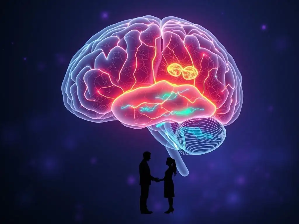
Ever wondered how therapy actually changes your brain? This article delves into the fascinating world of brain imaging therapy benefits, offering a unique perspective on how science is unveiling the tangible impact of mental health interventions. Join us as we explore the neuroscience behind healing and discover how imaging techniques are revolutionizing our understanding of the mind. Curious to see what's happening inside? Let's begin.
Tracking Therapy's Footprint in the Brain: An Imaging Guide
- Introduction: Unveiling Brain Changes Through Imaging After Therapy
- Why Use Brain Imaging to Evaluate Therapy Effectiveness?
-
Brain Imaging Techniques Used to Assess Therapy Outcomes
- Functional Magnetic Resonance Imaging (fMRI): Mapping Brain Activity Changes Post-Therapy
- Positron Emission Tomography (PET): Measuring Neurochemical Shifts and Receptor Binding
- Electroencephalography (EEG): Assessing Brainwave Patterns and Neural Synchronization
- Diffusion Tensor Imaging (DTI): Evaluating White Matter Integrity and Connectivity
- Specific Brain Regions Impacted by Different Therapies
- Interpreting Brain Imaging Results: What to Look For
- Case Studies: Real-World Examples of Brain Imaging in Therapy Evaluation
- The Future of Brain Imaging in Therapy: Personalized Treatment Approaches
- Ethical Considerations and Limitations of Brain Imaging in Therapy
- Conclusion: Harnessing Brain Imaging for Enhanced Mental Healthcare
-
Frequently Asked Questions
- What exactly is brain imaging in the context of therapy, and how does it relate to mental well-being?
- What are the potential benefits of using brain imaging to guide or personalize mental health therapy?
- Can brain imaging definitively diagnose mental health conditions, or is it more of a supplementary tool?
- How accessible and affordable is brain imaging for individuals seeking mental health treatment?
- What are the ethical considerations surrounding the use of brain imaging in mental health therapy?
- Final Reflection: Mapping the Future of Therapeutic Brain Change
- Unveil Your Brain's Healing Journey: Take Action Today!
Introduction: Unveiling Brain Changes Through Imaging After Therapy
The Growing Importance of Brain Imaging in Mental Health Treatment
The landscape of mental health treatment is continuously evolving, moving beyond purely behavioral observations and self-report measures to incorporate objective biological insights. Brain imaging techniques are playing an increasingly crucial role in this evolution, offering a window into the neural changes that occur as a result of therapeutic interventions.
This shift allows clinicians and researchers to better understand how therapy works at a fundamental level, identifying specific neural pathways and regions that are modulated or altered by different therapeutic approaches. The ability to visualize these changes provides empirical support for the effectiveness of various therapies and helps to demystify the process for both practitioners and patients.
This growing emphasis on objective measures like brain scans is transforming how we evaluate and discuss mental health treatments. By providing tangible evidence of therapy's impact on the brain, imaging studies contribute significantly to the understanding of brain imaging therapy benefits. They move the conversation from "does therapy work?" to "how does therapy change the brain, and for whom?". This objective data can help reduce stigma by framing mental health conditions as disorders with biological underpinnings that can be positively influenced by treatment, much like physical ailments.
Understanding Brain Changes and Long-Term Outcomes
Therapy is not just about learning new coping skills; it can instigate profound and lasting changes in brain structure and function. These changes are thought to underpin the long-term benefits experienced by individuals who successfully engage in treatment. Understanding which brain regions and networks are affected provides valuable clues about the mechanisms of recovery and resilience. For example, therapies aimed at managing anxiety might lead to reduced activity in fear-processing centers, while therapies focusing on emotional regulation could enhance connectivity within circuits involved in executive control.
Tracking these neural alterations over time using brain imaging allows researchers to correlate specific patterns of change with sustained improvements in symptoms and overall well-being. This offers insights into the durability of therapeutic effects and helps identify potential neural markers for long-term recovery. Studies using techniques like fMRI have shown that positive brain changes can persist months or even years after therapy concludes, highlighting the potential for lasting brain imaging therapy benefits. This underscores the value of therapy not just as a short-term solution but as a powerful tool for promoting long-term neural adaptation and mental health resilience.
Why Use Brain Imaging to Evaluate Therapy Effectiveness?
Objective Measurement of Therapeutic Progress: Beyond Subjective Reports
Evaluating the success of therapy has traditionally relied heavily on subjective reports from patients and clinical observations from therapists. While essential, these methods can be influenced by various factors, including recall bias, desire to please, or difficulty articulating internal states. Brain imaging offers an objective, biological lens through which to assess therapeutic progress. By measuring changes in brain activity, structure, or connectivity, imaging provides data that is independent of subjective perception.
This objective data complements and validates self-report measures, offering a more complete picture of an individual's response to treatment. For instance, a patient might report feeling less anxious, and an fMRI scan could provide objective support by showing decreased activity in the amygdala when processing fear-inducing stimuli. This convergence of subjective experience and objective neural data strengthens the evidence for the effectiveness of therapy and helps highlight the real brain imaging therapy benefits. This dual approach enhances the precision and reliability of treatment outcome evaluations.
Identifying Neural Correlates of Mental Health Improvements
Mental health conditions are associated with specific patterns of brain dysfunction, such as altered activity in certain regions or disrupted connectivity between brain networks. Effective therapy is believed to work, at least in part, by normalizing these dysfunctional patterns. Brain imaging allows researchers to identify the specific neural correlates that change in tandem with clinical improvements. For example, successful treatment for depression might be associated with increased activity in reward pathways or improved connectivity within mood-regulating circuits.
Pinpointing these neural correlates helps researchers understand the biological mechanisms underlying recovery. This knowledge is crucial for refining existing therapies and developing new, more targeted interventions. By observing which neural pathways are altered by a specific therapeutic approach, we gain insights into its mode of action. This process of mapping clinical improvement onto neural changes is fundamental to understanding the brain imaging therapy benefits and advancing the scientific basis of mental health treatment.
Distinguishing Between Different Therapy Modalities Through Brain Scans
Different therapeutic approaches, such as Cognitive Behavioral Therapy (CBT), psychodynamic therapy, or mindfulness-based interventions, operate through distinct theoretical frameworks and employ different techniques. While they may target similar conditions, they might exert their effects on the brain through different pathways. Brain imaging techniques can potentially reveal these modality-specific neural footprints. For example, one therapy might primarily impact prefrontal cortex function related to cognitive control, while another might focus more on modulating limbic system activity related to emotional processing.
Studies comparing the brain changes induced by different therapies for the same condition can help researchers understand their unique mechanisms of action and potentially identify which therapy might be best suited for an individual based on their specific neural profile. Although research in this area is still developing, the potential to differentiate the neural impact of various therapies through imaging is a significant step towards personalized mental healthcare, highlighting the diverse brain imaging therapy benefits offered by different approaches.
Brain Imaging Techniques Used to Assess Therapy Outcomes
Functional Magnetic Resonance Imaging (fMRI): Mapping Brain Activity Changes Post-Therapy
Functional Magnetic Resonance Imaging (fMRI) is one of the most widely used techniques for evaluating therapy outcomes in the brain. It measures brain activity by detecting changes in blood flow. When a brain region is more active, it requires more oxygenated blood, and fMRI can detect the magnetic properties of oxygenated versus deoxygenated hemoglobin. By having participants perform tasks (e.g., emotional regulation, cognitive tasks) or simply rest inside the scanner before and after therapy, researchers can identify how therapy has altered the patterns of brain activation in response to specific stimuli or during resting states.
For instance, studies on anxiety disorders treated with CBT have frequently used fMRI to show decreased activity in the amygdala (a region associated with fear processing) when individuals are exposed to anxiety triggers post-treatment. Conversely, activity might increase in prefrontal regions associated with cognitive control over emotions. This provides concrete evidence of the brain's adaptation following therapy, demonstrating significant brain imaging therapy benefits at the neural level. A typical finding might show a reduction in amygdala activation to fearful faces by around 20-30% after successful CBT compared to baseline.
Positron Emission Tomography (PET): Measuring Neurochemical Shifts and Receptor Binding
Positron Emission Tomography (PET) offers a different perspective by measuring metabolic activity or the distribution of specific molecules in the brain, such as neurotransmitters, receptors, or enzymes. This is achieved by injecting a small amount of a radioactive tracer that binds to the molecule of interest. PET scans can reveal how therapy impacts the neurochemical environment of the brain, providing insights into mechanisms related to neurotransmitter systems like serotonin, dopamine, or opioid receptors, which are implicated in various mental health conditions.
While less commonly used than fMRI for routine therapy outcome studies due to its invasiveness and cost, PET is invaluable for understanding the neurochemical brain imaging therapy benefits. For example, research using PET has investigated how therapies for addiction might alter dopamine receptor availability in reward pathways or how treatments for depression might affect serotonin transporter density. Studies have shown changes in serotonin transporter binding potential in specific brain regions, sometimes up to 15%, following successful antidepressant treatment, which can be complemented by therapy.
Electroencephalography (EEG): Assessing Brainwave Patterns and Neural Synchronization
Electroencephalography (EEG) measures the electrical activity of the brain using electrodes placed on the scalp. It provides excellent temporal resolution, capturing brain activity changes that occur in milliseconds. EEG can assess overall brainwave patterns (e.g., alpha, beta, theta, delta waves), which are associated with different states of consciousness and cognitive processes. It can also measure neural synchronization, or how different brain regions communicate with each other through coordinated electrical activity.
EEG is particularly useful for studying the dynamic aspects of brain function and how they change with therapy. For example, mindfulness-based therapies have been shown in EEG studies to increase alpha wave activity, associated with relaxed alertness, and improve neural synchronization between frontal and parietal regions, suggesting enhanced attentional control. Research might report an increase in alpha power in certain regions by approximately 10-25% after mindfulness training. These EEG findings provide insights into the temporal dynamics of the brain imaging therapy benefits.
Diffusion Tensor Imaging (DTI): Evaluating White Matter Integrity and Connectivity
Diffusion Tensor Imaging (DTI), a type of MRI, focuses on the white matter tracts in the brain, which are the bundles of nerve fibers that connect different brain regions. DTI measures the diffusion of water molecules along these tracts, providing information about their integrity and directionality. This allows researchers to assess the structural connectivity between brain areas – essentially, how well different parts of the brain are "wired" together. Changes in white matter pathways can reflect neuroplasticity, the brain's ability to reorganize itself in response to experience, including therapeutic interventions.
DTI studies can reveal whether therapy leads to improvements in the structural connections within relevant brain networks. For example, treatment for trauma might lead to increased integrity in pathways connecting the prefrontal cortex (involved in regulation) to the amygdala (involved in fear), suggesting improved top-down control over emotional responses. Research might show an increase in fractional anisotropy (a common DTI measure of white matter integrity) in specific tracts, sometimes around 5-10%, post-therapy, indicating enhanced connectivity. These structural changes are crucial for understanding the long-term brain imaging therapy benefits.
Specific Brain Regions Impacted by Different Therapies
Cognitive Behavioral Therapy (CBT) and Prefrontal Cortex Modulation
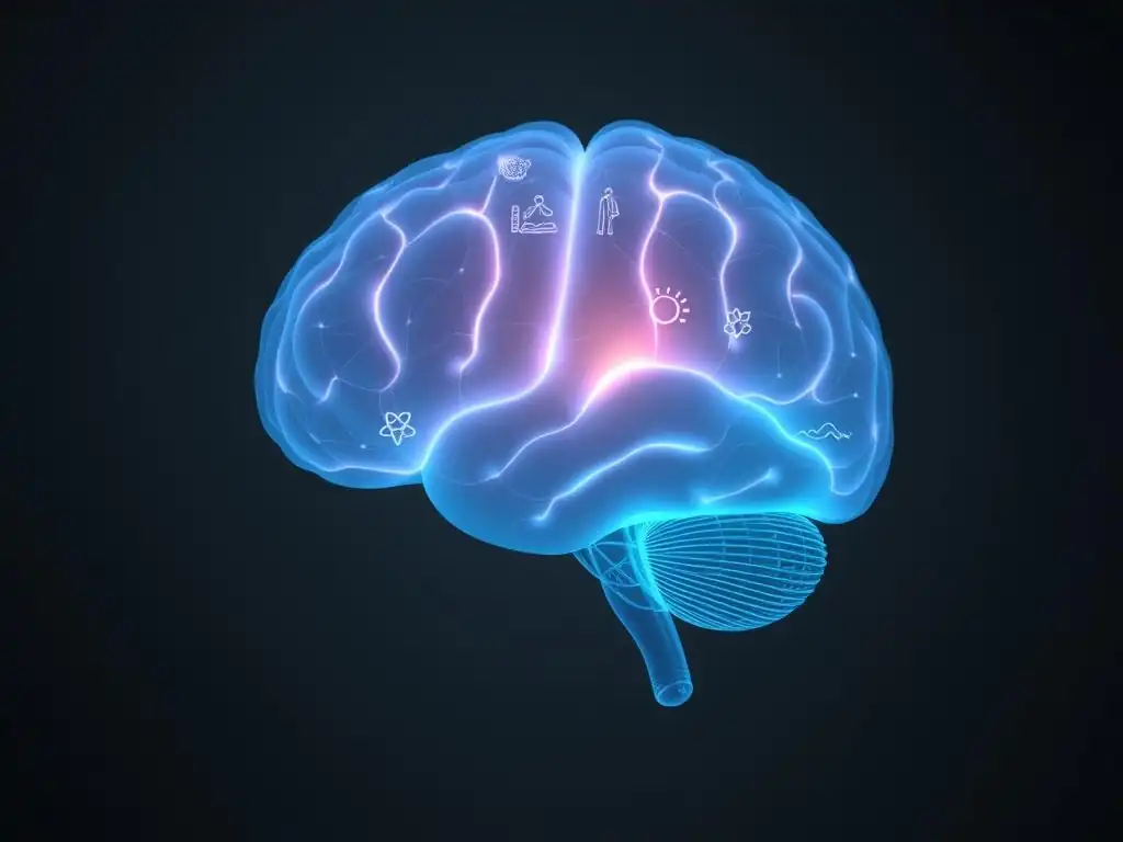
Cognitive Behavioral Therapy (CBT) is a widely used therapy that focuses on identifying and changing negative thought patterns and behaviors. Neuroimaging studies have consistently shown that CBT exerts significant influence on the prefrontal cortex (PFC), particularly areas involved in executive functions, decision-making, and emotional regulation, such as the dorsolateral and ventromedial PFC. Successful CBT for conditions like depression or anxiety is often associated with increased activity in these PFC regions, particularly when individuals are engaged in tasks that require cognitive control over emotional stimuli.
This modulation of the prefrontal cortex is thought to be a key mechanism behind CBT's effectiveness. By helping individuals challenge irrational thoughts and develop more adaptive responses, CBT strengthens the capacity of the PFC to regulate activity in subcortical emotional centers like the amygdala. Studies tracking brain changes before and after CBT commonly report enhanced functional connectivity between the PFC and the amygdala, indicating improved top-down control.
For example, research on social anxiety treated with CBT has shown increased coupling between the ventrolateral PFC and amygdala during social threat anticipation. These findings highlight the specific brain imaging therapy benefits related to cognitive control.
Psychodynamic Therapy and Amygdala Activity Changes
Psychodynamic therapy delves into unconscious processes, past experiences, and interpersonal relationships to understand current difficulties. While traditionally less studied with neuroimaging than CBT, emerging research suggests that psychodynamic approaches can significantly impact limbic system structures, particularly the amygdala, which is central to processing emotions like fear and anxiety. The focus on processing difficult emotions and understanding their roots may lead to a desensitization effect on these emotional centers.
Studies using fMRI have shown that psychodynamic therapy can lead to decreased reactivity in the amygdala, similar to findings in other emotion-focused therapies. For individuals with depression, for instance, psychodynamic psychotherapy has been associated with reduced amygdala activity when viewing sad stimuli. Furthermore, exploring past experiences and their emotional impact in therapy might alter how memories are processed and stored, potentially involving regions like the hippocampus and its connections to the amygdala. While specific percentage changes vary widely depending on the study and condition, the general trend points towards a normalization or reduction in exaggerated limbic responses.
Mindfulness-Based Therapies and Default Mode Network Connectivity
Mindfulness-Based Therapies, such as Mindfulness-Based Stress Reduction (MBSR) or Mindfulness-Based Cognitive Therapy (MBCT), cultivate present-moment awareness without judgment. Neuroimaging research suggests these therapies impact brain networks involved in self-referential processing and attention, notably the Default Mode Network (DMN). The DMN is typically active when the mind is wandering, reflecting on the past or future, or engaging in self-referential thought. Overactivity or dysregulation of the DMN is implicated in conditions like depression and anxiety.
Mindfulness training has been associated with decreased activity in the DMN and altered connectivity between the DMN and other networks, such as the Executive Control Network (ECN). This shift is thought to reflect a reduced tendency for mind-wandering and rumination, and an enhanced ability to intentionally direct attention. For example, studies have shown reduced functional connectivity between the medial prefrontal cortex (a key DMN hub) and the posterior cingulate cortex after MBSR.
Additionally, mindfulness can increase activity in regions associated with present-moment awareness, such as the insula. These changes in network dynamics represent significant brain imaging therapy benefits related to attention and self-awareness.
Exposure Therapy and Amygdala Habituation
Exposure Therapy is a core component of treating anxiety disorders, particularly phobias and PTSD, by gradually exposing individuals to feared stimuli in a safe environment. The underlying principle is habituation and extinction of the fear response. Neuroimaging studies using fMRI during exposure tasks show that successful exposure therapy leads to decreased activity in the amygdala and associated fear circuitry over time as individuals habituate to the stimulus.
Initially, encountering a feared stimulus activates the amygdala strongly. With repeated, safe exposure facilitated by therapy, this amygdala response diminishes. Furthermore, exposure therapy is thought to strengthen the inhibitory pathways from the prefrontal cortex to the amygdala, allowing for better regulation of the fear response. Studies have documented significant reductions in amygdala activation to phobic stimuli, sometimes dropping by 40% or more after a full course of exposure therapy compared to pre-treatment levels. This demonstrable change in a key fear-processing region provides compelling evidence of the brain imaging therapy benefits for anxiety reduction.
Interpreting Brain Imaging Results: What to Look For
Identifying Increased Brain Activity in Key Regions
When interpreting brain imaging results related to therapy outcomes, one key aspect is identifying changes in brain activity levels within specific regions known to be involved in the mental health condition being treated. For example, in depression, successful therapy might lead to increased activity in areas associated with positive emotion processing or cognitive control, such as parts of the prefrontal cortex or striatum, particularly when engaging in relevant tasks or during resting states. Conversely, therapies for anxiety disorders often aim to decrease activity in fear-related regions like the amygdala when confronted with triggers.
Observing such changes provides direct evidence that the therapy is impacting the neural circuits hypothesized to be dysfunctional. It's not just about finding any change, but changes in regions and directions (increase/decrease) that align with the expected mechanisms of the therapy and the nature of the condition. For instance, finding increased activity in the insula after mindfulness therapy aligns with the therapy's goal of enhancing body awareness. These specific directional changes in activity are crucial indicators of brain imaging therapy benefits.
Detecting Changes in Neural Connectivity Patterns
Beyond changes in activity within single regions, therapy can also alter how different brain regions communicate with each other – their functional or structural connectivity. Functional connectivity, often measured with fMRI, assesses how correlated the activity of different regions is over time, suggesting they are working together. Structural connectivity, measured with DTI, assesses the integrity of the white matter pathways connecting regions. Therapy can strengthen beneficial connections or weaken maladaptive ones.
For instance, successful therapy for PTSD might involve strengthening the functional and structural connections between the prefrontal cortex and the amygdala, indicating improved cognitive regulation of fear. Conversely, conditions like depression can show decreased connectivity in certain reward circuits, and successful treatment might restore these connections. Identifying these network-level changes is vital because mental health conditions often involve dysfunction across distributed brain networks, not just isolated regions. Changes in connectivity patterns are powerful indicators of the brain imaging therapy benefits at a systemic level.
Understanding the Significance of Neurotransmitter Levels
While fMRI and DTI primarily focus on activity and connectivity, PET imaging provides insights into the neurochemical effects of therapy, such as changes in neurotransmitter receptor availability or metabolic activity. Understanding the significance of these neurochemical shifts is crucial, as neurotransmitter imbalances are implicated in many mental health conditions. Therapy, even without medication, can influence neurotransmitter systems indirectly through changing neural activity patterns.
For example, behavioral activation therapy for depression might increase engagement in rewarding activities, which could indirectly influence dopamine pathways. While PET studies specifically on the neurochemical changes induced solely by therapy (without medication) are complex and less common than those involving medication, the principle remains: changes in the availability or binding of neurotransmitters or their receptors in relevant brain areas are strong indicators of therapy's impact on the underlying neurochemistry. Observing changes in, for example, serotonin transporter binding in mood-regulating regions after therapy would be a significant finding regarding brain imaging therapy benefits.
Addressing Potential False Positives and Negatives
Interpreting brain imaging results requires caution, as potential false positives and negatives exist. Brain imaging data is complex and requires sophisticated statistical analysis. Findings can sometimes be influenced by factors unrelated to the therapy itself, such as scanner noise, participant movement, or even the specific analytical methods used. A result showing a significant change might be a statistical artifact rather than a true biological effect (false positive). Conversely, a real therapeutic effect on the brain might be too subtle to detect with current techniques or study designs (false negative).
Researchers employ rigorous statistical methods, replication across studies, and careful control groups (e.g., waitlist control, placebo control) to mitigate these risks. It is also important to correlate imaging findings with clinical outcomes; a brain change is most meaningful if it is associated with actual symptom improvement. Understanding these limitations is crucial for accurate interpretation and prevents overstating the brain imaging therapy benefits based on preliminary or isolated findings. Imaging provides valuable correlational data, but establishing causation requires careful study design.
Case Studies: Real-World Examples of Brain Imaging in Therapy Evaluation
Case Study 1: fMRI Evidence of CBT's Impact on Anxiety Disorders
A classic example illustrating the brain imaging therapy benefits of CBT comes from studies on social anxiety disorder. Participants diagnosed with social anxiety disorder often show exaggerated activity in the amygdala when anticipating or experiencing social situations. One well-cited study involved participants undergoing a course of CBT specifically tailored for social anxiety. Before and after therapy, participants underwent fMRI scans while viewing faces with neutral or negative expressions or engaging in simulated social interactions.
The results consistently showed a significant reduction in amygdala activation when processing social threat cues after CBT compared to their pre-treatment scans. Furthermore, this decreased amygdala activity was often correlated with the degree of clinical improvement reported by the participants (e.g., reduction in social anxiety symptoms). Some studies also reported increased activity in prefrontal regions involved in emotion regulation. This fMRI evidence provides compelling objective data supporting the effectiveness of CBT in modulating the brain's fear response circuitry.
Case Study 2: PET Scans Showing the Effects of Medication and Therapy on Depression
While often used to study medication effects, PET imaging has also contributed to understanding how therapy influences the brain, sometimes in comparison or combination with pharmacotherapy. For instance, studies on major depressive disorder have used PET to examine changes in brain metabolism or neurotransmitter systems following treatment. One approach has been to measure glucose metabolism, an indicator of neural activity, across different brain regions.
Research comparing antidepressant medication and psychotherapy (like CBT or interpersonal therapy) for depression using PET has sometimes shown overlapping but also distinct patterns of metabolic change. For example, one study using FDG-PET (which measures glucose metabolism) found that both medication and psychotherapy led to increased metabolism in certain prefrontal areas and decreased metabolism in some limbic regions like the amygdala, suggesting a common pathway towards symptom reduction. However, therapy might preferentially impact certain cortical regions, while medication might have stronger effects on subcortical areas. These studies highlight the diverse ways interventions can confer brain imaging therapy benefits.
Case Study 3: EEG Analysis Revealing Enhanced Brainwave Coherence After Mindfulness Training
EEG studies offer insights into the dynamic changes in brain function following therapies like mindfulness training. One illustrative case involves participants undertaking an 8-week Mindfulness-Based Stress Reduction (MBSR) program. EEG recordings were taken before and after the program, often during meditation or resting states. Researchers analyze various aspects of the EEG signal, including power in different frequency bands and coherence, which measures the degree of synchronization between electrical signals from different brain regions.
Studies have reported changes such as an increase in alpha wave power, particularly in frontal and parietal regions, which is associated with relaxed attention and reduced rumination. More notably, some research has shown increased coherence between brain regions involved in executive control and attention, suggesting improved communication and coordination within these networks. For example, enhanced connectivity between frontal midline areas and parietal regions has been observed. These EEG findings provide temporal and network-level evidence for the brain imaging therapy benefits related to improved attentional control and altered states of consciousness cultivated by mindfulness practices.
The Future of Brain Imaging in Therapy: Personalized Treatment Approaches
Using Brain Scans to Predict Therapy Response
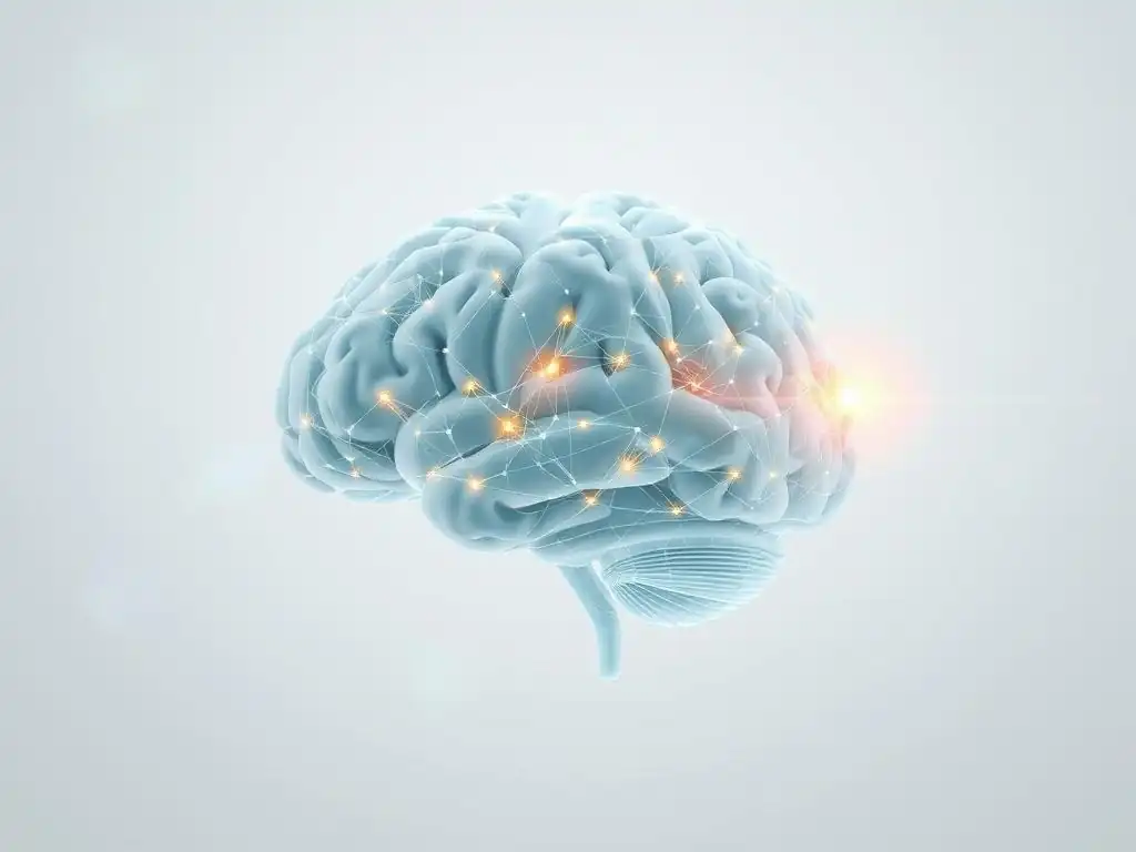
One of the most exciting potential applications of brain imaging in therapy is its use in predicting who will benefit from a particular treatment before it even begins. Just as individuals have different symptoms and life experiences, their brains also exhibit unique patterns of activity, connectivity, and structure. These baseline neural profiles might hold clues about how a person will respond to different therapeutic modalities. For example, a specific pattern of prefrontal cortex activity might predict a better response to CBT, while a different pattern might suggest that psychodynamic therapy or mindfulness is more likely to be effective.
Research is actively exploring potential neural biomarkers for treatment response. This involves scanning individuals before therapy and analyzing their brain data in relation to their eventual treatment outcomes. While still largely experimental, findings are beginning to emerge suggesting that baseline brain characteristics can predict the degree of symptom reduction or the likelihood of treatment completion. Imagine a future where a brain scan could help guide a patient towards the therapy most likely to yield significant brain imaging therapy benefits for them.
Tailoring Treatment Plans Based on Individual Brain Profiles
Building on the potential for prediction, the ultimate goal is to use brain imaging to tailor treatment plans to individual patients. If a patient's brain scan reveals a specific neural pattern associated with a particular type of dysfunction (e.g., exaggerated amygdala reactivity, poor connectivity in a specific network), this information could inform the choice of therapy. For instance, someone with prominent amygdala hyperactivity might be directed towards therapies known to effectively modulate this region, such as exposure therapy or certain forms of emotion-focused therapy.
This personalized approach moves away from a one-size-fits-all model and aims to match the treatment to the individual's unique biological and psychological profile. While significant research is needed to establish reliable links between specific brain profiles and optimal therapy choices, this represents a major frontier in mental healthcare. Integrating brain imaging data with clinical assessments, genetic information, and behavioral data could lead to highly individualized treatment strategies designed to maximize brain imaging therapy benefits.
Integrating Brain Imaging into Clinical Practice Guidelines
For brain imaging to truly revolutionize mental healthcare, it needs to move beyond the research lab and into clinical practice. This requires developing standardized protocols for acquiring and interpreting imaging data in a clinical context, establishing clear guidelines for when imaging is indicated, and training clinicians to understand and utilize this information. Currently, brain imaging is not a routine part of mental health diagnosis or treatment planning, primarily due to cost, accessibility, and the need for more robust evidence linking specific imaging findings to treatment decisions for individual patients.
However, as research continues to solidify the links between brain changes, therapy outcomes, and predictive biomarkers, there is a growing potential for integrating brain imaging into clinical practice guidelines. This could involve using imaging data to refine diagnoses, select the most appropriate therapy, monitor progress objectively, or even predict relapse risk. While challenges remain, the integration of imaging data holds the promise of enhancing the precision and effectiveness of mental health interventions and making the assessment of brain imaging therapy benefits a more routine part of care.
Ethical Considerations and Limitations of Brain Imaging in Therapy
Ensuring Patient Privacy and Data Security
The use of brain imaging in therapy raises important ethical considerations, particularly regarding patient privacy and data security. Brain imaging data is highly personal and potentially sensitive, revealing information about an individual's neural function and potentially their predisposition to certain conditions. As imaging data is collected, stored, and potentially shared (for research or clinical purposes), ensuring robust safeguards against unauthorized access, data breaches, and misuse is paramount.
Patients must be fully informed about how their imaging data will be used, who will have access to it, and for how long it will be stored. Obtaining informed consent is crucial, especially if data is to be used in research databases. Developing standardized protocols for anonymizing or de-identifying data where possible, and adhering to strict data protection regulations (like GDPR or HIPAA), are essential steps to build trust and protect patient confidentiality in the context of utilizing brain imaging therapy benefits research.
Addressing the Potential for Misinterpretation of Imaging Results
Brain imaging data is complex and requires expert interpretation. There is a significant risk of misinterpreting results, both by clinicians who may lack specialized training in neuroimaging and potentially by patients who might oversimplify or misunderstand the findings. A single brain scan is a snapshot and must be interpreted within the context of a person's overall clinical picture, history, and symptoms. Attributing complex psychological states or treatment outcomes solely to isolated findings on a scan can be misleading and potentially harmful.
For example, identifying a certain pattern of activity does not definitively diagnose a condition or guarantee a specific treatment outcome. Brain function is highly dynamic and influenced by many factors. Educating clinicians, patients, and the public about the capabilities and limitations of brain imaging is crucial. Emphasis should be placed on imaging as a tool to complement clinical assessment, not replace it, and on the probabilistic nature of findings rather than deterministic conclusions about brain imaging therapy benefits for an individual.
Acknowledging the Cost and Accessibility Challenges of Brain Imaging Technologies
Despite the significant potential of brain imaging in understanding and improving therapy, the technologies themselves are expensive and not universally accessible. MRI, PET, and even advanced EEG setups require specialized equipment, trained personnel (technicians, radiologists, neuroscientists), and significant infrastructure, limiting their availability primarily to large hospitals or research institutions. This creates disparities in access to potentially valuable insights into brain imaging therapy benefits based on geographical location or socioeconomic status.
Integrating these technologies into routine clinical practice on a large scale would require substantial investment in equipment, training, and healthcare system changes. Currently, the cost barrier prevents brain imaging from being a standard part of mental health assessment or treatment monitoring for most individuals. Addressing these cost and accessibility challenges is crucial for ensuring that the potential benefits of neuroimaging in mental healthcare can be realized equitably.
Conclusion: Harnessing Brain Imaging for Enhanced Mental Healthcare
The Potential of Brain Imaging to Revolutionize Therapy Evaluation
Brain imaging techniques are powerful tools that offer unprecedented insights into the neural effects of psychological therapies. By providing objective measures of changes in brain activity, connectivity, and neurochemistry, imaging is revolutionizing how we evaluate therapy effectiveness. It moves the assessment beyond subjective reports to reveal the tangible biological footprint that therapy leaves on the brain. This capability strengthens the evidence base for therapy, helps to understand its mechanisms of action, and highlights the profound brain imaging therapy benefits.
From demonstrating the modulation of fear circuits by exposure therapy to revealing changes in network dynamics following mindfulness, imaging studies provide empirical validation for the transformative power of therapy. This objective evidence is crucial for increasing public and professional confidence in psychological treatments and advocating for their importance within the broader healthcare system.
Advancing Our Understanding of Brain Changes Following Therapy
The ongoing application of brain imaging in therapy research continues to deepen our understanding of neuroplasticity – the brain's remarkable ability to change and adapt throughout life. Imaging studies are mapping the specific neural pathways and networks that are altered by different therapeutic interventions for a wide range of conditions. This growing body of knowledge is not only confirming long-held theories about how therapy might work but also uncovering novel mechanisms and brain regions involved in recovery and resilience.
Every new study contributes another piece to the complex puzzle of how psychological interventions translate into biological changes in the brain. This advancing understanding is fundamental to developing more effective, targeted, and personalized therapies in the future. It underscores that brain changes following therapy are real, measurable, and contribute significantly to long-term mental well-being, solidifying the concept of brain imaging therapy benefits.
A Call for Continued Research and Innovation in the Field
Despite the significant progress made, the field of brain imaging in therapy is still relatively young. Continued research is essential to refine imaging techniques, improve analytical methods, replicate findings across diverse populations, and establish clear links between specific brain changes and clinical outcomes. Further innovation is needed to make imaging technologies more accessible, affordable, and clinically applicable.
Investing in this research is crucial for unlocking the full potential of brain imaging to enhance mental healthcare. It holds the promise of enabling personalized treatment approaches, improving diagnostic precision, and ultimately leading to better outcomes for individuals struggling with mental health challenges. As research progresses, the insights gained from brain imaging will undoubtedly play an increasingly vital role in understanding and maximizing the brain imaging therapy benefits for all.
Frequently Asked Questions
What exactly is brain imaging in the context of therapy, and how does it relate to mental well-being?
Brain imaging, such as fMRI and PET scans, allows therapists to visualize brain activity during and after therapy. This can provide insights into how the brain is responding to treatment and contribute to a comprehensive understanding of mental well-being.
What are the potential benefits of using brain imaging to guide or personalize mental health therapy?
Brain imaging can help tailor therapy by identifying specific brain areas affected by a condition. This personalization can lead to more effective treatment plans and improved patient outcomes, ultimately maximizing brain imaging therapy benefits.
Can brain imaging definitively diagnose mental health conditions, or is it more of a supplementary tool?
Brain imaging is not a definitive diagnostic tool for most mental health conditions. It serves as a supplementary tool offering valuable insights alongside clinical assessments and patient history.
How accessible and affordable is brain imaging for individuals seeking mental health treatment?
The accessibility and affordability of brain imaging remain significant challenges. It is often expensive and not widely covered by insurance, limiting its availability for many seeking mental health treatment.
What are the ethical considerations surrounding the use of brain imaging in mental health therapy?
Ethical concerns include privacy of brain data, potential for misinterpretation of results, and the risk of over-reliance on imaging at the expense of other important aspects of care.
Final Reflection: Mapping the Future of Therapeutic Brain Change
The convergence of advanced brain imaging and therapeutic interventions marks a pivotal moment in our understanding and treatment of neurological and psychological conditions. This isn't just about observing brain activity; it's about actively shaping it for the better, offering hope where previously there was only limited understanding. The potential for personalized, targeted therapies based on individual brain responses is truly transformative.
The journey through this article has highlighted the profound impact brain imaging is having, not just on scientific understanding, but on the lived experiences of countless individuals. It's a testament to human ingenuity and our relentless pursuit of improved well-being. The ability to visualize the very mechanisms of change – to see how therapy reshapes neural pathways and strengthens connections – is incredibly powerful. The greatest discovery of all time is that a person can change his future by merely changing his attitude.
– Oprah Winfrey. This quote resonates deeply with the core message of this article: that through understanding, we can empower change.
Now, consider your own relationship with your mind and body. What if you could visualize the impact of your coping mechanisms, your mindfulness practices, or your therapeutic journey? I invite you to reflect on the potential of brain imaging, not just as a diagnostic tool, but as a pathway to self-discovery and empowerment. Embrace the possibilities, advocate for advancements in this field, and remember that the journey towards a healthier, more resilient brain is a journey worth taking, one that we are only beginning to truly understand.
Unveil Your Brain's Healing Journey: Take Action Today!
Thanks for exploring the fascinating world of brain imaging and therapy with us! You've now gained valuable insights into how we can track progress in mental health treatment. Let's use this knowledge to move forward:
- Dive deeper into personalized therapy: Research different types of therapy and find one that resonates with your specific needs and goals. Consider journaling about your experience to track your own progress.
- Share this knowledge: Discuss your newfound understanding of brain imaging and therapy with friends, family, or your therapist. Starting a conversation can be incredibly powerful.
- Challenge yourself: Ask your therapist about the possibility of utilizing brain imaging techniques to monitor your treatment's efficacy – even if it’s just a conversation starter.
Ready to embark on a more informed and empowered journey towards better mental wellbeing? What specific step will you take today to enhance your therapy experience?