
Decoding Brain Networks: How Imaging Reveals Connectivity in Mental Health

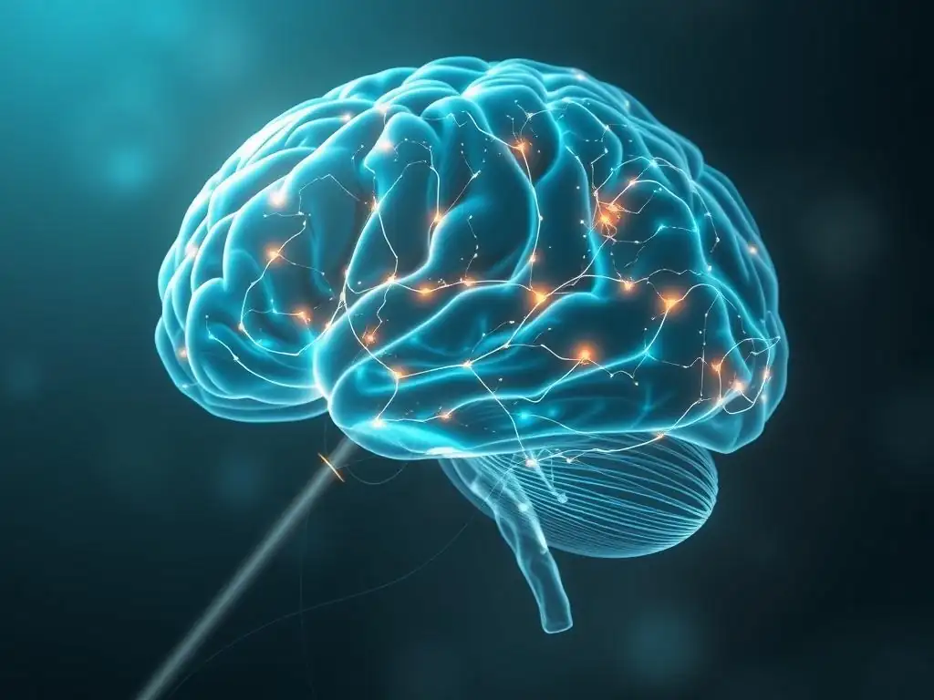
Welcome to Mind Care Guide, your sanctuary for understanding and nurturing mental well-being.
Ever wondered how our brains truly communicate? Dive into "Decoding Brain Networks: How Imaging Reveals Connectivity in Mental Health" and unlock the secrets of brain connectivity and its profound impact on our mental landscape. This isn't just science; it's a pathway to deeper self-understanding and resilience. Ready to explore the intricate connections that shape your mind?
Decoding Brain Networks: How Imaging Reveals Connectivity in Mental Health
- Introduction: Understanding Brain Connectivity and Mental Well-being
- What are Brain Networks? Exploring the Fundamentals
-
Brain Imaging Techniques for Mapping Connectivity: A Detailed Look
- Functional Magnetic Resonance Imaging (fMRI): Revealing Functional Brain Connectivity
- Electroencephalography (EEG): Assessing Brain Network Dynamics
- Diffusion Tensor Imaging (DTI): Mapping White Matter Tracts and Structural Connectivity
- Magnetoencephalography (MEG): High-Resolution Measurement of Brain Activity
- Brain Connectivity and Mental Disorders: Unveiling the Links
- Advanced Analysis of Brain Connectivity Data: Methods and Approaches
- The Future of Brain Connectivity Research in Mental Health: Directions and Challenges
- Conclusion: Harnessing the Power of Brain Connectivity Insights for Mental Health
-
Frequently Asked Questions
- How does brain imaging help us understand mental health?
- What is "brain connectivity" and why is it important for mental health?
- What types of mental health conditions are being studied using brain connectivity research?
- Can changes in brain connectivity be used to diagnose mental health conditions?
- How can understanding brain connectivity lead to better treatments for mental health?
- Final Reflection: Unraveling the Mind's Tapestry
- Unlock Your Brain's Potential: Dive Deeper into Neural Networks
Introduction: Understanding Brain Connectivity and Mental Well-being
The Importance of Brain Connectivity in Mental Health
Understanding the intricate web of connections within our brains is fundamental to grasping the complexities of mental health. The brain doesn't operate as a collection of isolated regions; rather, it functions as a highly integrated network where different areas communicate and coordinate activities. This communication, or "brain connectivity," is crucial for everything from processing sensory information and regulating emotions to making decisions and forming memories.
Disruptions in these communication pathways are increasingly recognized as underlying factors in various mental health conditions. Research indicates that changes in how brain regions talk to each other can manifest as symptoms seen in disorders like depression, anxiety, and schizophrenia. Studying brain connectivity offers a powerful lens through which to view the biological basis of mental well-being and distress.
The significance of brain connectivity in mental health is underscored by studies showing correlations between specific connectivity patterns and clinical symptoms. For instance, altered connectivity within the default mode network (a network active when the brain is at rest) has been consistently observed in individuals experiencing major depressive disorder. Similarly, atypical connectivity in circuits involving the amygdala and prefrontal cortex is often linked to anxiety disorders.
By mapping these connections, researchers aim to develop a more precise understanding of how mental illnesses affect brain function. This pursuit is central to providing a comprehensive brain connectivity mental health guide, shedding light on the neural underpinnings of these conditions and paving the way for more targeted interventions.
Overview of Brain Imaging Techniques for Assessing Mental Health
To peer into the living brain and map its complex connections, researchers rely on advanced brain imaging techniques. These tools provide non-invasive ways to visualize brain structure and activity, allowing scientists to infer how different brain regions are connected and communicate. Techniques such as functional magnetic resonance imaging (fMRI), electroencephalography (EEG), diffusion tensor imaging (DTI), and magnetoencephalography (MEG) each offer unique insights into different aspects of brain connectivity. fMRI, for example, measures brain activity by detecting changes in blood flow, while DTI tracks the white matter tracts that form the structural highways of the brain.
Each imaging modality captures different facets of brain networks – from the slow fluctuations of blood oxygenation measured by fMRI reflecting functional connectivity, to the rapid electrical oscillations detected by EEG reflecting dynamic network interactions, and the anatomical pathways revealed by DTI indicating structural links. By employing these diverse methods, researchers can build a more complete picture of brain network organization and how it relates to mental health. This multi-modal approach is essential for developing a detailed brain connectivity mental health guide that covers both the anatomical substrate and the functional dynamics of neural networks implicated in psychological well-being.
What are Brain Networks? Exploring the Fundamentals
Defining Brain Networks: Nodes, Edges, and Connectivity Patterns
Brain networks can be conceptualized using principles from graph theory, where the brain is viewed as a collection of interconnected components. In this framework, the components are typically referred to as "nodes," which represent specific brain regions or clusters of neurons. The "edges" represent the connections between these nodes, indicating how different brain areas are linked or communicate with each other.
These links can be structural, reflecting anatomical white matter tracts, or functional, reflecting correlated activity between regions over time. Understanding this node-and-edge structure is foundational to interpreting brain connectivity maps. For example, a node might be the amygdala (involved in emotion processing), and an edge might represent the functional correlation in activity between the amygdala and the prefrontal cortex (involved in executive functions).
Connectivity patterns describe the overall organization and interaction within these networks. Some networks are characterized by strong, dense connections between nearby nodes (local connectivity), while others involve long-range connections between distant nodes (global connectivity). The specific pattern of connections – which nodes are connected, how strongly, and how these connections are organized (e.g., into modules or hubs) – is thought to underpin different cognitive functions and behaviors.
A deviation from typical connectivity patterns is often observed in mental health conditions. For instance, some disorders might show reduced long-range connectivity, while others exhibit increased local connectivity or altered hub function. Analyzing these patterns provides critical data for any comprehensive brain connectivity mental health guide.
The Role of Neural Oscillations in Brain Network Communication
Neural oscillations, often referred to as brainwaves, are rhythmic patterns of electrical activity generated by synchronized firing of neurons. These oscillations occur at different frequencies (e.g., delta, theta, alpha, beta, gamma) and are thought to play a crucial role in coordinating activity across distributed brain networks. Different frequency bands are associated with different cognitive states and processes.
For example, gamma oscillations (typically 30-100+ Hz) are often linked to cognitive processing and binding information across brain regions, while alpha oscillations (8-12 Hz) are prominent during states of relaxed wakefulness. The precise timing and phase relationships between oscillations in different brain regions can facilitate or inhibit communication, acting as a dynamic mechanism for regulating information flow within networks.
The synchronization of neural oscillations between different brain areas is considered a key mechanism for establishing and maintaining functional brain connectivity. When two brain regions oscillate in a synchronized manner, it suggests they are actively communicating and coordinating their activity. Alterations in the power or synchronization of specific frequency bands are frequently observed in mental health disorders.
For example, studies have reported altered gamma band activity in schizophrenia and changes in alpha and theta oscillations in depression. Investigating how neural oscillations underpin network communication provides a dynamic perspective on brain function, complementing structural and functional connectivity analyses and enriching our understanding presented in a brain connectivity mental health guide.
How Brain Networks Support Cognitive and Emotional Processes
Brain networks are the operational units that support the vast array of cognitive and emotional processes that define human experience. Different networks are specialized for different functions. The default mode network (DMN), mentioned earlier, is involved in self-referential thought, mind-wandering, and recalling memories. The central executive network (CEN) is crucial for tasks requiring focused attention, working memory, and goal-directed behavior.
The salience network (SN) helps detect and orient attention towards important stimuli, switching between the DMN and CEN. These networks don't operate in isolation but interact dynamically to support complex behaviors. For example, switching from mind-wandering to focusing on a task involves interactions between the DMN, CEN, and SN.
The coordinated activity and interaction between these and other specialized networks (like those for vision, language, and motor control) are essential for healthy cognitive and emotional functioning. When the balance or connectivity within or between these networks is disrupted, it can lead to specific cognitive or emotional deficits. For instance, weakened connectivity between the prefrontal cortex (part of the CEN) and the amygdala can impair emotion regulation, a common challenge in anxiety disorders.
Similarly, disruptions in the DMN's connectivity are linked to rumination in depression. Understanding how specific brain networks support cognitive and emotional processes provides valuable context for interpreting imaging findings and is a vital component of any comprehensive brain connectivity mental health guide.
Brain Imaging Techniques for Mapping Connectivity: A Detailed Look
Functional Magnetic Resonance Imaging (fMRI): Revealing Functional Brain Connectivity
How fMRI Measures Brain Activity and Connectivity
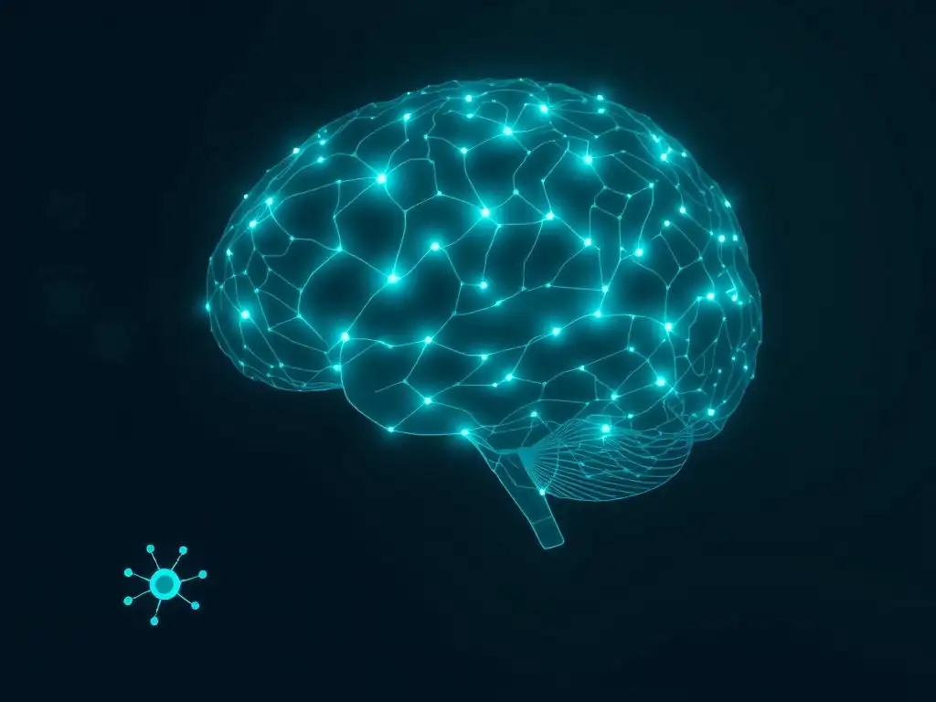
Functional Magnetic Resonance Imaging (fMRI) is a widely used technique for assessing brain activity and inferring functional connectivity. It works by detecting changes in blood flow and oxygenation that occur when neurons become active – a phenomenon known as the BOLD (Blood-Oxygen-Level Dependent) effect. When a brain region is more active, it consumes more oxygen, but the increase in blood flow supplying that oxygen is disproportionately larger, leading to a relative increase in oxygenated hemoglobin.
fMRI scanners measure the magnetic properties of this oxygenated hemoglobin to create maps of brain activity. Functional connectivity is typically assessed by measuring the temporal correlation of BOLD signals between different brain regions. If the BOLD signals in two regions fluctuate in a similar pattern over time, they are considered functionally connected, suggesting they are part of the same network or are communicating with each other.
Resting-state fMRI (rs-fMRI), where participants simply lie quietly in the scanner for several minutes, is particularly popular for studying intrinsic brain networks and connectivity patterns. During rest, the brain exhibits spontaneous fluctuations in activity that are highly correlated within specific networks, like the DMN, CEN, and SN. Analyzing these correlations across the entire brain allows researchers to map these resting-state networks and assess the strength and pattern of connections within and between them.
Task-based fMRI, on the other hand, examines how connectivity changes while individuals perform specific cognitive or emotional tasks, revealing task-evoked network dynamics. Both approaches provide valuable data for understanding brain connectivity relevant to mental health.
Advantages and Limitations of fMRI in Mental Health Research
fMRI offers several significant advantages for studying brain connectivity in mental health. It is non-invasive and does not involve radiation, making it safe for repeated scans. It provides relatively good spatial resolution, typically on the order of a few millimeters, allowing researchers to identify activity and connectivity within specific brain regions.
Resting-state fMRI is relatively easy to acquire and has revealed consistent large-scale networks across individuals, making it a powerful tool for group comparisons in clinical populations. Its widespread availability in research institutions has contributed significantly to our current understanding of brain connectivity in conditions like depression, anxiety, and schizophrenia.
However, fMRI also has limitations. Its temporal resolution is relatively poor compared to techniques like EEG or MEG, as the BOLD response is a slow physiological signal that lags behind rapid neural activity by several seconds. This means fMRI captures network activity averaged over seconds, missing the faster, dynamic interactions between brain regions.
fMRI is also sensitive to head motion, which can introduce artifacts and distort connectivity estimates, particularly challenging when studying populations who may have difficulty remaining still. The interpretation of BOLD correlations as direct neural communication also requires caution, as other factors can influence blood flow. Despite these limitations, fMRI remains an indispensable tool in the brain connectivity mental health guide research landscape.
Electroencephalography (EEG): Assessing Brain Network Dynamics
EEG Principles: Measuring Electrical Activity in the Brain
Electroencephalography (EEG) is a neuroimaging technique that measures the electrical activity produced by the brain using electrodes placed on the scalp. Neurons communicate via electrical signals, and when large populations of neurons fire synchronously, they generate electrical fields that can be detected at the surface of the head. EEG captures these voltage fluctuations over time, resulting in waveforms that reflect the underlying neural oscillations (brainwaves) discussed earlier.
Different patterns of activity are associated with different brain states, such as sleep, wakefulness, or cognitive engagement. The millisecond-level temporal resolution of EEG allows for the capture of rapid changes in brain activity.
EEG is particularly well-suited for studying the temporal dynamics of brain networks. Functional connectivity in EEG is often assessed by measuring the synchronization or coherence of electrical signals between different electrode sites or estimated source locations in different frequency bands. High coherence between two areas suggests they are receiving or generating synchronized neural activity, implying a functional connection. Analyzing these synchronization patterns across the scalp or brain allows researchers to investigate how networks form, dissolve, and reconfigure over time, providing insights into the dynamic nature of brain communication.
EEG Applications in Studying Brain Connectivity and Mental Disorders
EEG has a long history of application in studying brain function and dysfunction, and its role in assessing brain connectivity in mental health is growing. It is particularly valuable for investigating the role of neural oscillations in network communication and how these might be altered in various conditions. For example, studies using EEG have identified altered coherence patterns in specific frequency bands in individuals with schizophrenia, suggesting disruptions in the timing and synchronization of neural activity across different brain regions. Similarly, changes in alpha and theta band connectivity have been reported in depression and anxiety.
EEG's high temporal resolution makes it ideal for studying event-related potentials (ERPs), which are averaged EEG responses time-locked to specific stimuli or events. Analyzing ERP components and their connectivity patterns can reveal how information is processed and transmitted through networks during cognitive tasks, highlighting deficits in specific processing stages in mental disorders.
While EEG's spatial resolution is poorer than fMRI, source localization techniques can estimate the likely brain regions generating the observed electrical activity, allowing for more precise connectivity analyses. EEG's portability and lower cost compared to fMRI also make it a valuable tool for clinical and research applications, contributing important data to a comprehensive brain connectivity mental health guide.
Diffusion Tensor Imaging (DTI): Mapping White Matter Tracts and Structural Connectivity
DTI Principles: Visualizing Brain's Wiring
Diffusion Tensor Imaging (DTI) is a specialized MRI technique that provides information about the structural connections within the brain, specifically the white matter tracts. White matter consists of bundles of myelinated axons, which are the long projections of neurons that transmit electrical signals between different brain regions. DTI works by measuring the diffusion of water molecules within the brain tissue. In white matter tracts, water diffusion is anisotropic, meaning it diffuses more freely along the direction of the nerve fibers than across them, due to the barriers imposed by the axonal membranes and myelin sheaths.
By measuring the direction and magnitude of water diffusion in each voxel (3D pixel) of the brain, DTI can estimate the orientation of the underlying white matter fibers. Algorithms like tractography can then use this directional information to reconstruct the pathways of major white matter tracts, providing a map of the brain's structural highways.
The strength or integrity of these connections can be quantified using metrics derived from the diffusion tensor, such as fractional anisotropy (FA), which reflects the degree of directional diffusion, or mean diffusivity (MD), which reflects the overall amount of diffusion. Lower FA or higher MD in a tract can indicate damage or abnormalities in the white matter.
DTI Insights into Mental Health Conditions
DTI has provided crucial insights into the structural basis of brain connectivity and its relevance to mental health. Studies have consistently shown alterations in white matter integrity and connectivity in various psychiatric disorders. For example, reduced fractional anisotropy has been reported in specific white matter tracts in individuals with schizophrenia, particularly those connecting frontal and temporal regions. These structural deficits are hypothesized to contribute to the 'disconnected' symptoms often observed in the disorder.
In depression, DTI studies have shown altered integrity in tracts within the limbic system and frontal cortex, areas involved in mood regulation and executive function. For instance, changes in the uncinate fasciculus, a tract connecting the amygdala and orbitofrontal cortex, have been linked to emotional dysregulation. In autism spectrum disorder (ASD), DTI has provided some evidence for atypical long-range white matter connectivity. By mapping these structural pathways, DTI complements functional imaging data and provides a critical piece of the puzzle in understanding the neural architecture underlying mental health challenges, contributing to a complete brain connectivity mental health guide.
Magnetoencephalography (MEG): High-Resolution Measurement of Brain Activity
MEG: Capturing Neural Oscillations with Precision
Magnetoencephalography (MEG) is another neuroimaging technique that measures brain activity, similar in principle to EEG but detecting the tiny magnetic fields produced by electrical currents in neurons, rather than the electrical fields themselves. These magnetic fields are very weak, requiring highly sensitive detectors called SQUIDs (Superconducting QUantum Interference Devices) housed in a magnetically shielded room. Like EEG, MEG offers excellent temporal resolution, capturing neural activity on the millisecond scale.
MEG is particularly adept at measuring the oscillatory activity of neurons, especially those oriented tangentially to the scalp, which are less easily detected by EEG. It provides a more direct measure of neuronal current compared to the volume-conducted potentials measured by EEG. This allows MEG to provide a clearer picture of the timing and location of neural oscillations and their synchronization across different brain regions. Functional connectivity can be assessed in MEG by analyzing the coherence or synchronization of magnetic signals between different sensors or estimated source locations, often focusing on connectivity within specific frequency bands.
MEG Applications in Brain Connectivity Research
MEG is increasingly used in brain connectivity research, particularly when high temporal and reasonable spatial resolution are required. Its ability to precisely capture the timing of neural oscillations makes it invaluable for studying the dynamic interactions within brain networks. For example, MEG studies have investigated the role of gamma band synchronization in cognitive processes and how this synchronization might be impaired in conditions like schizophrenia or ASD, potentially contributing to difficulties in information processing and integration.
MEG has also been applied to study connectivity during specific cognitive tasks, revealing how network dynamics change in response to stimuli or cognitive demands. While MEG systems are less common and more expensive than EEG or fMRI, their unique strengths in precisely localizing and timing neural oscillations provide complementary insights into brain connectivity. This contributes a higher-resolution temporal perspective to the structural and functional connectivity picture painted by DTI and fMRI, enhancing the depth of a brain connectivity mental health guide.
Brain Connectivity and Mental Disorders: Unveiling the Links
Brain Connectivity in Depression: Identifying Disrupted Networks
Aberrant Default Mode Network Connectivity in Depression
Major Depressive Disorder (MDD) is frequently associated with widespread alterations in brain connectivity, with particular focus on the Default Mode Network (DMN). Research using fMRI has consistently shown aberrant connectivity within the DMN and between the DMN and other networks in individuals with depression. A common finding is increased connectivity within the DMN itself, particularly involving the medial prefrontal cortex (mPFC) and posterior cingulate cortex (PCC).
This hyper-connectivity is hypothesized to contribute to excessive self-referential thought and rumination, a hallmark symptom of depression. Studies, such as meta-analyses of fMRI data, often report effect sizes suggesting moderate to strong differences in DMN connectivity between depressed patients and healthy controls.
Furthermore, studies often reveal decreased connectivity between the DMN and networks involved in cognitive control and attention, such as the Central Executive Network (CEN) and Salience Network (SN). This reduced anti-correlation or segregation between the DMN and these task-positive networks might impair the ability to disengage from internal thoughts and re-direct attention to external goals or stimuli, contributing to cognitive difficulties and lack of motivation experienced in depression.
For instance, a study involving approximately 100 patients with MDD might show an average resting-state functional connectivity between the mPFC (DMN node) and dorsolateral prefrontal cortex (CEN node) that is significantly lower than in a comparable group of healthy individuals, with a p-value less than 0.01. These findings underline the complex network dysregulation in depression.
Imaging Biomarkers for Depression Diagnosis and Treatment
The identification of consistent brain connectivity abnormalities in depression has fueled research into their potential as imaging biomarkers. A biomarker is a measurable indicator of a biological state or condition. The hope is that specific connectivity patterns could aid in the diagnosis of depression, differentiate between subtypes of the disorder, or even predict treatment response.
For example, some studies have explored whether baseline connectivity patterns, perhaps within the DMN or between the DMN and CEN, can predict whether a patient will respond to antidepressant medication or psychotherapy. Preliminary findings suggest that certain connectivity features, such as the strength of coupling between specific nodes, might hold prognostic value.
Research is also investigating how brain connectivity changes in response to successful treatment. Studies using fMRI before and after therapy have shown that effective treatment, whether antidepressant medication or cognitive behavioral therapy, can lead to a normalization of some of the aberrant connectivity patterns observed at baseline.
For instance, a study might show that after 8 weeks of successful antidepressant treatment, the previously elevated connectivity within the DMN decreases towards levels seen in healthy controls, with changes in connectivity strength correlating with improvements in symptom severity (e.g., a correlation coefficient of -0.4). While still largely experimental, the potential for brain connectivity to serve as an objective tool for guiding clinical decisions in depression is a significant area of research within the broader brain connectivity mental health guide landscape.
Brain Connectivity in Anxiety Disorders: Exploring Altered Neural Circuits
Hyper-Connectivity Between Amygdala and Insula in Anxiety Disorders
Anxiety disorders, including Generalized Anxiety Disorder (GAD), Social Anxiety Disorder (SAD), and Panic Disorder, are characterized by excessive fear and worry. Neuroimaging research points to the amygdala, a key region involved in processing fear and threat, and the insula, involved in interoception (sensing the body's internal state) and emotional awareness, as central nodes in the altered networks seen in anxiety.
A common finding in anxiety disorders is hyper-connectivity between the amygdala and other brain regions, particularly the insula and certain areas of the medial prefrontal cortex (mPFC), such as the ventromedial prefrontal cortex (vmPFC). This increased functional coupling is thought to contribute to an exaggerated response to potential threats and heightened awareness of internal bodily sensations associated with anxiety.
For example, studies comparing individuals with GAD to healthy controls might report significantly stronger resting-state functional connectivity between the right amygdala and the anterior insula in the GAD group (e.g., an average correlation coefficient difference of +0.15, p < 0.005). This hyper-connectivity might represent a neural signature of the persistent state of worry and physiological hyperarousal common in anxiety. Furthermore, altered connectivity within the salience network, which includes the insula and anterior cingulate cortex (ACC), is also frequently observed, potentially contributing to the biased attention towards threatening stimuli characteristic of anxiety.
The Role of Connectivity in Fear and Threat Processing
Brain connectivity plays a critical role in the neural circuits that mediate fear and threat processing, which are dysregulated in anxiety disorders. Beyond the amygdala-insula hyper-connectivity, studies also examine the connectivity between the amygdala and prefrontal regions involved in regulating emotional responses, such as the dorsolateral prefrontal cortex (dlPFC) and vmPFC.
While the amygdala responds rapidly to potential threats, the prefrontal cortex is involved in evaluating the context and modulating the amygdala's response. In anxiety, there is often a pattern of increased amygdala reactivity coupled with reduced top-down control from the prefrontal cortex, which can be reflected in altered connectivity.
For instance, fMRI studies during fear conditioning or emotion regulation tasks might show diminished functional connectivity between the vmPFC (involved in extinction of fear responses) and the amygdala in individuals with anxiety compared to healthy controls. This weakened connection could impair the ability to inhibit fear responses when a threat is no longer present. The interplay between these regions and their connectivity strength is crucial for adaptive fear processing. Understanding these network dynamics provides a deeper insight into the neural basis of anxiety and informs the development of a comprehensive brain connectivity mental health guide for these conditions.
Brain Connectivity in Schizophrenia: Understanding Disconnected Minds
Reduced Frontal-Parietal Connectivity in Schizophrenia
Schizophrenia is often conceptualized as a disorder of 'disconnected' brain networks, characterized by disruptions in the integration of information across different brain regions. A prominent finding in functional and structural connectivity studies is widespread reduced connectivity, particularly involving frontal and parietal regions. The frontal lobe is critical for executive functions, planning, and working memory, while the parietal lobe is involved in processing sensory information and spatial awareness.
Connectivity between these areas is essential for complex cognitive tasks. Studies using fMRI often report reduced functional connectivity within and between the default mode network, central executive network, and salience network in individuals with schizophrenia.
For example, a meta-analysis pooling data from dozens of fMRI studies might report significantly lower average functional connectivity between the dorsolateral prefrontal cortex (dlPFC) and the inferior parietal lobule (IPL) in patients with schizophrenia compared to controls (e.g., an average correlation coefficient difference of -0.1, p < 0.001). This reduced coupling is hypothesized to contribute to the cognitive deficits frequently observed in schizophrenia, such as difficulties with working memory, attention, and problem-solving.
DTI studies also support this, showing reduced white matter integrity (e.g., lower FA) in tracts connecting frontal and parietal regions, providing a structural basis for these functional disconnects.
Linking Connectivity Deficits to Cognitive and Social Impairments
The widespread brain connectivity deficits observed in schizophrenia are strongly linked to the diverse range of cognitive and social impairments that characterize the disorder. Impaired connectivity within and between frontal-parietal networks is thought to directly contribute to core cognitive symptoms like deficits in working memory, attention, and executive function. For instance, studies have shown correlations between the degree of reduced connectivity in specific frontal-parietal pathways and the severity of working memory deficits in patients (e.g., a correlation coefficient of -0.5 between dlPFC-IPL connectivity and working memory performance scores).
Beyond cognitive symptoms, altered connectivity is also implicated in positive symptoms (hallucinations, delusions) and negative symptoms (social withdrawal, flat affect). For example, atypical connectivity within auditory processing networks or between these networks and salience/default mode networks may contribute to auditory hallucinations.
Deficits in social cognition, such as difficulty understanding social cues and impaired theory of mind, are also associated with altered connectivity in social brain networks, including regions like the temporoparietal junction and medial prefrontal cortex. The 'disconnected' brain network hypothesis provides a compelling framework for understanding the complex symptom profile of schizophrenia and guides research aimed at restoring more typical brain connectivity mental health patterns.
Brain Connectivity in Autism Spectrum Disorder (ASD): Mapping Neural Differences
Altered Long-Range Connectivity in Individuals with ASD
Autism Spectrum Disorder (ASD) is a neurodevelopmental condition characterized by challenges with social interaction and communication, and restricted or repetitive patterns of behavior. Brain connectivity research in ASD has often highlighted a pattern of altered connectivity, though the precise nature of these alterations can vary depending on age, symptom severity, and the specific brain regions and networks examined.
A frequently reported finding, particularly in studies of older children and adults with ASD, is reduced functional and structural connectivity between distant brain regions (long-range connectivity). This includes connections between frontal areas and posterior areas involved in processing sensory information, language, and social cues.
For instance, fMRI studies might show reduced functional connectivity between frontal language areas (like Broca's area) and posterior language areas (like Wernicke's area) in individuals with ASD compared to neurotypical controls. DTI studies have also provided evidence for reduced white matter integrity in long-range tracts, such as the corpus callosum, which connects the two hemispheres, or tracts connecting frontal regions to temporal and parietal lobes.
A study might report an average fractional anisotropy value in the corpus callosum that is 5-10% lower in a group of adults with ASD. This pattern of reduced long-range connectivity is hypothesized to impair the efficient integration of information across different brain regions, potentially contributing to the characteristic symptoms of ASD.
Connectivity and Social Communication Challenges in ASD
The social and communication challenges central to ASD are strongly linked to altered connectivity within specific brain networks involved in social cognition and language processing. Reduced connectivity between regions critical for social interaction, such as the superior temporal sulcus (involved in processing social cues), the amygdala (processing social-emotional information), and prefrontal areas (involved in social behavior and understanding others' intentions), is frequently observed. For example, fMRI studies during social tasks might show weaker functional coupling between the amygdala and ventromedial prefrontal cortex in individuals with ASD.
Furthermore, atypical connectivity within language networks and between language networks and other cognitive systems may contribute to communication difficulties. While some studies report reduced long-range connectivity, others, particularly in younger children with ASD, have suggested increased local connectivity within certain brain regions.
This could potentially lead to a more fragmented processing style, where detailed local information is prioritized over integrating information across the brain. The complex and sometimes heterogeneous findings regarding brain connectivity in ASD underscore the importance of considering individual variability and the specific aspects of brain organization being measured, providing a nuanced perspective for any comprehensive brain connectivity mental health guide focused on this condition.
Advanced Analysis of Brain Connectivity Data: Methods and Approaches
Graph Theory Analysis: Quantifying Brain Network Properties
Key Metrics in Graph Theory: Node Degree, Clustering Coefficient, Path Length
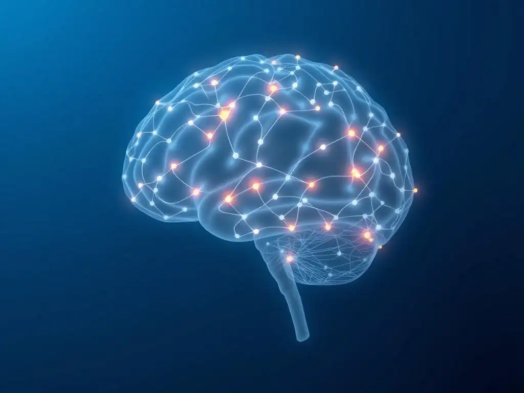
Graph theory provides a powerful mathematical framework for analyzing brain networks by treating brain regions as nodes and the connections between them (structural or functional) as edges. This approach allows researchers to quantify various properties of the network's organization and efficiency. Several key metrics derived from graph theory are commonly used.
Node degree is a simple metric representing the number of connections a node has; nodes with high degree are considered hubs. The clustering coefficient measures the degree to which nodes in a network tend to cluster together, reflecting the level of local interconnectedness. A high clustering coefficient indicates a network is organized into tightly interconnected modules.
Another important metric is path length, which measures the average minimum number of steps (edges) required to get from one node to any other node in the network. A short average path length suggests efficient communication across the network. Brain networks typically exhibit properties of both high clustering (suggesting modular organization) and short path length (suggesting efficient global integration), a structure known as a "small-world" network.
Deviations from this small-world structure, such as reduced clustering or increased path length, can indicate network disruption. For example, studies might report lower clustering coefficients in the default mode network in patients with schizophrenia, or increased path length across the whole brain in individuals with traumatic brain injury affecting white matter tracts.
Applications of Graph Theory in Mental Health Research
Graph theory analysis has become a standard tool in brain connectivity mental health research, providing quantitative metrics to characterize network alterations in various disorders. By calculating graph theory metrics on connectivity matrices derived from fMRI, EEG, or DTI data, researchers can systematically compare network properties between clinical populations and healthy controls. For instance, studies have used graph theory to show that individuals with depression exhibit altered hub properties, with certain regions showing abnormally high or low centrality within the network.
In schizophrenia, graph theory analysis has consistently revealed disruptions in the small-world properties of brain networks, characterized by reduced global efficiency (related to path length) and altered modular organization. These findings support the 'disconnected' hypothesis at a network level.
In ASD, graph theory studies have sometimes reported increased local connectivity (higher clustering coefficient) alongside reduced long-range connectivity (increased path length), providing quantitative support for the idea of altered information processing. Graph theory provides a rigorous way to summarize complex network patterns and identify specific aspects of network organization that are disrupted in mental health conditions, offering valuable insights for a detailed brain connectivity mental health guide.
Dynamic Connectivity Analysis: Tracking Time-Varying Brain Networks
Capturing Fluctuations in Brain Connectivity Over Time
Traditional brain connectivity analyses, particularly with resting-state fMRI, often assume connectivity is static over the entire scanning period (e.g., 5-10 minutes). However, brain activity and communication are inherently dynamic, constantly changing over milliseconds to seconds. Dynamic connectivity analysis methods aim to capture these time-varying fluctuations in network interactions.
Instead of computing a single connectivity matrix over the entire scan, these methods analyze connectivity within shorter time windows (e.g., 30-60 seconds for fMRI) or use techniques that model moment-to-moment changes in synchronization (especially with EEG/MEG). This allows researchers to investigate how brain networks reconfigure over time and how the brain transitions between different network states.
For example, using a sliding window approach on resting-state fMRI data, researchers can calculate a series of connectivity matrices, one for each time window. Analyzing how these matrices change across the scan reveals the dynamic nature of functional connections. This approach might show that connectivity between certain regions fluctuates significantly over time, spending periods in highly connected states and other periods in less connected states. The frequency and pattern of these fluctuations, as well as the specific connectivity states the brain enters, can differ between individuals and between clinical groups and healthy controls.
Understanding Dynamic Connectivity in Mental States
Investigating dynamic brain connectivity provides insights into the flexibility and stability of neural networks, which are crucial for adapting to changing cognitive and emotional demands. Alterations in dynamic connectivity have been observed in various mental health conditions. For example, studies have suggested that individuals with schizophrenia exhibit reduced variability or flexibility in brain network configurations compared to healthy controls, potentially reflecting impaired ability to switch between different cognitive states.
In depression, dynamic connectivity analysis might reveal prolonged periods in states characterized by strong connectivity within the DMN, consistent with persistent rumination. Conversely, they might show reduced time spent in states associated with effective cognitive control.
For instance, a study might report that patients with MDD spend on average 20% more time in a specific DMN-hyperconnected state than healthy controls (p < 0.05). Understanding these dynamic patterns adds a new layer of complexity to the brain connectivity mental health guide, suggesting that not just the average connectivity, but also its temporal variability and the stability of different network states, are important for mental well-being.
Machine Learning and Brain Connectivity: Predicting Mental Health Outcomes
Using Machine Learning to Classify Mental Disorders Based on Connectivity Data
Machine learning algorithms are increasingly being applied to brain connectivity data with the goal of developing objective tools for classifying mental disorders. Instead of comparing group averages, which may obscure individual differences, machine learning can analyze complex patterns across many connectivity features (e.g., the strength of connections between hundreds or thousands of region pairs) within individual brains.
Algorithms are trained on large datasets of connectivity data from individuals with and without a specific disorder, learning to identify patterns that distinguish the two groups. Once trained, the algorithm can then be used to predict the diagnosis of a new, unseen individual based solely on their brain connectivity profile.
For example, using resting-state fMRI connectivity data, a machine learning model might be trained to classify individuals as having depression or not. The model might achieve a classification accuracy of 70-80% in distinguishing patients from controls in a test dataset, based on a complex pattern of hyper- and hypo-connectivity across multiple networks.
While these accuracies are not yet sufficient for standalone clinical diagnosis, they demonstrate the potential for connectivity data to provide objective markers of mental health conditions. Machine learning can also be used to identify which specific connectivity features are most important for classification, highlighting key network alterations associated with the disorder.
Predictive Modeling for Treatment Response
Beyond diagnosis, a major area of interest is using brain connectivity data and machine learning to predict how an individual will respond to a particular treatment. This aligns with the goal of personalized medicine, where treatment choices are tailored to individual characteristics. For instance, studies are exploring whether baseline brain connectivity patterns can predict whether a patient with depression is more likely to respond to a specific antidepressant medication, psychotherapy, or neuromodulation technique like transcranial magnetic stimulation (TMS).
A machine learning model might analyze connectivity data from patients before treatment and correlate these patterns with their treatment outcomes (e.g., reduction in symptom severity). For example, a study might find that patients with a specific pattern of connectivity between the prefrontal cortex and subcortical regions are significantly more likely to respond to TMS than those without this pattern (e.g., 75% response rate vs. 30% response rate).
While this research is still in its early stages and requires validation in larger clinical trials, the promise of using brain connectivity as a predictor of treatment response is significant. It offers the potential to move away from trial-and-error approaches and select the most effective treatment from the outset, a key area for advancement in the practical application of a brain connectivity mental health guide.
The Future of Brain Connectivity Research in Mental Health: Directions and Challenges
Emerging Technologies for Brain Connectivity Mapping
The field of brain connectivity mapping is constantly evolving with the development of new technologies and refinements of existing ones. One exciting direction is the integration of different imaging modalities to obtain a more complete picture of brain function and connectivity. Simultaneous EEG-fMRI, for example, allows researchers to record both the rapid electrical activity measured by EEG and the slower hemodynamic responses measured by fMRI concurrently.
This combination leverages the high temporal resolution of EEG and the relatively good spatial resolution of fMRI, providing a richer dataset for analyzing dynamic network interactions than either technique alone. For instance, simultaneous EEG-fMRI can be used to investigate how rapid changes in oscillatory power or phase synchronization (from EEG) are related to fluctuations in fMRI functional connectivity, offering insights into the neural events underlying fMRI network dynamics.
Other emerging technologies include advancements in MRI hardware allowing for higher spatial resolution or faster acquisition, as well as novel computational methods for analyzing complex, multi-modal data. For example, ultra-high field MRI scanners (7 Tesla and above) can provide finer spatial detail, potentially allowing for connectivity analysis within smaller brain structures or across cortical layers.
Developments in diffuse optical imaging (DOI) and functional near-infrared spectroscopy (fNIRS), which measure changes in blood oxygenation using light, are also being explored for brain connectivity studies, offering potentially more portable and less expensive alternatives to fMRI, although with limitations in spatial resolution and depth penetration. These technological advancements promise to provide even more detailed and dynamic maps of brain networks, enhancing the future depth of a brain connectivity mental health guide.
Simultaneous EEG-fMRI and its potential
Simultaneous EEG-fMRI holds significant potential for advancing our understanding of brain connectivity in mental health. By capturing neural activity at different temporal scales, it allows researchers to bridge the gap between rapid neuronal events and slower network dynamics. For example, researchers can investigate how transient bursts of specific frequency band activity measured by EEG (e.g., gamma oscillations during cognitive processing) are coupled with changes in fMRI functional connectivity. This could provide more direct evidence for how oscillatory synchronization contributes to network communication and how this coupling is altered in disorders like schizophrenia or ASD.
Furthermore, simultaneous EEG-fMRI can be used to study brain activity during states that are difficult to capture with fMRI alone, such as sleep or epileptic seizures. In mental health research, it could be particularly valuable for studying the neural basis of symptoms that have rapid fluctuations, such as panic attacks or transient psychotic experiences.
For instance, researchers could use EEG to detect the onset of a panic-like state and then analyze the concurrent fMRI data to identify the network reconfigurations associated with that state. While challenging to implement due to technical complexities and artifact removal, the ability to simultaneously capture electrical and hemodynamic signals offers a powerful avenue for future brain connectivity research relevant to a comprehensive brain connectivity mental health guide.
Personalized Medicine and Brain Connectivity: Tailoring Treatments
Using Brain Connectivity to Predict Treatment Efficacy
One of the most exciting future directions for brain connectivity research is its potential to facilitate personalized medicine in mental health. Currently, choosing the right treatment for an individual with a mental health condition often involves a process of trial and error, which can be time-consuming and distressing.
The hope is that objective measures of brain connectivity could help predict which treatment is most likely to be effective for a particular patient. This could involve using baseline connectivity patterns to predict response to specific types of psychotherapy (e.g., CBT vs. psychodynamic therapy), different classes of antidepressant medications, or neuromodulation techniques like TMS or electroconvulsive therapy (ECT).
For example, research is exploring whether the strength of connectivity within certain brain circuits involved in reward processing or emotion regulation can predict response to specific antidepressants. Some preliminary studies suggest that patients with a particular pattern of connectivity might be more likely to respond to SSRIs, while those with a different pattern might benefit more from alternative medications or therapies. Similarly, connectivity patterns might predict who will respond to TMS treatment for depression, which targets specific brain regions.
While still in development, using brain connectivity as a predictive biomarker could revolutionize clinical practice, allowing clinicians to select the most promising treatment from the outset, improving outcomes and reducing the burden of ineffective treatments. This personalized approach is a key future goal for the application of a brain connectivity mental health guide.
Ethical Considerations in Brain Connectivity Research
Data Privacy and Informed Consent
As brain connectivity research advances and generates increasingly detailed information about individual brains, important ethical considerations come to the forefront. One major concern is data privacy. Brain imaging data, particularly high-resolution connectivity maps, are inherently unique to an individual and could potentially be used to identify them.
Furthermore, connectivity data might reveal information about an individual's predispositions, cognitive abilities, or mental health status that they may not wish to share widely. Ensuring robust data anonymization techniques and secure storage is paramount. As large datasets are collected and shared across research institutions, establishing clear guidelines for data access and usage is essential to protect participant privacy.
Informed consent is another critical ethical consideration. Participants in brain connectivity studies must be fully informed about the nature of the research, what their data will be used for, how it will be stored and protected, and any potential risks or benefits. Given the complexity of brain imaging and connectivity concepts, explaining these aspects in an understandable way can be challenging.
Researchers must take care to ensure potential participants genuinely understand what they are consenting to, including the potential for their data to be used in future research or shared in de-identified form. As the potential for using connectivity data in clinical or even non-clinical settings grows, discussions about the ethical implications of interpreting and using this information for diagnosis, prognosis, or prediction become increasingly important. This ongoing ethical dialogue is a necessary part of developing responsible applications of brain connectivity mental health guide insights.
Conclusion: Harnessing the Power of Brain Connectivity Insights for Mental Health
Summarizing the Role of Brain Imaging in Understanding Mental Health
Brain imaging techniques have revolutionized our ability to peer inside the living human brain and understand its complex organization. By providing tools like fMRI, EEG, DTI, and MEG, researchers can map both the structural pathways and the dynamic functional interactions that constitute brain networks. These techniques have been instrumental in revealing that mental health conditions are not simply disorders of isolated brain regions, but rather involve widespread disruptions in how different parts of the brain communicate and coordinate activity.
From altered connectivity within specific networks like the DMN in depression, to widespread disconnectivity in schizophrenia, and atypical structural connections in ASD, brain imaging has provided compelling evidence for the network-level basis of mental illness. This has fundamentally changed our understanding, moving towards a view of mental disorders as "connectopathies."
The detailed insights provided by these imaging modalities, analyzed with sophisticated methods like graph theory and dynamic connectivity analysis, have illuminated specific aspects of network dysfunction associated with various symptoms. For example, hyper-connectivity between the amygdala and insula is linked to heightened anxiety, while reduced frontal-parietal connectivity is associated with cognitive deficits in schizophrenia.
These findings underscore the power of brain connectivity research in providing a deeper, biologically grounded understanding of mental health challenges. This knowledge forms the bedrock of a comprehensive brain connectivity mental health guide, explaining how the brain's wiring and communication patterns are related to well-being and distress.
The Promise of Brain Connectivity Research for Improved Diagnosis and Treatment of Mental Disorders.
The ultimate promise of brain connectivity research in mental health lies in its potential to translate these fundamental insights into improved clinical practice. By identifying specific connectivity biomarkers associated with different disorders or symptom profiles, researchers hope to develop more objective and precise diagnostic tools. This could complement or even enhance current diagnostic approaches, which rely heavily on subjective symptom reports. Furthermore, understanding the specific network disruptions in an individual could lead to the development of novel, targeted interventions designed to restore more typical connectivity patterns, such as specific forms of neuromodulation or network-informed psychotherapy.
Perhaps most significantly, brain connectivity research holds immense promise for guiding personalized treatment decisions. By using baseline connectivity data and machine learning, clinicians may one day be able to predict which treatment modality or medication is most likely to be effective for a specific patient, avoiding lengthy periods of ineffective trials.
As technology continues to advance and our understanding of network dynamics deepens, the insights gained from mapping brain connectivity are poised to transform the diagnosis, treatment, and ultimately, the prevention of mental health disorders, offering a future where a brain connectivity mental health guide is not just descriptive, but truly predictive and prescriptive.
Frequently Asked Questions
How does brain imaging help us understand mental health?
Brain imaging techniques, like fMRI, allow us to visualize brain activity and connectivity patterns. This helps researchers and clinicians identify neurological differences associated with various mental health conditions.
What is "brain connectivity" and why is it important for mental health?
Brain connectivity refers to how different regions of the brain communicate with each other. Disrupted connectivity can lead to impaired cognitive functions and contribute to the development or worsening of mental health disorders.
What types of mental health conditions are being studied using brain connectivity research?
Research is exploring the role of brain connectivity in a wide range of conditions, including depression, anxiety disorders, schizophrenia, and ADHD. Understanding these connections can pave the way for targeted treatments.
Can changes in brain connectivity be used to diagnose mental health conditions?
While brain connectivity patterns can offer valuable insights, they are not currently used as a standalone diagnostic tool. They are typically used in conjunction with clinical assessments and other data.
How can understanding brain connectivity lead to better treatments for mental health?
By identifying specific connectivity abnormalities, researchers can develop targeted therapies such as brain stimulation techniques or medications aimed at restoring healthy brain network function. This offers the potential for more effective and personalized treatments.
Final Reflection: Unraveling the Mind's Tapestry
Understanding brain connectivity is no longer a futuristic aspiration; it's a crucial tool in the present-day fight for better mental healthcare. The intricate web of neural pathways, as revealed through advanced imaging techniques, offers unprecedented insights into the origins and treatments of a vast array of mental health conditions. This knowledge holds the potential to revolutionize diagnosis, personalize therapies, and ultimately, improve the lives of millions struggling with mental illness.
The journey into the brain's complex network has profoundly shifted our understanding of mental health, moving beyond simplistic models of localized dysfunction. It's a humbling realization that the mind's well-being isn't solely determined by individual brain regions, but by the dynamic interplay between them. This interconnectedness underscores the holistic nature of mental health, emphasizing the importance of considering the whole person, not just isolated symptoms. The brain is a network, not a collection of independent parts.
This simple statement encapsulates the paradigm shift that brain connectivity research has brought about.
Now, consider your own mental landscape. How might a deeper understanding of brain connectivity influence your approach to self-care, stress management, or even seeking professional help? Take a moment to reflect on the interconnectedness of your thoughts, emotions, and behaviors. This article serves as a starting point; your journey to understanding your own brain's intricate network is just beginning. Embrace the power of this knowledge to advocate for yourself and others, and to contribute to a future where mental health is understood and treated with the precision and compassion it deserves.
Unlock Your Brain's Potential: Dive Deeper into Neural Networks
Thanks for exploring the fascinating world of brain connectivity and its impact on mental health! We hope you gained valuable insights into how brain imaging helps us understand these complex networks. Now, it's time to take action and deepen your understanding:
- Explore further: Research specific brain regions mentioned in the article and their roles in mental well-being. Consider looking up recent studies on neuroimaging techniques used to map brain networks. This will help solidify your understanding of the topic!
- Share your thoughts: What surprised you most about the relationship between brain networks and mental health? Share your insights in the comments below – let's spark a conversation!
- Challenge your understanding: Try to explain the concepts of functional and structural connectivity to a friend or family member. Can you simplify these ideas for those unfamiliar with neuroscience?
What innovative applications of brain imaging do you think will shape the future of mental health treatment?