
RestingState fMRI: Probing the Brain's Idle Activity for Mental Health Clues

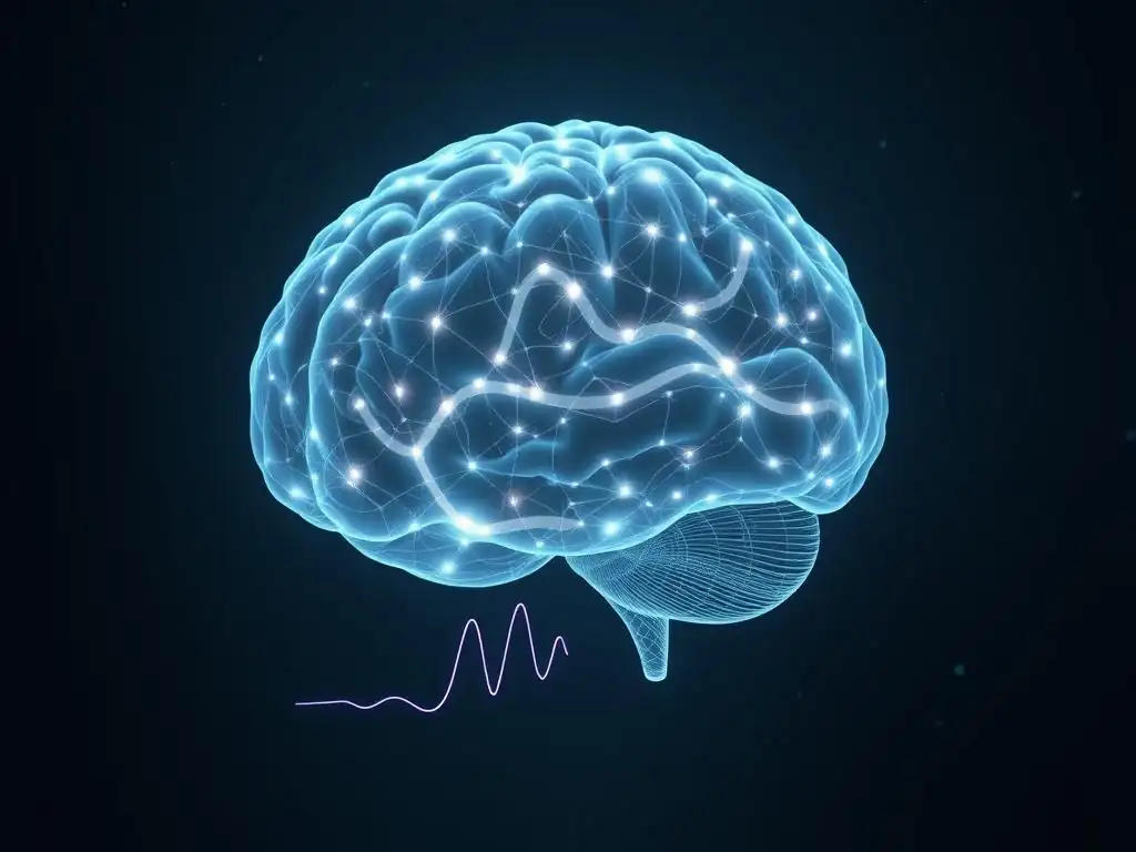
Welcome to Mind Care Guide.
Feeling overwhelmed by the complexities of mental well-being? You're not alone. We're here to explore the intricate pathways of the mind, offering insights and support on your journey to better mental health. Dive into our exploration of Restingstate fMRI mental health – a fascinating area where neuroscience is unlocking new clues about how our brains work, even when at rest. What secrets does your brain hold when it's seemingly doing nothing? Let's find out together.
Resting-State fMRI: Probing the Brain's Idle Activity for Mental Health Clues
- Introduction to Resting-State fMRI and Mental Health
- What is Resting-State fMRI (rs-fMRI)? A Detailed Explanation
- rs-fMRI Techniques: Exploring Brain Networks in Mental Health
-
Applications of Resting-State fMRI in Understanding Mental Disorders
- rs-fMRI in Depression Research: Identifying Biomarkers and Neural Correlates
- Investigating Anxiety Disorders with Resting-State fMRI: From GAD to PTSD
- rs-fMRI Studies in Schizophrenia: Exploring Disrupted Functional Connectivity
- Exploring Autism Spectrum Disorder (ASD) through Resting-State Brain Activity
- rs-fMRI and ADHD: Examining Attention Deficit and Hyperactivity
- Advantages and Limitations of Resting-State fMRI in Mental Health Studies
- Future Directions in Resting-State fMRI and Mental Health
- rs-fMRI: A Powerful Tool in the Neuroscience of Mental Health
-
Frequently Asked Questions
- What is Resting-State fMRI and how does it relate to mental health?
- How can rs-fMRI help in understanding mental illnesses?
- What are the potential benefits of using rs-fMRI in clinical practice?
- What are some limitations of using rs-fMRI for mental health assessment?
- How does rs-fMRI differ from traditional task-based fMRI when studying mental health?
- Final Reflection: Unlocking the Silent Symphony of the Brain
- Unlock Your Brain's Potential: Explore Resting-State fMRI
Introduction to Resting-State fMRI and Mental Health
Understanding Resting-State Brain Activity
Even when we are not actively engaged in a specific task – whether reading a book, solving a math problem, or talking to a friend – our brain is far from silent. Instead, it is engaged in what is known as "resting-state" activity. This activity involves spontaneous fluctuations in neuronal firing that, while seemingly random, are highly synchronized across spatially separated brain regions.
These synchronous fluctuations reflect intrinsic functional connectivity, essentially how different parts of the brain "talk" to each other when the mind is at rest. Understanding this baseline communication pattern is crucial because it represents the brain's fundamental organizational structure and how information flows through its networks.
Research into resting-state brain activity has revealed that these spontaneous fluctuations are not merely noise but form consistent patterns and networks that are present across individuals. These networks, such as the default mode network (involved in introspection and mind-wandering), the salience network (involved in detecting important stimuli), and the executive control network (involved in complex cognitive tasks), remain active and interact even when no external task is being performed.
Studying these intrinsic networks provides a unique window into the brain's fundamental operational state, offering insights that task-based studies might miss. For instance, altered connectivity within these networks has been observed in various neurological and psychiatric conditions, suggesting that disruptions in this "idle" communication can have significant implications for mental well-being.
The Relevance of Resting-State fMRI in Mental Health Research
Resting-state functional magnetic resonance imaging (rs-fMRI) has emerged as a powerful non-invasive tool to probe these intrinsic brain networks and their potential links to mental health. By measuring changes in blood oxygenation levels (BOLD signal) over time while a participant simply lies still in the scanner without performing a specific task, rs-fMRI allows researchers to map the functional connectivity between different brain regions.
This approach provides a unique perspective on how brain circuits are organized and how their communication patterns might be disrupted in various mental disorders. Unlike task-based fMRI, which focuses on activity elicited by specific stimuli or cognitive demands, rs-fMRI captures the brain's inherent functional architecture, which is believed to underpin various cognitive and emotional processes.
The application of restingstate fMRI mental health research has led to significant discoveries regarding the neural correlates of conditions such as depression, anxiety disorders, schizophrenia, and autism spectrum disorder. Studies have consistently shown that individuals with these conditions often exhibit altered patterns of functional connectivity within and between key brain networks compared to healthy individuals.
For example, disruptions in the default mode network and its interactions with other networks are frequently observed in depression. By identifying these characteristic connectivity changes, rs-fMRI holds promise not only for enhancing our understanding of the underlying neurobiology of mental illness but also potentially for developing biomarkers for diagnosis, predicting treatment response, and monitoring disease progression.
Mind Care Guide's Focus on Brain Imaging and Mental Well-being
At Mind Care Guide, we recognize the profound impact that advancements in neuroscience and brain imaging have on our understanding of mental well-being. Our platform is dedicated to providing comprehensive information and resources related to mental health, and brain imaging techniques like restingstate fMRI mental health studies are integral to this mission.
We aim to bridge the gap between complex scientific research and accessible knowledge for individuals seeking to understand the biological underpinnings of mental health challenges. By exploring the tools and techniques used in neuroscience, including cutting-edge methods like rs-fMRI, we empower our readers with insights into how the brain functions and how these functions can be affected by mental health conditions.
Our focus extends to explaining how these advanced neuroimaging techniques contribute to clinical practice and research, ultimately leading to better diagnostic tools, more targeted therapies, and improved outcomes for individuals facing mental health issues. We delve into how studies utilizing resting-state brain activity analysis are helping to redefine our understanding of psychiatric disorders, moving beyond symptom-based diagnoses to encompass the complex network dysfunctions observed in the brain.
Through our content, we strive to highlight the potential of techniques like rs-fMRI to pave the way for a future where mental health care is more personalized, effective, and rooted in a deep understanding of the brain's intricate workings.
What is Resting-State fMRI (rs-fMRI)? A Detailed Explanation
fMRI Basics: How Functional MRI Works
Functional Magnetic Resonance Imaging (fMRI) is a non-invasive neuroimaging technique that measures brain activity by detecting changes in blood flow. The basic principle relies on the fact that when a brain region becomes more active, it requires more oxygen. This increased demand is met by sending oxygenated blood to that area. Hemoglobin, the protein in red blood cells that carries oxygen, has different magnetic properties depending on whether it is carrying oxygen (oxyhemoglobin) or not (deoxyhemoglobin). Deoxyhemoglobin is paramagnetic, meaning it distorts the surrounding magnetic field, while oxyhemoglobin is diamagnetic and does not.
An fMRI scanner uses a strong magnetic field and radio waves to detect these subtle differences in magnetic properties. When a brain region is active, the influx of oxygenated blood is typically more than is immediately used, leading to a relative increase in the concentration of oxyhemoglobin and a decrease in deoxyhemoglobin in that area.
This change alters the local magnetic field, which is detected by the scanner and translated into a signal known as the BOLD (Blood-Oxygen-Level Dependent) signal. By repeatedly measuring the BOLD signal across the entire brain over time, fMRI creates maps of brain activity, showing which areas are more or less active and how their activity changes in response to stimuli or during different cognitive states.
The Concept of "Resting-State": Defining Idle Brain Activity
In the context of fMRI, the "resting-state" refers to a condition where the participant is instructed to simply lie quietly in the scanner, typically with their eyes closed or fixated on a cross, and to refrain from engaging in any specific cognitive task, such as counting or problem-solving. The goal is to capture the brain's spontaneous, intrinsic activity patterns that occur when not driven by external stimuli or goal-directed behavior. While the participant is at rest, their brain is still highly active, exhibiting complex patterns of synchronized fluctuations in the BOLD signal across different regions.
These synchronized fluctuations are thought to reflect ongoing communication and coordination between brain areas that form functional networks. These networks are often referred to as "intrinsic connectivity networks" because they appear to be fundamental organizational principles of the brain, present even during sleep or light anesthesia.
The analysis of these spontaneous fluctuations in resting-state fMRI allows researchers to study the strength and patterns of functional connectivity between brain regions, providing insights into the brain's baseline organization and how different parts of the brain interact when not specifically challenged by a task. This idle activity, far from being trivial, represents the brain's default mode of operation and is crucial for maintaining cognitive function and emotional regulation.
How rs-fMRI Differs from Task-Based fMRI
The primary difference between resting-state fMRI (rs-fMRI) and task-based fMRI lies in the experimental design and the type of brain activity being measured. In task-based fMRI, participants are given specific tasks to perform (e.g., pressing a button when seeing a certain image, memorizing a list of words, performing a mathematical calculation). The analysis then focuses on identifying brain regions whose activity levels increase or decrease in response to the task compared to a baseline or control condition. This approach is excellent for mapping the brain regions involved in specific cognitive processes or responses to stimuli.
In contrast, rs-fMRI does not involve any explicit task. Participants simply rest in the scanner, and the data collected reflects the brain's spontaneous activity. The analysis focuses on measuring the temporal correlations of the BOLD signal between different brain regions.
High correlations between the activity time courses of two areas suggest that they are functionally connected, meaning they tend to activate and deactivate together as part of the same network. This allows researchers to identify and characterize intrinsic functional networks. While task-based fMRI shows where the brain is active during a specific cognitive function, rs-fMRI reveals how different brain regions are intrinsically connected and organized, providing a complementary view of brain function that is particularly valuable for understanding the brain's underlying architecture and its disruptions in restingstate fMRI mental health studies.
rs-fMRI Techniques: Exploring Brain Networks in Mental Health
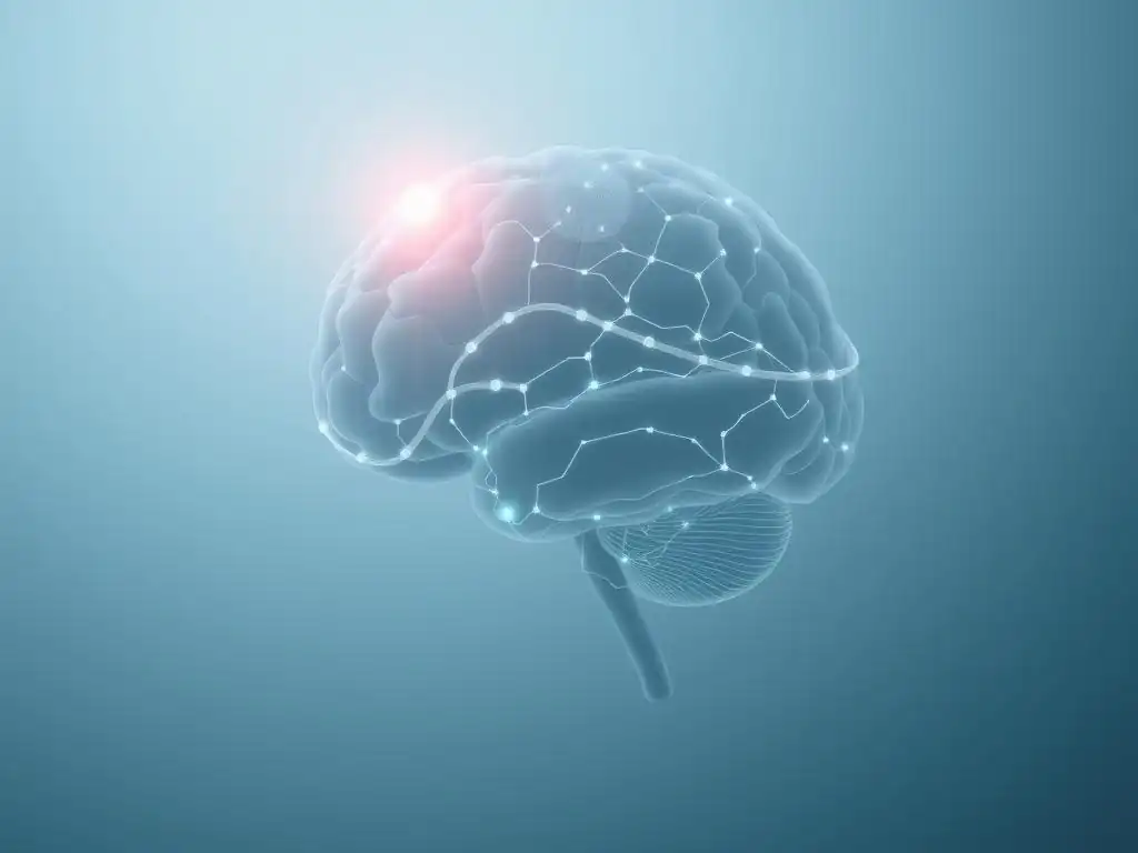
Seed-Based Correlation Analysis: Identifying Functional Connectivity
Seed-based correlation analysis is one of the most straightforward and widely used techniques in resting-state fMRI mental health research to investigate functional connectivity. This method requires the researcher to select a specific brain region of interest, known as a "seed" region. The time series of the BOLD signal is extracted from this seed region.
Then, the correlation coefficient between this seed region's time series and the time series of every other voxel (3D pixel) in the brain is calculated. A high correlation indicates that the activity in that voxel fluctuates in sync with the activity in the seed region, suggesting a strong functional connection.
The output of a seed-based analysis is a correlation map, showing the strength of functional connectivity between the seed region and all other parts of the brain. For example, if the posterior cingulate cortex (a key node in the default mode network) is chosen as a seed, the resulting map will typically show high correlations with other regions known to be part of the DMN, such as the medial prefrontal cortex and angular gyrus.
Studies applying this technique in mental health have frequently used seeds in areas like the amygdala (involved in emotion) or the anterior cingulate cortex (involved in cognitive control and emotion regulation) to examine how their connectivity to other brain regions is altered in conditions like anxiety or depression.
For instance, research published around 2016 using amygdala seeds in patients with generalized anxiety disorder often reported increased connectivity between the amygdala and prefrontal regions compared to healthy controls.
Independent Component Analysis (ICA): Discovering Intrinsic Brain Networks
Independent Component Analysis (ICA) is a data-driven technique used in rs-fMRI that does not require the researcher to pre-select specific seed regions. Instead, ICA attempts to decompose the entire rs-fMRI dataset into a set of statistically independent spatial maps, each associated with a specific time course. These spatial maps often correspond to known intrinsic connectivity networks, such as the default mode network, salience network, executive control network, visual network, and auditory network. ICA is powerful because it can identify these networks without prior assumptions about their location or connectivity patterns.
After applying ICA, researchers examine the spatial maps and their associated time courses. Spatial maps that represent plausible brain networks are identified, and their corresponding time courses are analyzed for properties like amplitude, frequency, or connectivity between different components. This technique has been instrumental in revealing disruptions in the integrity and interaction of these large-scale brain networks in various mental health conditions.
For example, a landmark study using ICA in schizophrenia research, published in the early 2000s, was among the first to demonstrate reduced functional connectivity within the default mode network in patients compared to controls. Subsequent studies have used ICA to show altered connectivity between networks, such as reduced anti-correlation between the default mode network and task-positive networks (like the executive control network) in conditions like ADHD and depression, affecting restingstate fMRI mental health findings.
Graph Theory Analysis: Mapping Brain Network Properties in Mental Disorders
Graph theory provides a mathematical framework to model the brain as a network and quantify its organizational properties using rs-fMRI data. In this approach, brain regions are treated as "nodes" or "vertices," and the functional connections between them (typically measured by the correlation of their rs-fMRI time series) are represented as "edges."
The resulting brain network can then be analyzed using various graph theory metrics to characterize its structure and efficiency. These metrics can describe properties at the level of individual nodes (e.g., how connected a specific region is, its "degree" or "centrality") or the entire network (e.g., how efficiently information can flow across the network, its "global efficiency," or the degree to which it is organized into interconnected clusters, its "modularity").
Applying graph theory to restingstate fMRI mental health data allows researchers to identify specific ways in which brain network architecture is altered in mental disorders. For instance, studies have used graph theory to show that the brains of individuals with schizophrenia often exhibit reduced "small-worldness," a property reflecting a balance between local clustering and global integration, suggesting a less efficiently organized network.
In autism spectrum disorder, graph theory studies have frequently reported increased local connectivity ("clustering") but reduced long-range connectivity, particularly in specific networks. Metrics like nodal centrality can also pinpoint specific brain regions that act as "hubs" (highly connected nodes) whose importance or connectivity patterns are disrupted in conditions like bipolar disorder or depression, offering a quantitative way to describe complex network changes observed with rs-fMRI.
Applications of Resting-State fMRI in Understanding Mental Disorders
rs-fMRI in Depression Research: Identifying Biomarkers and Neural Correlates
Resting-state fMRI has significantly advanced our understanding of Major Depressive Disorder (MDD) by revealing characteristic alterations in brain connectivity patterns. Studies using rs-fMRI in depression have consistently pointed to dysfunctions within and between key intrinsic networks. For example, numerous studies, including meta-analyses involving thousands of participants, have reported increased functional connectivity within the Default Mode Network (DMN) in depressed individuals compared to healthy controls. This hyperconnectivity within the DMN is hypothesized to relate to excessive self-referential thought and rumination, common symptoms of depression.
Furthermore, research frequently highlights altered interactions between the DMN and other networks, particularly the Salience Network (SN) and the Executive Control Network (ECN). Normally, these networks show a degree of anti-correlation, meaning that when one is active, the other is less so. However, studies in depression often show a reduced anti-correlation between the DMN and ECN, suggesting a failure to disengage from internal thoughts when cognitive control is required.
Additionally, altered connectivity of the amygdala, a region central to processing emotions, with other brain areas has been a consistent finding. For instance, a study published around 2017 examining over 1,100 individuals found altered amygdala-prefrontal connectivity patterns that correlated with symptom severity. Identifying these specific resting-state fMRI mental health markers offers potential avenues for developing objective biomarkers for diagnosis and tracking treatment response.
Investigating Anxiety Disorders with Resting-State fMRI: From GAD to PTSD
Resting-state fMRI has provided valuable insights into the neural underpinnings of various anxiety disorders, including Generalized Anxiety Disorder (GAD), Social Anxiety Disorder (SAD), Panic Disorder, and Post-Traumatic Stress Disorder (PTSD). A common theme across these conditions is dysregulation of brain circuits involved in processing fear and emotion, particularly the amygdala and its connections to prefrontal regulatory regions. rs-fMRI allows researchers to examine the baseline connectivity patterns in these circuits when individuals are not actively processing a threat, revealing enduring alterations.
In GAD, studies often report increased functional connectivity within the Salience Network, particularly involving the anterior insula and amygdala, suggesting a heightened state of vigilance even at rest. Research, such as a study from 2015 involving dozens of GAD patients, found increased resting-state connectivity between the amygdala and dorsal medial prefrontal cortex, which correlated with worry severity. In PTSD, rs-fMRI studies have consistently shown altered connectivity within the fear circuit, including heightened amygdala connectivity to the hippocampus and disruptions in prefrontal control over the amygdala.
For instance, a large-scale study published around 2019 involving hundreds of veterans with PTSD revealed widespread alterations in connectivity, including reduced connectivity within the DMN and altered connections involving the ventral medial prefrontal cortex, a region implicated in fear extinction. These restingstate fMRI mental health findings help pinpoint the specific network dysfunctions characteristic of different anxiety subtypes.
rs-fMRI Studies in Schizophrenia: Exploring Disrupted Functional Connectivity
Schizophrenia is characterized by severe disruptions in thought processes, perception, and emotion, and rs-fMRI has been a crucial tool in uncovering the widespread brain network dysconnectivity associated with the disorder. Studies in schizophrenia consistently report abnormalities in functional connectivity across multiple intrinsic networks. One of the most robust findings is reduced connectivity within the Default Mode Network (DMN), often interpreted as a failure to maintain coherent internal mentation. Furthermore, altered connectivity between the DMN and task-positive networks is frequently observed, potentially contributing to difficulties distinguishing internal thoughts from external reality (hallucinations and delusions).
Beyond the DMN, rs-fMRI research in schizophrenia has revealed dysconnectivity in other key networks, including the Salience Network (SN), Executive Control Network (ECN), and sensory networks. For example, studies using Independent Component Analysis (ICA) have shown disruptions in the integrity of the SN, which plays a role in detecting and responding to salient stimuli, potentially contributing to symptoms like paranoia and disorganized thinking.
Graph theory analyses have often demonstrated a less integrated and efficient global brain network organization in patients with schizophrenia compared to controls. A comprehensive meta-analysis published around 2017 synthesizing data from over 2,000 patients confirmed widespread reductions in functional connectivity, particularly affecting frontal and temporal regions, highlighting the complex network-level changes captured by restingstate fMRI mental health investigations in this severe mental illness.
Exploring Autism Spectrum Disorder (ASD) through Resting-State Brain Activity
Resting-state fMRI has provided significant insights into the atypical brain connectivity patterns observed in individuals with Autism Spectrum Disorder (ASD). While findings can be heterogeneous, a common theme in rs-fMRI studies of ASD is altered long-range and short-range functional connectivity. Many studies, particularly in younger individuals with ASD, have reported reduced long-range connectivity between distant brain regions, especially those involved in social cognition and language processing, such as connections between frontal and temporal areas. This is sometimes accompanied by increased local connectivity within certain brain regions.
Conversely, some studies in adults or those with less severe symptoms have reported increased long-range connectivity in certain networks. The heterogeneity in findings likely reflects the wide spectrum of ASD presentations, as well as differences in age, cognitive ability, and data analysis methods. Despite this variability, rs-fMRI has consistently shown altered connectivity within core networks implicated in social processing, communication, and repetitive behaviors, such as the DMN, salience network, and reward network.
For example, a large multi-site study published around 2014 involving hundreds of participants found reduced functional connectivity between the medial prefrontal cortex and the posterior cingulate cortex within the DMN in individuals with ASD, a pattern that correlated with social impairment severity. These restingstate fMRI mental health studies are crucial for understanding the neural basis of the diverse symptoms in ASD.
rs-fMRI and ADHD: Examining Attention Deficit and Hyperactivity
Resting-state fMRI has been highly effective in characterizing the brain network dysfunctions associated with Attention-Deficit/Hyperactivity Disorder (ADHD). Key findings from rs-fMRI studies in ADHD often point to altered connectivity within and between networks involved in attention, cognitive control, and reward processing. A prominent finding is reduced functional connectivity within the Executive Control Network (ECN), which is crucial for planning, decision-making, and inhibiting impulsive behaviors. Additionally, altered connectivity within the Default Mode Network (DMN) and its relationship with task-positive networks is frequently observed.
Specifically, many studies report weaker anti-correlation between the DMN and the ECN in individuals with ADHD compared to typically developing controls. This reduced anti-correlation is thought to reflect a difficulty in suppressing internal thoughts (DMN activity) when attention is needed for external tasks (ECN activity), contributing to distractibility and inattention. Altered connectivity within the reward circuit, involving regions like the striatum and prefrontal cortex, is also commonly reported, potentially underlying issues with motivation and impulsivity.
A meta-analysis published around 2016 summarizing data from numerous studies consistently showed reduced connectivity within fronto-parietal networks (part of the ECN) and altered connectivity between the DMN and other networks in individuals with ADHD. These restingstate fMRI mental health findings help explain the core symptoms of inattention and impulsivity in ADHD at the level of brain network function.
Advantages and Limitations of Resting-State fMRI in Mental Health Studies
Strengths of rs-fMRI: Non-Invasive, Task-Free, and Clinically Relevant
One of the significant advantages of resting-state fMRI is its non-invasive nature. It does not involve radiation, making it safe for repeated use in the same individuals, which is crucial for longitudinal studies tracking disease progression or treatment effects.
The procedure simply requires the participant to lie still in the scanner, making it relatively easy to implement, even with clinical populations who might have difficulty performing complex tasks or following instructions consistently. This task-free nature is particularly beneficial when studying disorders characterized by cognitive impairments, motivational deficits, or difficulty with task compliance, such as severe depression, schizophrenia, or young children with developmental disorders.
Furthermore, rs-fMRI directly probes the brain's intrinsic functional architecture, which is thought to represent a fundamental organizational property that underlies various cognitive functions and behaviors. By examining the brain's baseline connectivity patterns, rs-fMRI can reveal enduring network abnormalities that might not be fully captured during specific task performance.
The findings from restingstate fMRI mental health studies have shown clinical relevance, identifying connectivity patterns that correlate with symptom severity, predict treatment response, or differentiate patient groups from healthy controls. This potential for providing clinically meaningful information makes rs-fMRI a valuable tool for both research and potential future clinical applications in mental health.
Limitations of rs-fMRI: Data Interpretation, Motion Artifacts, and Individual Variability
Despite its strengths, resting-state fMRI also presents several limitations. Interpreting the biological meaning of functional connectivity derived from rs-fMRI can be challenging. While high correlations in BOLD signal time series suggest that brain regions are functionally connected, this does not necessarily imply direct anatomical connections or causal relationships. The BOLD signal itself is an indirect measure of neural activity, reflecting hemodynamic responses that can be influenced by physiological factors unrelated to neuronal firing.
Another significant challenge in rs-fMRI is susceptibility to motion artifacts. Even small head movements during the scan can introduce spurious correlations or reduce true correlations, significantly distorting connectivity estimates. This is particularly problematic when studying clinical populations, such as individuals with ADHD or certain forms of anxiety, who may find it difficult to remain perfectly still.
While sophisticated data processing techniques exist to mitigate motion effects, completely eliminating them remains an active area of research. Furthermore, there is considerable individual variability in resting-state connectivity patterns, even among healthy individuals, which can make it difficult to identify consistent biomarkers for specific disorders and requires large sample sizes for robust findings in restingstate fMRI mental health studies.
Addressing Challenges in rs-fMRI Data Acquisition and Analysis
Researchers are actively working to address the limitations of rs-fMRI to improve its reliability and clinical utility. Significant efforts are being made to develop more robust data acquisition protocols that minimize motion artifacts, such as using prospective motion correction techniques or providing explicit feedback to participants about their head movement. Post-processing techniques have also become increasingly sophisticated, including methods for identifying and removing motion-related noise from the data, such as ICA-based denoising or regression of motion parameters.
Standardization of data analysis pipelines is another crucial area of development. Different processing steps and analytical choices (e.g., filtering parameters, spatial smoothing, network definition) can influence the results, making it challenging to compare findings across studies. Initiatives promoting data sharing and the use of standardized analysis platforms are helping to improve reproducibility.
Furthermore, researchers are exploring ways to account for individual variability by using more personalized approaches to network definition or by focusing on deviations from typical connectivity patterns. Advances in machine learning are also being applied to rs-fMRI data to improve the classification of individuals based on their connectivity profiles, moving towards potentially using restingstate fMRI mental health data for predictive or diagnostic purposes.
Future Directions in Resting-State fMRI and Mental Health
Combining rs-fMRI with Other Neuroimaging Techniques (e.g., EEG, PET)
The future of using resting-state fMRI in mental health research lies partly in integrating it with other neuroimaging and physiological measurement techniques. Each modality offers a unique perspective on brain function. For example, electroencephalography (EEG) and magnetoencephalography (MEG) provide excellent temporal resolution, capturing brain activity on the millisecond scale, which is much faster than the seconds-long resolution of fMRI's BOLD signal. Combining rs-fMRI with simultaneous EEG allows researchers to correlate slow BOLD fluctuations with faster electrical brain rhythms (like alpha, theta, or gamma waves) in the resting state, potentially linking specific network activity to underlying neural oscillations.
Positron Emission Tomography (PET), on the other hand, allows for the measurement of specific neurochemical processes, such as neurotransmitter activity (e.g., dopamine, serotonin) or metabolic rates (glucose uptake). Combining rs-fMRI with PET can link functional connectivity patterns to underlying neurochemical profiles, providing a more comprehensive understanding of how brain network dysfunction relates to specific molecular abnormalities implicated in mental disorders.
For instance, a study might use PET to measure dopamine receptor availability in the striatum and rs-fMRI to assess striatal connectivity, exploring how neurochemistry influences network function in conditions like addiction or schizophrenia. This multimodal approach promises a richer, more detailed picture of the complex biological factors contributing to mental health conditions.
Using rs-fMRI for Personalized Mental Health Treatment: Predicting Treatment Response
A major goal in mental health care is to move towards more personalized treatments, tailoring interventions to individual patient needs rather than relying on trial-and-error. Resting-state fMRI holds significant promise in contributing to this goal by potentially identifying biomarkers that can predict how an individual patient will respond to a specific therapy, such as medication, psychotherapy, or neuromodulation techniques like transcranial magnetic stimulation (TMS). Different connectivity patterns observed at rest might reflect underlying biological differences that influence treatment efficacy.
For example, research is exploring whether specific rs-fMRI connectivity profiles in patients with depression can predict whether they are more likely to respond to a selective serotonin reuptake inhibitor (SSRI) versus cognitive behavioral therapy (CBT). A study published around 2017 identified resting-state connectivity patterns within the subgenual anterior cingulate cortex that predicted response to antidepressant medication.
Similarly, studies are investigating whether baseline resting-state connectivity can predict who will benefit most from TMS for depression, with some preliminary findings suggesting that connectivity between the stimulation target and specific brain networks is predictive. By identifying these predictive restingstate fMRI mental health markers, clinicians could potentially make more informed decisions about treatment selection, saving patients time and reducing exposure to ineffective therapies.
Exploring the Potential of rs-fMRI in Early Detection and Prevention of Mental Illness
Beyond diagnosis and treatment selection, rs-fMRI is being explored for its potential role in the early detection and even prevention of mental illness. Many mental disorders have prodromal phases or risk factors that can be identified before the full onset of symptoms. Changes in resting-state brain connectivity might serve as early indicators of increased vulnerability. Longitudinal studies are using rs-fMRI to track brain development and connectivity patterns in individuals at high genetic or clinical risk for developing conditions like schizophrenia, bipolar disorder, or depression.
For instance, studies following adolescents with a family history of schizophrenia are using rs-fMRI to look for subtle deviations in network connectivity that might precede the development of psychotic symptoms. Similarly, research on individuals experiencing subthreshold symptoms of depression or anxiety is investigating whether resting-state connectivity patterns can predict which individuals will later develop a full-blown disorder.
Identifying such early restingstate fMRI mental health markers could open doors for targeted early interventions aimed at preventing or mitigating the severity of illness onset. While still largely in the research phase, this application highlights the exciting potential of rs-fMRI to shift the paradigm towards proactive, rather than reactive, mental health care.
rs-fMRI: A Powerful Tool in the Neuroscience of Mental Health
Summarizing the Key Findings and Implications of rs-fMRI Research
Resting-state fMRI has revolutionized the study of the human brain's functional organization and its relevance to mental health. By allowing researchers to map intrinsic functional connectivity, rs-fMRI has consistently revealed that mental disorders are associated with characteristic disruptions in large-scale brain networks. Key findings across various conditions, including depression, anxiety disorders, schizophrenia, ASD, and ADHD, point to altered connectivity within and between networks such as the Default Mode Network, Salience Network, and Executive Control Network. These network abnormalities appear to underlie the diverse cognitive, emotional, and behavioral symptoms observed in these disorders.
The implications of these findings are profound. They shift the focus from localized brain abnormalities to understanding mental illness as a disorder of brain circuits and network communication. This network perspective offers a more holistic view of psychopathology and provides a framework for developing new theoretical models of mental disorders.
Furthermore, the identification of specific resting-state connectivity patterns associated with different conditions or symptom dimensions suggests the potential for rs-fMRI to contribute to objective diagnostics and personalized treatment approaches in the future. While challenges remain, the wealth of data generated by restingstate fMRI mental health studies is rapidly expanding our knowledge base.
The Role of Resting-State fMRI in Advancing Mental Health Care
Resting-state fMRI plays a crucial role in advancing mental health care by providing a non-invasive window into the living brain's functional architecture. Its ability to characterize intrinsic network connectivity, even in individuals who cannot perform complex tasks, makes it uniquely valuable for studying diverse clinical populations. By identifying the specific network dysfunctions associated with different mental disorders, rs-fMRI research contributes directly to understanding the biological basis of these conditions. This enhanced understanding is essential for developing more effective interventions.
Looking ahead, the integration of rs-fMRI findings into clinical practice could potentially lead to significant improvements in diagnosis, prognosis, and treatment selection. While still primarily a research tool, ongoing advancements in data analysis, standardization, and the combination with other modalities are bringing rs-fMRI closer to clinical translation.
The promise of using resting-state fMRI mental health data to predict treatment response or identify individuals at risk for developing illness represents a paradigm shift towards more precise and preventative mental health care. As research continues, rs-fMRI is poised to remain a cornerstone technique in the neuroscience of mental health, driving forward our ability to understand, diagnose, and treat psychiatric conditions.
Further Resources on Brain Imaging and Mental Health from Mind Care Guide
Understanding the complex relationship between brain function and mental well-being is a core focus at Mind Care Guide. We provide a wealth of information on various neuroimaging techniques, including fMRI, PET, EEG, and MEG, and how they are applied in mental health research and clinical settings. Our resources delve into the specific brain regions and networks implicated in different mental disorders, explaining how tools like restingstate fMRI mental health studies help us map these intricate connections.
Explore our articles and guides to learn more about:
- How different brain imaging techniques work.
- The specific brain areas involved in conditions like depression, anxiety, and schizophrenia.
- The concept of brain networks and their importance in mental health.
- The latest research findings from neuroimaging studies.
- The potential future applications of brain imaging in diagnosis and treatment.
Visit our dedicated sections on Brain Imaging in Mental Health Research and The Neuroscience of Mental Health for in-depth information designed to inform and empower your understanding of the brain's role in mental well-being.
Frequently Asked Questions
What is Resting-State fMRI and how does it relate to mental health?
Resting-state fMRI (rs-fMRI) is a neuroimaging technique that measures brain activity while a person is at rest, not performing any specific task. This can reveal patterns of neural connectivity that are linked to various mental health conditions.
How can rs-fMRI help in understanding mental illnesses?
Rs-fMRI can identify abnormal patterns of brain connectivity in individuals with mental illnesses. These patterns can serve as biomarkers for diagnosis, treatment monitoring, and predicting treatment response.
What are the potential benefits of using rs-fMRI in clinical practice?
Rs-fMRI offers a non-invasive way to assess brain function and potentially personalize treatment plans. It could lead to earlier and more accurate diagnoses, as well as more effective interventions for mental health disorders.
What are some limitations of using rs-fMRI for mental health assessment?
Rs-fMRI data interpretation can be complex and requires specialized expertise. The technique is also sensitive to motion artifacts and variations in individual brain anatomy, requiring careful data processing.
How does rs-fMRI differ from traditional task-based fMRI when studying mental health?
Unlike task-based fMRI, which measures brain activity during specific tasks, rs-fMRI captures the brain's baseline activity, revealing intrinsic functional connectivity that may be disrupted in mental health disorders, even in the absence of external stimuli.
Final Reflection: Unlocking the Silent Symphony of the Brain
The exploration of resting-state fMRI (rs-fMRI) is no longer a niche scientific pursuit; it's a rapidly evolving frontier with the potential to revolutionize mental health diagnosis and treatment. Understanding the brain's "idle" activity offers unprecedented insights into the complex neural networks underlying conditions like depression, anxiety, and schizophrenia, paving the way for more precise and personalized interventions. This is not simply about technological advancement; it's about unlocking the silent symphony of the brain to improve countless lives.
The impact of rs-fMRI research extends far beyond the laboratory, subtly reshaping our understanding of mental illness itself. By revealing the intricate patterns of brain connectivity, this technology challenges traditional diagnostic methods and encourages a more holistic view of mental health. It moves us away from simplistic categorizations and towards a more nuanced appreciation of the individual neural landscape. The brain is a complex system, and understanding its dynamics requires looking beyond simple averages and focusing on the intricate interplay of its various parts.
This quote encapsulates the transformative power of rs-fMRI in shifting our perspective from a reductionist approach to a more systems-based understanding.
Consider the implications of this research for your own life. Perhaps you know someone struggling with a mental health condition. Or perhaps you've grappled with your own mental well-being. This technology offers hope for earlier and more accurate diagnoses, leading to more effective treatments and ultimately, improved quality of life. Take a moment to reflect on the potential of rs-fMRI – not just as a scientific tool, but as a beacon of hope in the ongoing quest for better mental health care. The future of mental health lies, in part, in understanding the silent whispers of the resting brain.
Unlock Your Brain's Potential: Explore Resting-State fMRI
Thanks for taking the time to delve into the fascinating world of resting-state fMRI and its implications for mental health! You've gained valuable insights into how brain activity during rest can reveal crucial information about our well-being. Now, let's put that knowledge to work.
- Schedule a consultation with a neurologist or psychiatrist: This is your primary step towards potentially utilizing the latest advancements in brain imaging for a personalized mental health assessment. Discuss your interest in resting-state fMRI and explore if it's a suitable option for you.
- Share this article with someone who might benefit: Spread the word about this groundbreaking technology and its potential to revolutionize mental health care.
- Research local facilities offering resting-state fMRI: Explore resources available in your area and find out more about this exciting field.
Ready to take the next step toward understanding your brain’s incredible potential? What questions do you have about accessing or using resting-state fMRI for mental health insights?