
Predicting Recovery: How Brain Scans Guide Mental Health Treatment Approaches

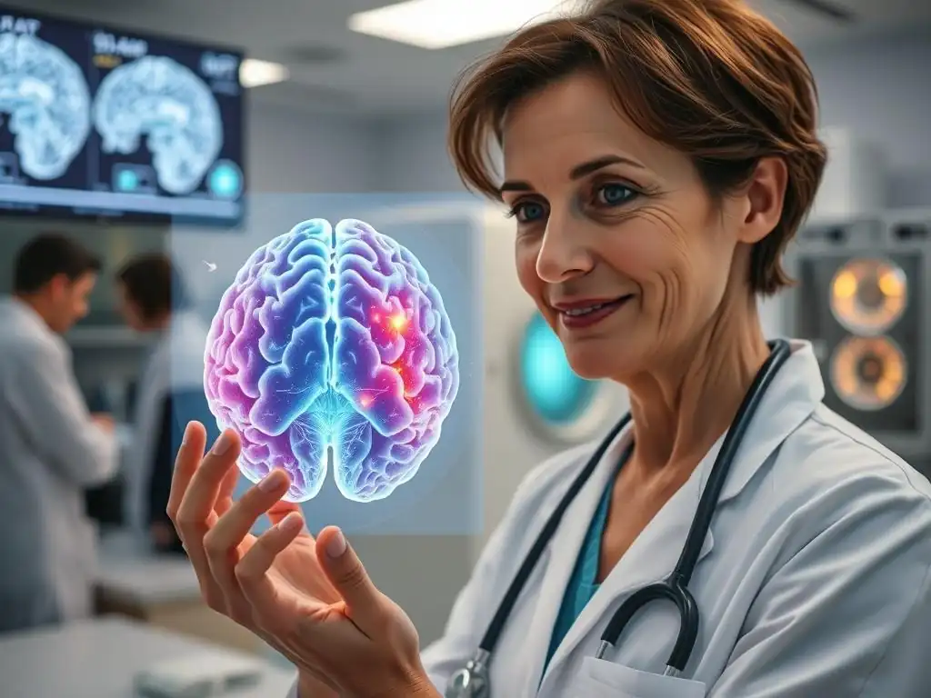
Welcome to Mind Care Guide, a sanctuary dedicated to understanding and nurturing your mental well-being. Feeling overwhelmed by the complexities of mental health treatment? You're not alone. Delve into our latest article, where we explore the fascinating world of brain scan recovery prediction and how cutting-edge neuroscience is revolutionizing personalized mental health approaches. Curious about how your brain's unique activity can inform your path to recovery? Let's explore the possibilities together...
- Predicting Recovery: How Brain Scans Guide Mental Health Treatment Approaches
- Brain Scan Technology for Predicting Mental Health Outcomes
- How Brain Scan Recovery Prediction Works: A Detailed Explanation
- Specific Mental Health Conditions and Brain Scan Applications
- Benefits of Brain Scan-Guided Mental Health Treatment
- Limitations and Challenges of Brain Scan Recovery Prediction
- The Future of Brain Scans in Mental Health: Advancements and Possibilities
- Conclusion: Brain Scans as a Powerful Tool for Guiding Mental Health Recovery
-
Frequently Asked Questions
- How can brain scans help predict recovery in mental health treatment?
- What types of brain scans are used to predict recovery?
- What are the benefits of using brain scan recovery prediction in mental health treatment?
- Is brain scan recovery prediction a foolproof method?
- How accessible is brain scan recovery prediction for mental health patients?
- Final Reflection: The Dawn of Personalized Mental Healthcare
- Unlock Your Brain's Potential: Take Charge of Your Mental Wellbeing
Predicting Recovery: How Brain Scans Guide Mental Health Treatment Approaches
Understanding the Role of Brain Imaging in Mental Healthcare
Brain imaging has emerged as a powerful tool in the field of mental healthcare, moving beyond simple anatomical visualization to explore the complex functional and structural underpinnings of psychiatric conditions. Techniques like MRI, fMRI, PET, and EEG allow researchers and clinicians to gain insights into brain activity patterns, connectivity, and volume changes associated with various disorders, from depression and anxiety to schizophrenia and bipolar disorder.
This deeper understanding is crucial because mental health conditions are inherently disorders of the brain, involving intricate neural circuits and biochemical processes. By visualizing these processes, brain scans offer objective data that can complement subjective clinical assessments, potentially leading to more accurate diagnoses and a better understanding of individual patient profiles.
Furthermore, brain imaging contributes significantly to the development of new therapeutic strategies. By identifying specific neural targets or pathways involved in a disorder, researchers can design interventions, whether pharmacological or psychotherapeutic, that are more precisely aimed at correcting the underlying brain dysfunction.
This shift towards a neurobiological perspective in mental health treatment is paving the way for more personalized and effective care. The ability to see how a treatment affects the brain, or why a patient might not be responding, opens up possibilities for adjusting approaches in real-time, moving away from traditional trial-and-error methods that can be time-consuming and discouraging for patients.
The Growing Need for Personalized Mental Health Treatment
Mental health conditions are highly heterogeneous, meaning that even within the same diagnostic category, patients can experience vastly different symptoms, severity levels, and responses to treatment. A therapy or medication that works well for one individual with depression might be completely ineffective for another.
This variability highlights the urgent need for more personalized treatment approaches that consider the unique biological and psychological profile of each patient. The current standard of care often involves a process of trying different treatments sequentially until one is found to be effective, a process that can take months or even years, during which time a patient continues to suffer.
Personalized mental health treatment aims to predict which intervention is most likely to be effective for a specific individual before treatment begins. This is where the potential of brain scan recovery prediction becomes particularly exciting. If brain imaging can reveal biomarkers or patterns that correlate with a higher likelihood of responding to cognitive behavioral therapy versus a specific antidepressant, clinicians could make more informed decisions from the outset. This not only accelerates the path to recovery but also minimizes exposure to ineffective treatments and their potential side effects, ultimately improving patient outcomes and optimizing healthcare resources.
Brain Scan Technology for Predicting Mental Health Outcomes
Overview of Different Brain Scan Techniques Used in Psychiatry
Various brain imaging techniques offer unique perspectives on brain structure and function, each contributing valuable data to the study of mental health and the prediction of treatment outcomes. These technologies differ in their spatial and temporal resolution, what aspects of brain activity or structure they measure, and their accessibility and cost.
Researchers often combine data from multiple modalities to build a more comprehensive picture of the brain's state. The choice of technology depends on the specific research question or clinical goal, whether it's identifying areas of reduced brain volume, measuring metabolic activity, tracking electrical signals, or observing blood flow as a proxy for neural activity.
Understanding the strengths and limitations of each technique is essential for interpreting findings related to brain scan recovery prediction. While some methods provide detailed anatomical images, others excel at capturing the dynamic nature of brain activity. Integrating these different types of data is a significant challenge but also holds immense promise for developing robust predictive models. The ongoing refinement of these technologies, coupled with advancements in data analysis, continues to expand their potential applications in psychiatric research and clinical practice.
fMRI (Functional Magnetic Resonance Imaging) and Recovery Prediction
fMRI measures brain activity by detecting changes in blood flow. When a brain region is more active, it requires more oxygenated blood, and fMRI can pick up on the altered magnetic properties of oxygenated versus deoxygenated hemoglobin. This allows researchers to see which parts of the brain are engaged during specific tasks or in a resting state.
In the context of brain scan recovery prediction, fMRI is used to identify patterns of connectivity or activity in specific neural circuits that might predict a patient's response to therapy or medication. For instance, studies have shown that connectivity differences in the default mode network or the salience network can predict whether a patient with depression is likely to respond to psychotherapy.
Research using fMRI has provided compelling evidence linking specific brain activity patterns to treatment outcomes. For example, some studies suggest that individuals with higher connectivity between the subgenual anterior cingulate cortex and other brain regions before treatment are less likely to respond to certain antidepressant medications. Conversely, different patterns of connectivity might predict a better response to cognitive behavioral therapy. This type of data helps build models that can potentially guide treatment selection, offering a non-invasive way to peer into the neural landscape influencing recovery potential.
EEG (Electroencephalography) and its Applications in Mental Health Prognosis
EEG measures electrical activity in the brain using electrodes placed on the scalp. It has excellent temporal resolution, meaning it can capture rapid changes in brain activity, though its spatial resolution is lower than fMRI or PET. EEG patterns, such as brainwave frequencies (alpha, beta, theta, delta), evoked potentials (brain responses to specific stimuli), and connectivity measures, can reflect different states of brain function and dysfunction. Its relatively lower cost and portability compared to MRI make it a more accessible tool in some settings.
In mental health prognosis, EEG is being explored for its ability to predict treatment response, particularly in conditions like depression and schizophrenia. Certain EEG markers, such as frontal alpha asymmetry or specific patterns of evoked potentials elicited by emotional stimuli, have been associated with the likelihood of responding to antidepressant treatment or transcranial magnetic stimulation (TMS). While perhaps not providing the detailed anatomical insights of MRI, EEG offers a window into the brain's dynamic electrical communication, which is fundamentally altered in many psychiatric disorders, making it a valuable component in the suite of tools for brain scan recovery prediction.
PET (Positron Emission Tomography) and Insights into Brain Function
PET imaging uses radioactive tracers injected into the bloodstream to measure metabolic activity, blood flow, or the distribution of specific molecules like neurotransmitter receptors in the brain. It provides unique insights into the biochemical processes underlying mental health conditions. For example, PET can measure glucose metabolism, indicating how active different brain regions are, or quantify the density of dopamine or serotonin receptors, which are targets for many psychiatric medications.
PET scanning has been instrumental in understanding the neurochemical basis of disorders and predicting response to treatments targeting specific neurotransmitter systems. Studies using PET have shown altered glucose metabolism in certain brain regions (like the prefrontal cortex or amygdala) in conditions like depression and anxiety, and these metabolic patterns can sometimes correlate with treatment outcomes. Furthermore, measuring the occupancy of neurotransmitter receptors by a medication using PET can help determine if a drug is hitting its target effectively in a patient, providing insights into why someone might not be responding to treatment and potentially guiding medication adjustments.
How Brain Scan Recovery Prediction Works: A Detailed Explanation
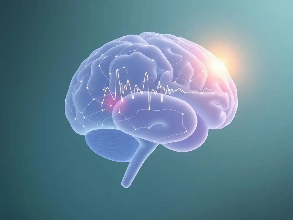
Identifying Biomarkers Through Brain Scans: Altered Amygdala Activity, Reduced Hippocampal Volume
Brain scan recovery prediction often begins with identifying neural biomarkers – specific features of brain structure, function, or chemistry observable through imaging that are associated with a particular condition or predict a future outcome, such as response to treatment. For example, research consistently shows that individuals with depression often exhibit altered activity in the amygdala, a brain region involved in processing emotions. Studies have found that the degree of this altered activity, or its connectivity with other brain regions, prior to treatment can predict the likelihood of remission with certain therapies.
Similarly, reduced volume of the hippocampus, a brain structure important for memory and emotional regulation, is frequently observed in chronic depression and PTSD. This reduced hippocampal volume has been linked to poorer treatment outcomes and a higher risk of relapse, serving as a potential biomarker for prognosis.
Beyond structural and activity markers, brain scans can identify biomarkers related to brain chemistry. For instance, PET scans can measure dopamine receptor availability in specific brain regions, which has been explored as a predictor of response to antipsychotic medications in schizophrenia. Identifying these specific neural signatures provides objective data points that can be integrated with clinical information to build predictive models. It's not usually a single biomarker, but rather a combination of several markers across different brain regions and imaging modalities, that offers the most robust predictive power for brain scan recovery prediction.
Mapping Brain Activity Patterns to Predict Treatment Response
Beyond isolated biomarkers, predictive models increasingly focus on patterns of activity and connectivity across entire brain networks. The brain operates as a complex system of interconnected regions, and disruptions in these networks are thought to underlie many mental health conditions. Functional MRI, in particular, is well-suited to study these networks by examining how activity in different regions is correlated (functional connectivity), both during tasks and in a resting state. Researchers analyze these functional connectivity patterns before treatment to see if they can predict whether a patient will respond to a specific intervention.
For example, studies on predicting antidepressant response using fMRI have explored connectivity within the default mode network, which is often hyperactive in depression, or connectivity between emotional processing regions like the amygdala and regulatory regions like the prefrontal cortex. Different patterns within these networks have been shown to differentiate between patients who achieve remission and those who do not after a course of medication or psychotherapy. This approach moves beyond looking at single brain areas to understanding the broader neural circuitry involved in mental health and its response to treatment, enhancing the sophistication of brain scan recovery prediction.
Using Machine Learning to Enhance Brain Scan Analysis for Recovery Prediction
The sheer volume and complexity of data generated by modern brain scans make traditional statistical analysis challenging for building highly accurate predictive models. This is where machine learning algorithms come into play. Machine learning can analyze intricate patterns across thousands or millions of data points from brain scans (e.g., voxel-wise activity, connectivity matrices) that might be invisible to the human eye or simpler statistical methods. These algorithms can learn from large datasets of brain scans and corresponding treatment outcomes to identify complex combinations of features that best predict recovery for individual patients.
Machine learning models can integrate data from different imaging modalities (fMRI, EEG, PET) and even combine imaging data with clinical information (symptom severity, history, genetics) to improve predictive accuracy. For instance, an algorithm might identify that a specific pattern of functional connectivity in the prefrontal cortex combined with a certain level of metabolic activity in the amygdala and a particular demographic factor is highly predictive of a good response to a specific therapy for PTSD.
This sophisticated pattern recognition is key to unlocking the full potential of brain scan data for personalized medicine and advancing the field of brain scan recovery prediction towards clinical applicability.
Specific Mental Health Conditions and Brain Scan Applications
Predicting Recovery from Depression with Brain Imaging
Major depressive disorder is a prime area where brain imaging is being extensively researched for predicting treatment response. The variability in how patients respond to antidepressants and psychotherapies is a major clinical challenge. Brain scans are helping to identify neural signatures associated with different subtypes of depression and different likelihoods of recovery with specific interventions.
For instance, studies have used fMRI to examine resting-state functional connectivity patterns, finding that connectivity within the subgenual anterior cingulate cortex and its connections to other limbic and prefrontal areas can predict remission rates. One study indicated that a particular pattern of connectivity in this region predicted non-response to an SSRI antidepressant with roughly 70% accuracy.
PET scans have also contributed valuable insights, particularly regarding metabolic activity and neurotransmitter systems. Reduced glucose metabolism in certain prefrontal regions has been linked to a lower likelihood of response to psychotherapy. Furthermore, studies examining serotonin transporter availability using PET have explored its role in predicting response to SSRIs. The goal is to move towards a scenario where, after a diagnosis of depression, a brain scan could help guide the clinician in selecting the most appropriate initial treatment, saving patients from potentially ineffective trials.
Identifying Neural Correlates of Treatment Response in Depression
Identifying the specific neural correlates that predict treatment response in depression is an active area of research. Beyond the subgenual cingulate and prefrontal cortex, studies have implicated regions like the insula, amygdala, and hippocampus, as well as the connectivity between these areas, as potential predictors. For instance, altered connectivity between the amygdala and the medial prefrontal cortex, regions involved in emotion processing and regulation, has been repeatedly linked to treatment outcomes. Some research suggests that stronger baseline connectivity in certain circuits is associated with better response to psychotherapy, while different connectivity patterns might favor medication.
Moreover, changes in brain activity or structure during treatment are also being investigated as early indicators of eventual recovery. For example, some studies using fMRI have shown that early changes in connectivity patterns after just a few sessions of therapy or a week or two on medication can predict whether a patient will eventually achieve remission. These findings underscore the dynamic nature of the brain's response to treatment and highlight the potential for brain scans to not only predict initial response but also to monitor progress and inform treatment adjustments.
Brain Scans in Predicting Outcomes for Anxiety Disorders
Anxiety disorders, including generalized anxiety disorder, panic disorder, social anxiety disorder, and phobias, also show promise for brain scan recovery prediction. These conditions are characterized by dysregulation in brain circuits involved in fear processing and emotional regulation, particularly involving the amygdala, prefrontal cortex, and hippocampus.
Brain imaging can help identify the specific neural patterns associated with different anxiety subtypes and predict response to treatments like cognitive behavioral therapy (CBT) or anxiolytic medications. For example, individuals with panic disorder often show heightened activity in the amygdala when anticipating a panic attack, and the degree of this activity or connectivity patterns might predict response to CBT targeting fear extinction.
Research has explored how baseline brain activity and connectivity relate to treatment outcomes in anxiety. For instance, studies in social anxiety disorder have found that activity levels in the amygdala and insula during social situations, or connectivity between these regions and the prefrontal cortex, can predict the degree of symptom reduction with CBT. While the research is still evolving compared to depression, these findings suggest that brain scans can provide valuable prognostic information for tailoring interventions to the specific neural profile of an individual with an anxiety disorder, improving the likelihood of successful recovery.
Using Brain Scans to Tailor Treatment for PTSD
Post-traumatic stress disorder (PTSD), a severe anxiety-related condition, is another area benefiting from brain imaging research focused on predicting treatment response. PTSD is characterized by persistent alterations in brain regions involved in processing trauma, fear, and memory, including the amygdala, hippocampus, and medial prefrontal cortex.
Individuals with PTSD often exhibit hyper-responsivity of the amygdala to trauma-related stimuli and reduced volume and activity in the medial prefrontal cortex and hippocampus. These neural differences are being investigated as potential predictors of how well a patient will respond to trauma-focused therapies like Prolonged Exposure or EMDR, or to antidepressant medications often used to treat PTSD symptoms.
Studies using fMRI in PTSD patients have shown that patterns of connectivity between the amygdala and the ventromedial prefrontal cortex, a region crucial for regulating fear responses, can predict symptom improvement following trauma-focused therapy. For example, one study found that stronger baseline connectivity in a specific circuit involving these regions was associated with greater symptom reduction after therapy.
Furthermore, hippocampal volume has been explored as a predictor, with some findings suggesting that greater volume loss might be associated with poorer treatment outcomes. By identifying these neural markers, brain scans offer the potential to match patients with the therapy most likely to help them process trauma effectively and achieve recovery.
Schizophrenia and Brain Scan Recovery Prediction: Reduced Gray Matter Volume in the Prefrontal Cortex Predicts Poorer Outcomes
Schizophrenia is a complex psychiatric disorder characterized by significant alterations in brain structure and function, including reductions in gray matter volume, particularly in the prefrontal cortex and temporal lobes, and changes in white matter connectivity. These structural and functional abnormalities are central to the symptoms of schizophrenia, such as hallucinations, delusions, disorganized thinking, and cognitive deficits. Predicting the course of the illness and response to antipsychotic medication is challenging, but brain imaging offers potential insights.
Research has consistently shown that the degree of gray matter volume reduction in certain brain regions, notably the prefrontal cortex, is associated with symptom severity and functional outcomes in schizophrenia. Furthermore, several studies suggest that greater reduction in prefrontal gray matter volume at the time of diagnosis or in early stages of the illness predicts poorer long-term functional recovery and less robust response to treatment. While antipsychotic medications are the cornerstone of treatment, response varies, and brain scans are being used to investigate neural predictors of medication response and side effects, potentially allowing for more personalized prescribing strategies.
Early Detection and Intervention Strategies Based on Brain Scan Data
The potential for brain scans to contribute to early detection and intervention in individuals at high risk for developing schizophrenia is a significant area of research. Studies of individuals experiencing prodromal symptoms (early, milder changes that may precede the full onset of psychosis) have used MRI and other imaging techniques to identify structural and functional brain differences compared to healthy controls. These differences, such as subtle reductions in gray matter volume in specific areas or altered functional connectivity, are being explored as potential markers to identify individuals who are most likely to transition to full-blown psychosis.
If brain imaging can reliably identify individuals at very high risk, it could pave the way for preventative interventions aimed at delaying or even preventing the onset of the most debilitating symptoms. For those already diagnosed, brain scan recovery prediction could help stratify patients based on their likely trajectory and response to different treatment modalities, including medication, cognitive remediation, and psychosocial therapies. While still largely in the research phase, the application of brain imaging in schizophrenia holds promise for earlier, more targeted interventions that could significantly alter the course of this severe illness.
Benefits of Brain Scan-Guided Mental Health Treatment
Enhanced Treatment Precision and Personalization
One of the most significant benefits of incorporating brain scan recovery prediction into mental healthcare is the potential for dramatically enhanced treatment precision and personalization. Instead of relying solely on symptom checklists and patient self-report, which can be subjective and overlap across different conditions, clinicians could use objective brain data to inform their treatment decisions.
This means potentially moving away from a one-size-fits-all approach to one tailored specifically to the individual's unique neural profile. For a patient with depression, for example, a brain scan might indicate a neural pattern that strongly predicts a positive response to CBT, guiding the therapist to prioritize this modality.
This precision could extend to medication management as well. While pharmacogenomic testing (genetic testing related to drug metabolism) offers some guidance, brain imaging could provide complementary information about the underlying brain dysfunction. If specific patterns of metabolic activity or receptor availability are linked to better responses to certain classes of antidepressants or antipsychotics, brain scans could help select the most promising medication from the outset, optimizing dosage, and potentially predicting likelihood of side effects associated with brain activity changes. This level of personalization represents a paradigm shift towards truly individualized mental health care.
Reduced Trial-and-Error in Medication and Therapy
The current trial-and-error approach to mental health treatment is a major source of patient suffering and frustration. It can take months or even years to find an effective treatment, during which time symptoms persist, and the patient's quality of life remains impaired. Each failed trial of medication or therapy can be discouraging and may even worsen symptoms or lead to new problems due to side effects. Brain scan recovery prediction offers a powerful tool to significantly reduce this arduous process.
By providing data that predicts the likelihood of success for different interventions, brain scans could help clinicians select the most appropriate first or second-line treatment with greater confidence. This could mean fewer ineffective prescriptions, fewer wasted therapy sessions, and a faster path to symptom relief and recovery. Reducing the trial-and-error phase is not just about efficiency; it's deeply empathetic, alleviating the burden on patients who are already struggling with mental health challenges and helping them access effective care more quickly.
Improved Patient Outcomes and Quality of Life
Ultimately, the goal of brain scan-guided treatment is to improve patient outcomes and enhance their quality of life. Faster access to effective treatment leads to quicker symptom remission, reduced severity and duration of illness episodes, and a decreased risk of relapse. When patients receive treatments that are well-matched to their needs from the start, they are more likely to engage fully in the therapeutic process and achieve better long-term results.
Improved outcomes translate directly into a better quality of life for individuals living with mental health conditions. This means regaining the ability to function at work or school, maintaining healthy relationships, pursuing hobbies, and experiencing overall well-being. By enabling more precise and efficient care, brain scan recovery prediction has the potential to not only alleviate suffering but also empower individuals to live fuller, more productive lives, fundamentally changing the trajectory of recovery for many.
Limitations and Challenges of Brain Scan Recovery Prediction
Cost and Accessibility of Brain Imaging Technology
Despite the exciting potential, significant limitations and challenges must be addressed before brain scan recovery prediction becomes a routine part of clinical mental healthcare. One major hurdle is the high cost and limited accessibility of advanced brain imaging technologies like fMRI and PET. MRI scanners require substantial investment to purchase and maintain, and PET scanners are even more expensive due to the cyclotron needed to produce radioactive tracers. Running these machines requires highly trained technicians and medical physicists.
Consequently, these technologies are primarily available at large academic medical centers or specialized clinics, often concentrated in urban areas. This limits access for many individuals, particularly those in rural or underserved communities, or those with limited financial resources or insurance coverage. For brain scan recovery prediction to be a truly equitable tool, its accessibility needs to expand significantly, or less expensive, more portable technologies need to be developed or leveraged more effectively.
Ethical Considerations in Using Brain Scans for Prediction
The use of brain scans to predict future outcomes raises complex ethical considerations. Predictive information, while potentially helpful, could also be misused or misinterpreted. There are concerns about privacy and data security, as brain scan data is highly personal and potentially identifiable. How should this sensitive data be stored, shared, and protected? Furthermore, predictive information could lead to issues of stigmatization or discrimination, for example, in insurance coverage or employment, if individuals are labeled as "high risk" based on their brain scan results.
Another ethical concern relates to the potential for deterministic thinking. While brain scans can identify probabilities of response or risk, they do not represent a fixed destiny. Focusing too heavily on prediction could lead to overlooking individual agency, resilience, and the potential for change through various interventions. It's crucial to use predictive information responsibly, ensuring patients understand the probabilistic nature of the predictions and that the information is used to empower them and guide treatment, not to limit opportunities or instill hopelessness. Clear guidelines, informed consent processes, and robust data governance are essential.
Need for Standardized Protocols and Data Interpretation
For brain scan recovery prediction to be reliably applied in clinical practice, there is a critical need for standardized protocols for data acquisition and interpretation across different sites and studies. Currently, there is considerable variability in how brain scans are conducted (e.g., scanner type, sequence parameters), how data is processed, and how results are analyzed. This variability makes it challenging to compare findings across studies and to translate research insights into consistent clinical tools. Without standardization, a predictive model developed at one research center might not work reliably on data acquired at a different clinic.
Furthermore, the interpretation of complex brain imaging data requires specialized expertise. There is a need for more trained professionals who can accurately analyze and interpret brain scan findings in the context of mental health conditions. Developing user-friendly software and decision-support tools for clinicians, as well as establishing clear guidelines for interpreting predictive scan results, are crucial steps towards integrating this technology into routine care. Addressing these challenges is vital for ensuring the validity, reliability, and responsible implementation of brain scan recovery prediction.
The Future of Brain Scans in Mental Health: Advancements and Possibilities
Integrating Brain Scan Data with Other Clinical Information
The future of brain scan recovery prediction likely lies in integrating imaging data with a wide range of other clinical and biological information. While brain scans provide invaluable insights into the brain's structure and function, they represent just one piece of the puzzle. Combining brain imaging data with genetic information (e.g., polygenic risk scores), clinical history, symptom profiles (perhaps captured via passive sensing from smartphones), cognitive testing results, and even lifestyle factors could lead to significantly more accurate and nuanced predictive models.
Advanced machine learning algorithms are particularly well-suited to handle and find patterns within such diverse, multi-modal datasets. By integrating these different layers of information, researchers can build a more holistic picture of an individual's mental health state and their likely trajectory. This integrated approach promises to move beyond predicting response to a single treatment to predicting overall illness course, risk of relapse, or even the likelihood of developing a condition, paving the way for more comprehensive and proactive mental healthcare strategies.
Developing More Sophisticated Algorithms for Prediction
The accuracy and reliability of brain scan recovery prediction are heavily dependent on the sophistication of the algorithms used to analyze the data. Future advancements will involve developing machine learning models that can handle the high dimensionality and complexity of brain imaging data more effectively, identify subtle but powerful predictive patterns, and generalize well across different patient populations and data acquisition sites. This includes exploring deep learning techniques, which are particularly adept at learning hierarchical features directly from raw data.
Furthermore, future algorithms will need to move beyond simple prediction to provide insights into why a particular prediction is being made. Explainable AI (XAI) is a growing field focused on making complex machine learning models more transparent. In the context of brain scan recovery prediction, XAI could help clinicians understand which specific brain features or patterns were most influential in a patient's prediction, providing valuable mechanistic insights that could further inform treatment planning and potentially lead to the discovery of new therapeutic targets.
The Potential for Preventative Mental Healthcare Through Brain Imaging
Perhaps the most transformative potential of brain imaging in the future is its role in preventative mental healthcare. If brain scans, combined with other data, can reliably identify individuals at high risk for developing certain mental health conditions before symptoms fully manifest, interventions could be implemented earlier. For instance, identifying young people with neural markers associated with a high risk of psychosis or severe depression could allow for targeted preventative strategies, such as stress reduction techniques, cognitive training, or early psychological support.
This shift from reactive treatment to proactive prevention could significantly reduce the incidence and severity of mental illness across populations. While research in this area is still in its early stages and raises important ethical questions about screening and potential stigma, the possibility of using brain imaging to identify vulnerability and intervene early represents a hopeful future where mental health challenges are addressed before they become debilitating, fundamentally changing how we approach mental well-being.
Conclusion: Brain Scans as a Powerful Tool for Guiding Mental Health Recovery
Embracing Innovation in Mental Health Treatment
The journey towards more effective and personalized mental healthcare is accelerating, and brain imaging is poised to play a pivotal role. Brain scan recovery prediction, while still an evolving field, offers a glimpse into a future where treatment decisions are guided by objective neural data, complementing traditional clinical assessments.
Techniques like fMRI, EEG, and PET are providing unprecedented insights into the complex brain processes underlying mental health conditions, revealing biomarkers and network patterns associated with different treatment responses and illness trajectories. Embracing these technological innovations is essential for moving beyond the current limitations of trial-and-error and providing patients with the most appropriate care from the outset, accelerating their path to recovery.
The potential benefits are significant: enhanced treatment precision, reduced time and suffering associated with ineffective interventions, and ultimately, improved patient outcomes and quality of life. As research progresses, integrating brain scan data with other clinical and biological information, coupled with advancements in machine learning, will refine predictive models and expand their applicability. This innovation holds the promise of transforming mental healthcare from a reactive system to one that is more predictive, personalized, and preventative.
The Importance of Responsible Implementation of Brain Scan Technology
As the potential of brain scan recovery prediction grows, so too does the importance of responsible implementation. Addressing the challenges of cost, accessibility, standardization, and ethical considerations is crucial to ensure that this powerful technology benefits all who need it, without exacerbating disparities or leading to unintended negative consequences. Robust ethical frameworks must be developed to guide the use of predictive brain data, protecting patient privacy, ensuring informed consent, and preventing potential misuse or discrimination.
Collaboration between researchers, clinicians, ethicists, policymakers, and patient advocates is necessary to navigate these complexities. The goal is to integrate brain imaging into clinical practice in a way that is not only scientifically sound and clinically useful but also equitable, ethical, and patient-centered. By proceeding thoughtfully and responsibly, we can harness the power of brain scans to guide mental health recovery, offering hope for more effective, personalized, and compassionate care in the years to come.
Frequently Asked Questions
How can brain scans help predict recovery in mental health treatment?
Brain scans can reveal biological markers associated with different mental health conditions. This allows clinicians to predict treatment response and tailor interventions for better outcomes, leading to improved recovery prediction.
What types of brain scans are used to predict recovery?
Commonly used brain scans include fMRI (functional magnetic resonance imaging), which measures brain activity, and PET scans (positron emission tomography), which show metabolic processes. These scans help identify neural patterns linked to recovery probabilities.
What are the benefits of using brain scan recovery prediction in mental health treatment?
Using brain scans for recovery prediction enables personalized treatment plans, potentially reducing trial-and-error approaches. This can lead to faster and more effective interventions, ultimately improving patient outcomes and reducing healthcare costs.
Is brain scan recovery prediction a foolproof method?
No, brain scan recovery prediction is not foolproof. It's a tool to aid clinicians, not a definitive predictor. Other factors like patient history, environmental influences, and treatment adherence also play crucial roles in recovery.
How accessible is brain scan recovery prediction for mental health patients?
Currently, brain scan recovery prediction is not widely accessible due to cost and availability of specialized equipment and expertise. Research is ongoing to improve accessibility and make it a more common practice in mental health treatment.
Final Reflection: The Dawn of Personalized Mental Healthcare
The ability to predict mental health recovery using brain scans is no longer a futuristic fantasy; it's a rapidly evolving reality with the potential to revolutionize treatment. This groundbreaking development promises a future where personalized interventions, tailored to individual brain responses, become the standard of care, dramatically improving outcomes and reducing the suffering caused by mental illness. The implications are vast, extending far beyond the clinic and into the lives of millions.
The journey towards understanding the brain's intricate relationship with mental health has been long and arduous, but the progress made is undeniably inspiring. The shift from a one-size-fits-all approach to a more nuanced, personalized model is a testament to the power of scientific innovation and collaborative research. This progress underscores the importance of continued investment in neuroimaging technologies and the development of sophisticated predictive models. The greatest advances in medicine have come from understanding the underlying mechanisms of disease,
and this holds true for mental health as well. This understanding, facilitated by brain scans, allows us to move beyond symptom management and towards targeted interventions that address the root causes of mental illness.
Consider the potential impact on your own life, or the lives of your loved ones. Imagine a future where early detection and personalized treatment plans become the norm, reducing the time spent navigating ineffective therapies and accelerating the path to recovery. This is not merely a scientific advancement; it's a beacon of hope, promising a future where mental health treatment is more effective, more efficient, and ultimately, more humane. Embrace this progress, advocate for continued research, and remember that the journey towards a healthier mind begins with a deeper understanding of the brain itself.
Unlock Your Brain's Potential: Take Charge of Your Mental Wellbeing
Thanks for taking the time to explore the fascinating world of brain scans and mental health treatment! You've learned how cutting-edge technology is revolutionizing our understanding of the mind. Now, let's put this knowledge into action.
- Schedule a consultation with your doctor or therapist: Discuss your mental health journey and explore whether brain imaging could be beneficial for your specific situation. This is the most impactful step you can take to personalize your treatment.
- Share this article with someone you care about: Help spread awareness about the exciting advancements in mental health care.
- Explore reputable online resources: Research organizations dedicated to mental health research and treatment to deepen your understanding of brain-based approaches.
Ready to embark on this journey towards improved mental wellbeing? What initial step will you take to better understand your own brain and its potential for healing?