
Tracking Neurotransmitters: How PET Aids Understanding of Mental Disorders

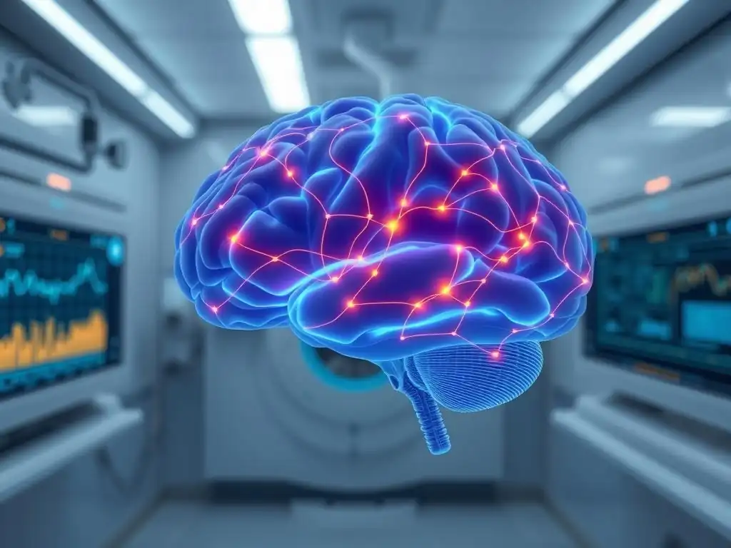
Welcome to Mind Care Guide, a space dedicated to understanding and nurturing your mental well-being.
Ever wondered how scientists truly see what's happening inside the brain of someone struggling with a mental health condition? Our exploration into the neuroscience of mental health begins with a fascinating look at PET scans and their vital role in deciphering the complexities of mental disorders. Join us as we delve into how this powerful imaging technique is helping us track neurotransmitters and gain a deeper understanding of the brain. Are you ready to uncover the secrets hidden within?
Tracking Neurotransmitters: How PET Aids Understanding of Mental Disorders
- Introduction: PET Scans in Mental Health - A Window into the Brain
- Understanding Neurotransmitters and Mental Illness
- PET Scans: A Powerful Tool for Imaging Neurotransmitters
- PET Scans and the Study of Specific Mental Disorders
- The Future of PET Scans in Mental Health: Advanced Applications
- Limitations of PET Scans in Mental Health Research
- Conclusion: PET Scans - Advancing Our Understanding of Mental Health
-
Frequently Asked Questions
- What role do PET scans play in understanding mental health?
- How do PET scans differ from other brain imaging techniques like MRI or CT scans?
- Can PET scans diagnose specific mental illnesses?
- What are the potential risks associated with PET scans?
- How can the information from PET scans improve mental health treatment?
- Final Reflection: The Evolving Landscape of Mental Health Through PET
- Unlock Your Brain's Potential: Explore the World of Neurotransmitters
Introduction: PET Scans in Mental Health - A Window into the Brain
Positron Emission Tomography, commonly known as PET, stands as a sophisticated brain imaging technique that provides researchers and clinicians with a unique window into the living brain's biochemical activity. Unlike structural imaging methods like MRI or CT scans, which show the brain's anatomy, PET scans mental health applications focus on visualizing and quantifying specific molecular processes. This capability is particularly vital in neuroscience research, allowing for the study of brain function at a chemical level, which is often disrupted in mental health conditions.
The ability of PET scans to track the movement, binding, and availability of neurotransmitters is profoundly important for advancing our understanding of mental disorders. Neurotransmitters are the chemical messengers that nerve cells use to communicate, and their intricate balance is essential for healthy brain function, mood regulation, and cognitive processes. By visualizing these chemicals in action, PET scanning offers invaluable insights into the underlying biological mechanisms of conditions like depression, schizophrenia, and anxiety, paving the way for more targeted diagnostic approaches and therapeutic interventions.
Understanding Neurotransmitters and Mental Illness
The Crucial Role of Neurotransmitters in Mental Health
Neurotransmitters are fundamental to the brain's complex signaling network, mediating communication between neurons across synapses. Key neurotransmitters like serotonin, dopamine, norepinephrine, glutamate, and GABA play diverse roles, influencing everything from mood, motivation, and cognition to sleep, appetite, and stress response. For instance, serotonin is widely associated with mood regulation and well-being, while dopamine is central to reward, motivation, and motor control pathways. Glutamate is the primary excitatory neurotransmitter, crucial for learning and memory, while GABA is the main inhibitory neurotransmitter, helping to calm neural activity and prevent over-excitation.
When the delicate balance or function of these neurotransmitter systems is disturbed, it can significantly impact mental health. Imbalances can manifest as altered levels of the neurotransmitter itself, issues with how it is released or reabsorbed, or problems with the receptors it binds to on receiving neurons. These disruptions are not merely theoretical; they are increasingly recognized as biological substrates contributing to the symptoms observed in various mental disorders. Understanding these specific dysfunctions is a key goal, and PET scans mental health research is instrumental in this pursuit.
Common Mental Disorders Linked to Neurotransmitter Dysfunction
Depression is often linked to dysfunctions in the monoamine neurotransmitter systems, particularly serotonin and norepinephrine. While the initial "chemical imbalance" hypothesis has evolved, research using tools like PET scans has provided tangible evidence of altered serotonin transporter (SERT) availability and norepinephrine signaling in individuals with depression compared to healthy controls. Studies have shown, for example, that some individuals with depression exhibit reduced SERT binding in certain brain regions, potentially leading to lower synaptic serotonin levels, although the picture is complex and varies among individuals.
Schizophrenia research has historically focused heavily on dopamine, specifically the hyperactivity of dopamine signaling in certain brain pathways. PET scans have been crucial in demonstrating altered dopamine D2/D3 receptor density and dopamine synthesis capacity in the brains of individuals with schizophrenia. For example, studies have consistently shown increased dopamine release in response to stimuli in patients experiencing psychosis. Furthermore, the efficacy of many antipsychotic medications is directly related to their ability to block dopamine D2 receptors, a mechanism confirmed and quantified using PET imaging.
Anxiety disorders involve complex neural circuits, but the inhibitory neurotransmitter GABA (gamma-aminobutyric acid) is strongly implicated. GABA's role is to reduce neuronal excitability, and disruptions in GABAergic signaling can lead to heightened states of arousal and fear. PET studies have explored GABA-A receptor availability in individuals with panic disorder or generalized anxiety disorder. While findings can be variable, some research suggests reduced binding or availability of GABA-A receptors in specific brain areas like the amygdala or prefrontal cortex in anxious individuals, potentially contributing to excessive worry and panic responses.
PET Scans: A Powerful Tool for Imaging Neurotransmitters
How PET Scans Work: Visualizing Brain Activity
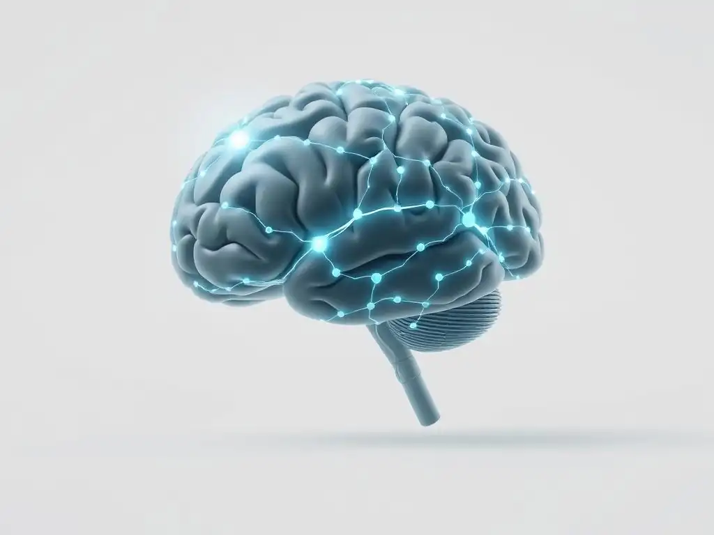
Positron Emission Tomography operates by detecting gamma rays emitted indirectly by a tracer, which is a radioactive substance introduced into the body, typically intravenously. This tracer is often linked to a molecule that mimics a natural substance in the body, such as glucose (used in FDG-PET to measure metabolic activity) or, critically for mental health research, molecules that bind specifically to neurotransmitters, their transporters, or receptors. Once injected, the tracer accumulates in areas of the brain where the target molecule is present or being utilized.
As the tracer decays, it emits positrons, which quickly encounter electrons in the surrounding tissue. This annihilation event produces a pair of gamma rays traveling in opposite directions.
Detectors surrounding the patient's head register these gamma rays, and sophisticated computer algorithms reconstruct a 3D image showing the distribution and concentration of the tracer within the brain. By using tracers designed to target specific components of neurotransmitter systems – known as radioligands – PET scans can effectively map the location and quantify the density or activity of these crucial chemical players in the brain, providing invaluable data for PET scans mental health studies.
Advantages of PET Imaging in Mental Health Research
One of the primary advantages of PET imaging is its exceptional sensitivity and specificity in detecting and quantifying specific molecular targets, including neurotransmitter receptors, transporters, and enzymes. Unlike functional MRI (fMRI), which measures blood flow as an indirect marker of neural activity, PET directly probes biochemical processes. This allows researchers to measure, for instance, the density of dopamine D2 receptors in the striatum or the availability of serotonin transporters in the brainstem with high precision, offering a level of molecular detail unmatched by other non-invasive imaging techniques.
Furthermore, PET scans allow for the quantification of neurotransmitter levels and receptor binding in vivo, meaning within the living brain. This capability is paramount for understanding the dynamic nature of neurotransmitter systems in health and disease. Researchers can perform studies to compare receptor densities between patient groups and healthy controls, assess how pharmaceutical treatments affect receptor occupancy (e.g., how well an antipsychotic drug is binding to dopamine receptors), or even measure changes in neurotransmitter release in response to stimuli. This quantitative data is essential for building robust models of brain function and dysfunction underlying mental disorders.
PET Scans and the Study of Specific Mental Disorders
PET Imaging in Depression Research: Serotonin Transporter Availability
Studies utilizing PET imaging have extensively investigated the serotonin transporter (SERT) in depression. The SERT is responsible for reuptaking serotonin from the synaptic cleft back into the presynaptic neuron, effectively regulating the duration and intensity of serotonin signaling. Using radioligands like [11C]DASB or [11C]MADAM, researchers can measure SERT binding potential, which is often interpreted as an index of SERT density or availability.
Numerous PET studies comparing individuals with major depressive disorder (MDD) to healthy controls have yielded complex but informative results. While some studies report reduced SERT binding in specific brain regions like the brainstem, limbic areas, or prefrontal cortex in depressed patients, others find no significant differences or even increased binding in certain subgroups or brain areas.
For instance, a meta-analysis pooling data from multiple studies might suggest an average reduction in SERT binding in the brainstem of around 10-20% in unmedicated depressed individuals compared to controls. These findings, though not universally consistent across all studies and patient populations, provide biological evidence of altered serotonin system function in depression and help inform the mechanisms targeted by selective serotonin reuptake inhibitors (SSRIs), medications that block the SERT.
Investigating Schizophrenia with PET: Dopamine Receptor Activity
PET imaging has been a cornerstone in the study of dopamine dysfunction in schizophrenia, particularly focusing on dopamine D2/D3 receptors, the primary targets of most antipsychotic medications. Radioligands such as [11C]raclopride or [18F]fallypride allow researchers to measure the density and occupancy of these receptors in key brain areas like the striatum. Studies have consistently shown elevated dopamine synthesis capacity and release in the striatum of patients with schizophrenia, especially during acute psychotic episodes.
For example, studies using [18F]FDOPA PET have estimated increased striatal dopamine synthesis capacity in patients with schizophrenia, sometimes reported to be elevated by 20-30% compared to healthy individuals. Furthermore, PET scans are routinely used in clinical trials to measure how effectively antipsychotic drugs block D2 receptors. Achieving optimal clinical response in schizophrenia is often associated with D2 receptor occupancy levels typically ranging from 65% to 80% in the striatum, a metric directly quantifiable through PET imaging. This provides a powerful tool for understanding drug mechanisms and potentially optimizing dosing strategies.
PET Scans for Anxiety Disorders: GABA Receptor Studies
Research into anxiety disorders using PET imaging has often focused on the GABAergic system, particularly GABA-A receptors, which are the targets of benzodiazepines and play a critical role in regulating neuronal excitability. Radioligands like [11C]flumazenil or [11C]Ro15-4513 have been employed to measure GABA-A receptor binding potential in various brain regions. The goal is to identify potential abnormalities in receptor density or function that could contribute to the excessive fear and worry characteristic of anxiety disorders.
Studies in panic disorder, for example, have explored GABA-A receptor availability in regions like the insula, amygdala, and prefrontal cortex. Some research has suggested reduced GABA-A receptor binding in certain cortical or limbic areas in individuals with panic disorder compared to healthy controls, although findings are not always conclusive and can vary depending on the specific anxiety subtype and radioligand used.
While the link between GABA system dysfunction and anxiety is complex and involves multiple brain circuits, PET imaging offers a unique approach to directly assess components of this inhibitory system in the living human brain, complementing findings from other research modalities.
The Future of PET Scans in Mental Health: Advanced Applications
Novel Radioligands for Targeting Specific Brain Targets
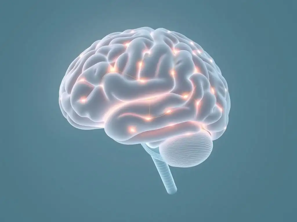
The field of PET imaging is continuously evolving, driven by the development of novel radioligands that target a wider array of specific brain molecules relevant to mental health. Beyond the well-established tracers for dopamine, serotonin transporters, and GABA-A receptors, researchers are developing ligands for other crucial systems, such as glutamate receptors (e.g., NMDA or mGluR5 receptors), cannabinoid receptors (CB1), opioid receptors, and various neuropeptide systems.
The availability of these new tracers allows for a much finer-grained analysis of the complex molecular landscape underlying mental disorders. For example, tracers targeting inflammatory markers like TSPO are being explored to investigate the role of neuroinflammation in conditions like depression or schizophrenia. Developing ligands with improved specificity, affinity, and pharmacokinetic properties is a major focus, promising to unlock new insights into previously inaccessible targets and refine our understanding of disease mechanisms at a molecular level.
Combining PET with Other Brain Imaging Techniques
To gain a more comprehensive picture of brain function and structure, PET imaging is increasingly being combined with other neuroimaging modalities. Integrated PET-MRI scanners, for instance, allow for simultaneous acquisition of PET data (molecular activity) and MRI data (structure, functional connectivity, diffusion) within the same scanning session. This co-registration provides highly accurate anatomical localization of the molecular findings from PET and allows researchers to correlate biochemical measures with structural integrity or functional network activity.
Combining PET with electroencephalography (EEG) is another powerful approach. While PET offers high spatial resolution for molecular targets, EEG provides excellent temporal resolution for electrical brain activity. Simultaneous PET-EEG recordings allow researchers to investigate how specific molecular processes measured by PET (e.g., dopamine receptor availability) relate to dynamic changes in neural oscillations and brain states measured by EEG. This multi-modal approach leverages the strengths of different techniques to provide a richer understanding of the interplay between molecular mechanisms, neural circuits, and the complex symptoms of mental disorders.
Personalized Medicine and PET Scans: Tailoring Treatment Strategies
A significant future application of PET scans in mental health lies in the realm of personalized medicine. By providing quantitative data on specific neurotransmitter systems or receptor availability in an individual patient's brain, PET imaging holds the potential to help predict response to specific treatments or guide medication selection and dosing. For example, research is ongoing to determine if pre-treatment PET scans assessing serotonin transporter availability could help predict which patients with depression are most likely to respond to SSRI therapy.
Similarly, in schizophrenia, PET scans can measure dopamine D2 receptor occupancy to ensure that an adequate dose of antipsychotic medication is being administered to achieve the therapeutically relevant occupancy range (often cited as 65-80%). This could potentially help optimize dosing, reduce side effects associated with unnecessarily high doses, and improve patient outcomes. While still largely a research tool for this purpose, the vision is that PET imaging could become a valuable part of clinical decision-making, moving towards more tailored and effective mental health care based on an individual's unique neurobiological profile.
Limitations of PET Scans in Mental Health Research
Cost and Availability of PET Imaging
Despite its powerful capabilities, PET imaging is a relatively expensive technique, posing significant limitations on its widespread use in both research and clinical settings. The scanners themselves are costly to acquire and maintain, often running into the millions of dollars. Furthermore, the radioactive tracers used in PET have very short half-lives (some decaying significantly within minutes or hours) and often need to be produced on-site using a cyclotron, adding substantial infrastructure and operational costs.
These high costs mean that PET scans are not readily available at all medical centers or research institutions. Access to PET imaging facilities capable of using specific radioligands relevant to mental health research is often limited to major academic medical centers or specialized research institutes. This restricted availability can make it challenging to conduct large-scale studies, recruit diverse patient populations, and translate findings into routine clinical practice outside of specialized centers.
Radiation Exposure and Ethical Considerations
A key limitation of PET imaging is that it involves the administration of a radioactive tracer, which exposes the participant or patient to ionizing radiation. While the dose is typically low and generally considered safe within established guidelines for research or clinical use, it is not without risk, particularly for vulnerable populations or with repeated scans. The amount of radiation exposure is similar to or slightly higher than that of a standard CT scan, depending on the specific tracer and protocol.
Due to this radiation exposure, ethical considerations are paramount when conducting PET scans, especially in research involving healthy volunteers or individuals with mental health conditions who may be more vulnerable. Strict ethical protocols and review boards are required to ensure that the potential benefits of the research outweigh the risks of radiation exposure. This inherent risk limits the number of scans an individual can undergo and necessitates careful justification for each study, distinguishing PET from non-ionizing imaging techniques like MRI.
Conclusion: PET Scans - Advancing Our Understanding of Mental Health
PET scans have undeniably revolutionized our ability to study the living human brain at a molecular level, offering unprecedented insights into the complex world of neurotransmitters and their roles in mental health. By allowing researchers to visualize and quantify specific components of neurotransmitter systems, PET imaging has provided crucial biological evidence for the involvement of dopamine, serotonin, GABA, and other chemical messengers in conditions ranging from schizophrenia and depression to anxiety disorders. This capability moves beyond simply observing symptoms to exploring the underlying biological mechanisms.
The future of PET scans in mental health research is bright, with ongoing advancements in radioligand development, the integration of PET with other sophisticated imaging modalities like MRI and EEG, and the exciting potential for using PET data to inform personalized treatment strategies. While challenges related to cost, availability, and radiation exposure remain, continued innovation promises to enhance the utility and accessibility of PET imaging. Ultimately, PET scans mental health research continues to deepen our understanding of these complex conditions, paving the way for more effective diagnostics, targeted therapies, and improved outcomes for individuals struggling with mental health challenges.
Frequently Asked Questions
What role do PET scans play in understanding mental health?
PET scans offer a non-invasive way to visualize and measure brain activity, specifically the function of neurotransmitters. This helps researchers and clinicians understand how neurochemical imbalances contribute to mental disorders.
How do PET scans differ from other brain imaging techniques like MRI or CT scans?
While MRI and CT scans provide structural information about the brain, PET scans focus on functional activity. They use radioactive tracers to detect neurotransmitter levels and receptor binding, offering insights into brain chemistry.
Can PET scans diagnose specific mental illnesses?
PET scans are not typically used for routine diagnosis. They are more often used in research settings to understand the underlying biological mechanisms of mental illnesses and to develop new treatments.
What are the potential risks associated with PET scans?
The primary risk is exposure to a small amount of radiation from the radioactive tracer. Allergic reactions to the tracer are also possible, but rare. Benefits typically outweigh the risks in research settings.
How can the information from PET scans improve mental health treatment?
By revealing specific neurotransmitter abnormalities, PET scans can help researchers develop more targeted medications and therapies. This personalized approach aims to improve treatment outcomes and reduce side effects.
Final Reflection: The Evolving Landscape of Mental Health Through PET
The ability to visualize the brain's intricate chemical processes using PET scans represents a monumental leap forward in our understanding and treatment of mental disorders. This technology, once a futuristic dream, is now actively shaping the diagnostic and therapeutic landscape, offering unprecedented insights into the complex interplay of neurotransmitters and mental illness. The implications for personalized medicine and improved patient outcomes are profound and far-reaching.
The journey of understanding the brain's chemistry has been long and arduous, but PET scans have undeniably accelerated our progress. This technology allows us to move beyond simply observing symptoms and delve into the underlying biological mechanisms driving conditions like depression, anxiety, and schizophrenia. This shift in perspective is crucial, fostering a more nuanced and empathetic approach to mental healthcare. The greatest advances in medicine have come from understanding the underlying mechanisms of disease, and PET scans are providing us with that crucial understanding in the field of mental health.
This quote encapsulates the transformative power of this technology, moving us towards a future where treatment is tailored to the individual's unique neurochemical profile.
Consider the implications of this technology for your own life, and the lives of those around you. What if we could diagnose mental health conditions earlier, with greater accuracy? What if treatments could be personalized to maximize efficacy and minimize side effects?
This isn't just about scientific advancement; it's about improving the human experience, fostering a future where mental health is treated with the same level of understanding and care as physical health. Embrace the potential of PET scans and other advancements in neuroimaging, and advocate for continued research and development in this vital field. The future of mental healthcare is being written, and you can be a part of it.
Unlock Your Brain's Potential: Explore the World of Neurotransmitters
Thanks for diving deep into the fascinating world of neurotransmitters and how PET scans are revolutionizing our understanding of mental health! You've gained valuable insights into complex brain processes – now let's put that knowledge into action.
- Share your key takeaway: What surprised you most about the role of neurotransmitters in mental disorders? Discuss it on social media using #NeurotransmitterInsights and #MindCareGuide! Let's spark a conversation!
- Learn more about PET scans: Research different types of brain imaging techniques and their applications in mental health care. This deeper dive will further enrich your understanding.
- Consider your mental well-being: Reflect on your personal health and consider seeking professional help if you feel you need support. Knowledge is power, and taking proactive steps for your mental health is crucial.
What's one small step you can take today to prioritize your mental wellness, based on what you learned?