
Diffusion Imaging (DTI/NODDI): Mapping Brain Fiber Pathways in Mental Health

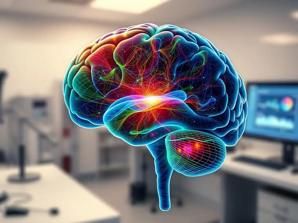
Welcome to Mind Care Guide, a place dedicated to understanding and nurturing your mental well-being. Ever wondered how cutting-edge science is revealing the intricate connections within your brain and their impact on mental health? This article delves into the fascinating world of DTI mental health research, exploring how diffusion imaging helps us map the very fiber pathways that influence our thoughts and emotions. Intrigued? Let's unravel the mysteries of the brain together and discover what this means for understanding and addressing mental health challenges.
Diffusion Imaging (DTI/NODDI): Mapping Brain Fiber Pathways in Mental Health
- Introduction to Diffusion Imaging in Mental Health
- DTI and NODDI: Principles and Techniques
-
Applications of DTI in Mental Health Research
- DTI Studies in Schizophrenia: Investigating White Matter Abnormalities
- Examining Brain Connectivity in Major Depressive Disorder with DTI
- DTI and Anxiety Disorders: Identifying Neural Correlates
- Exploring White Matter Integrity in Post-Traumatic Stress Disorder (PTSD) using DTI
- DTI Research in Autism Spectrum Disorder: Mapping Brain Connectivity Patterns
- NODDI's Contribution to Understanding Mental Health
- Advantages and Limitations of DTI and NODDI in Mental Health Research
- Interpreting DTI and NODDI Results in the Context of Mental Health
- The Future of Diffusion Imaging in Mental Health
- Conclusion: DTI, NODDI, and the Future of Mental Healthcare
-
Frequently Asked Questions
- What is Diffusion Tensor Imaging (DTI) and how does it relate to mental health research?
- How can DTI contribute to DTI mental health research and its progress?
- What types of mental health challenges are being studied using DTI/NODDI?
- What are the limitations of using DTI in mental health research?
- Where can I find more information about DTI mental health research and resources for mental well-being?
- Final Reflection: Charting the Untamed Territory of the Mind
- Unveil Your Brain's Hidden Pathways: Take the Next Step
Introduction to Diffusion Imaging in Mental Health
Understanding the Role of Brain Fiber Pathways in Mental Health Disorders
The human brain is an incredibly complex network, and its proper functioning relies heavily on efficient communication between different regions. This communication occurs primarily through white matter pathways, bundles of myelinated axons that act like the brain's high-speed internet cables. These pathways connect grey matter areas, where processing happens, enabling integrated thought, emotion, and behavior. When these connections are disrupted or altered, it can significantly impact mental processes, contributing to the symptoms observed in various mental health disorders.
Research into the structural integrity and connectivity of these white matter pathways is crucial for understanding the underlying neurobiology of conditions like schizophrenia, depression, anxiety, and autism spectrum disorder. By mapping and characterizing these fiber tracts, scientists gain insights into how communication breakdowns might manifest as psychiatric symptoms. This area of inquiry, often powered by techniques like DTI mental health research, seeks to identify specific white matter abnormalities associated with different conditions, potentially leading to better diagnostic markers and targeted therapeutic interventions.
What is Diffusion Tensor Imaging (DTI)? A Primer
Diffusion Tensor Imaging (DTI) is a non-invasive magnetic resonance imaging (MRI) technique specifically designed to study the brain's white matter pathways. It works by measuring the diffusion of water molecules within brain tissue. Water molecules in the brain are not free to move randomly in all directions; their movement is restricted by cellular structures like cell membranes, axons, and myelin sheaths. In white matter, water tends to diffuse more freely along the direction of the axon fibers than across them.
DTI captures this directional preference of water diffusion. By applying magnetic field gradients in multiple directions, the scanner can measure how water moves in different orientations at each point (voxel) in the brain. This information is then used to calculate a "diffusion tensor," a mathematical representation of the diffusion process. From this tensor, various metrics can be derived that provide insights into the integrity and orientation of the underlying white matter fibers, making DTI a foundational tool in DTI mental health research.
Beyond DTI: Exploring Neurite Orientation Dispersion and Density Imaging (NODDI)
While DTI provides valuable information about the overall directionality and magnitude of water diffusion, it has limitations, particularly in areas where fiber bundles cross or fan out. DTI models assume a single primary direction of diffusion within each voxel, which isn't always accurate in complex white matter architecture. This limitation spurred the development of more advanced diffusion models, such as Neurite Orientation Dispersion and Density Imaging (NODDI).
NODDI is an advanced diffusion MRI technique that attempts to provide a more detailed picture of brain microstructure than DTI. It models the diffusion signal as arising from distinct compartments: water within neurites (axons and dendrites), water in the extracellular space, and potentially water in the cerebrospinal fluid.
By separating these components, NODDI can estimate parameters like neurite density and orientation dispersion (how spread out the neurite orientations are within a voxel), offering a richer and potentially more specific understanding of tissue properties. This newer approach complements DTI mental health research by providing finer-grained insights into the cellular changes underlying mental health conditions.
DTI and NODDI: Principles and Techniques
How DTI Works: Measuring Water Diffusion in the Brain
The core principle behind DTI is the sensitivity of MRI signals to the movement of water molecules. In a standard MRI sequence, radiofrequency pulses and magnetic field gradients are used to excite water protons and then measure the signal they emit as they relax. DTI adds diffusion-sensitizing gradients to this sequence. These gradients are applied before and after the main radiofrequency pulse. If water molecules move between the application of these two gradients, their signal is attenuated. The amount of signal attenuation is proportional to how far the water molecules have moved.
By repeating this process with diffusion-sensitizing gradients applied along many different directions (typically 30 or more), DTI builds a picture of water diffusion in three dimensions at each point in the brain. In areas with highly organized white matter tracts, water diffuses preferentially along the direction of the fibers, resulting in high signal attenuation when gradients are applied perpendicular to the fibers and low attenuation when applied parallel to them.
In areas with less organized structure, like grey matter or cerebrospinal fluid, diffusion is more isotropic (equal in all directions). Analyzing these diffusion patterns allows the calculation of the diffusion tensor, which characterizes the shape and orientation of the diffusion ellipsoid in each voxel.
Understanding Fractional Anisotropy (FA), Mean Diffusivity (MD), and Other DTI Metrics
From the diffusion tensor calculated by DTI, several key metrics are derived that are widely used in DTI mental health research:
- Fractional Anisotropy (FA): This is perhaps the most commonly used DTI metric. FA measures the degree to which water diffusion is anisotropic (directional). A value of 0 indicates perfectly isotropic diffusion (equal in all directions), like in pure water or CSF. A value of 1 indicates perfectly anisotropic diffusion, where water only moves along one direction. In white matter, higher FA generally suggests greater structural integrity, organization, and myelination of fiber bundles. Lower FA can indicate damage, disorganization, or demyelination.
- Mean Diffusivity (MD): Also known as Apparent Diffusion Coefficient (ADC), MD is the average magnitude of water diffusion across all directions. It reflects the overall restrictiveness of the tissue environment to water movement. Higher MD can indicate conditions like edema (swelling) or tissue damage where barriers to diffusion are reduced. Lower MD might suggest increased cellularity or restricted space.
- Axial Diffusivity (AD): Measures the diffusion along the principal direction of diffusion (usually assumed to be along the fiber). Changes in AD can sometimes reflect axonal damage.
- Radial Diffusivity (RD): Measures the average diffusion perpendicular to the principal direction. Changes in RD are often associated with myelin damage or loss, as myelin typically restricts diffusion radially.
Researchers often examine these metrics in specific white matter tracts identified through tractography or by averaging values within predefined regions of interest. Alterations in FA, MD, AD, and RD in particular pathways are key findings reported in many DTI mental health research studies.
NODDI: A More Detailed View of Brain Microstructure
NODDI goes beyond the simple tensor model of DTI by incorporating a more biophysically realistic model of brain tissue. It divides the signal from each voxel into different compartments:
- Intra-neurite compartment: Represents water confined within axons and dendrites. Diffusion here is highly restricted perpendicular to the neurite orientation but relatively free along the neurite.
- Extra-neurite compartment: Represents water in the space outside the neurites. Diffusion here is hindered by the presence of neurites but is less restricted directionally than within the neurites.
- Isotropic compartment: Represents water in free spaces like cerebrospinal fluid or large extracellular spaces, where diffusion is equal in all directions.
By fitting the diffusion signal to this multi-compartment model, NODDI estimates parameters that are thought to be more specific to underlying tissue properties than DTI metrics.
Key NODDI metrics include:
- Neurite Density Index (NDI): Estimates the volume fraction of the intra-neurite compartment. This is thought to reflect the density of axons and dendrites in a voxel.
- Orientation Dispersion Index (ODI): Measures the degree of angular dispersion of neurites within a voxel. A low ODI indicates highly aligned neurites (like in a coherent fiber bundle), while a high ODI indicates neurites oriented in many different directions (like in crossing fibers or grey matter).
- Isotropic Volume Fraction (ISOVF): Estimates the volume fraction of the isotropic compartment, representing free water.
These parameters offer a more nuanced perspective on tissue structure than DTI's single tensor model. For instance, a reduction in FA in DTI could be due to reduced neurite density, increased orientation dispersion, or demyelination. NODDI's NDI and ODI can potentially help disentangle these different microstructural changes, providing more specific insights in DTI mental health research.
DTI vs. NODDI: Key Differences and Advantages
The fundamental difference between DTI and NODDI lies in their underlying mathematical models and the microstructural properties they are designed to capture.
- DTI: Uses a simple tensor model assuming a single dominant diffusion direction per voxel. It provides metrics like FA and MD, which are sensitive to overall diffusion anisotropy and magnitude but can be ambiguous in regions with complex fiber architecture (crossing, fanning).
- NODDI: Uses a multi-compartment model that attempts to separate diffusion within neurites, outside neurites, and in free water. It provides metrics like NDI and ODI, aiming for more specific measures of neurite density and orientation dispersion, which are potentially less affected by fiber crossings and fanning.
| Feature | DTI (Diffusion Tensor Imaging) | NODDI (Neurite Orientation Dispersion and Density Imaging) |
|---|---|---|
| Model | Single tensor | Multi-compartment (Intra-neurite, Extra-neurite, Isotropic) |
| Primary Metrics | FA, MD, AD, RD | NDI, ODI, ISOVF |
| Information Type | Overall anisotropy & magnitude of diffusion | Neurite density, orientation dispersion, free water fraction |
| Sensitivity | Sensitive to fiber orientation, integrity, myelination | More specific to neurite density and angular organization |
| Complexity (Model) | Simpler | More complex |
| Data Requirements | Fewer diffusion directions sufficient (e.g., 6-30) | Typically requires more diffusion directions and multiple b-values |
| Handling Crossings | Poorly handles fiber crossings/fanning | Better at resolving complex fiber orientations |
NODDI's primary advantage in DTI mental health research is its potential for improved specificity. By disentangling different factors that influence the diffusion signal, NODDI metrics might provide a clearer picture of the underlying cellular pathology. For example, a reduction in FA in a DTI study could be interpreted more specifically if a corresponding NODDI analysis shows reduced NDI (suggesting fewer axons) versus increased ODI (suggesting less organized fibers).
However, NODDI requires more complex data acquisition (multiple b-values) and processing than standard DTI. While DTI remains a cornerstone of DTI mental health research due to its relative simplicity and widespread availability, NODDI is increasingly used to provide complementary and potentially more specific microstructural insights.
Applications of DTI in Mental Health Research
DTI Studies in Schizophrenia: Investigating White Matter Abnormalities
Schizophrenia is a severe mental illness characterized by disruptions in thought processes, perceptions, emotional responsiveness, and social interactions. For decades, post-mortem studies hinted at widespread white matter abnormalities in the brains of individuals with schizophrenia. DTI mental health research has provided a non-invasive way to investigate these abnormalities in living patients. Numerous DTI studies have consistently reported reduced fractional anisotropy (FA) in various white matter tracts in individuals with schizophrenia compared to healthy controls.
Specific tracts frequently implicated include the corpus callosum (connecting the two hemispheres), the cingulum bundle (involved in emotion and cognition), the uncinate fasciculus (connecting temporal and frontal lobes, relevant to memory and emotion), and tracts within the frontal and temporal lobes. For instance, a meta-analysis published in Schizophrenia Bulletin in 2010, aggregating data from dozens of DTI studies, confirmed widespread FA reductions across multiple white matter regions, with effect sizes often ranging from 0.3 to 0.6 (Cohen's d) in tracts like the corpus callosum.
These findings support the hypothesis that schizophrenia involves a disruption in the brain's structural connectivity, potentially contributing to the fragmented experiences and cognitive deficits characteristic of the disorder. Further DTI mental health research continues to explore the relationship between the severity of white matter changes and specific symptom profiles or treatment responses.
Examining Brain Connectivity in Major Depressive Disorder with DTI
Major Depressive Disorder (MDD) is characterized by persistent sadness, loss of interest, and other emotional and cognitive symptoms. While MDD is often viewed through the lens of altered neurotransmitter function or activity in specific brain regions, DTI mental health research has increasingly focused on disruptions in the structural connections between these regions. Studies using DTI have reported white matter abnormalities in circuits involved in emotion regulation, reward processing, and cognitive control in individuals with MDD.
Common findings include altered FA and MD in tracts connecting areas like the prefrontal cortex, amygdala, hippocampus, and anterior cingulate cortex. For example, studies have frequently reported reduced FA in the cingulum bundle, particularly its subgenual part, a region strongly implicated in mood regulation.
Some research suggests that FA values in certain tracts, such as the uncinate fasciculus, might correlate with the severity of depressive symptoms or predict treatment response. While findings can vary depending on study population, methodology, and subtype of depression, the consistent implication of specific white matter pathways in MDD highlights the importance of disrupted structural connectivity in the pathophysiology of this common mental health condition, driving further DTI mental health research in this area.
DTI and Anxiety Disorders: Identifying Neural Correlates
Anxiety disorders, including Generalized Anxiety Disorder (GAD), Social Anxiety Disorder, and Panic Disorder, are characterized by excessive fear and worry. Neurobiological models often point to dysfunction in brain circuits involving the amygdala, prefrontal cortex, and hippocampus. DTI mental health research has explored whether structural connectivity abnormalities underlie these functional alterations.
Studies have investigated white matter integrity in tracts connecting these key regions. For example, research has examined the uncinate fasciculus, which connects the amygdala (a key region for processing fear and threat) to the ventromedial prefrontal cortex (involved in regulating emotional responses). Some studies have reported reduced FA in the uncinate fasciculus in individuals with anxiety disorders, suggesting potential disruptions in the pathway that allows the prefrontal cortex to modulate amygdala activity.
Findings, however, are not always consistent across different anxiety disorder subtypes and studies. For instance, one study published in Psychiatry Research: Neuroimaging reported lower FA in the uncinate fasciculus in patients with generalized anxiety disorder compared to controls, while other studies focusing on social anxiety have found different patterns. This variability underscores the need for larger, well-controlled DTI mental health research studies to pinpoint specific white matter signatures for different anxiety conditions.
Exploring White Matter Integrity in Post-Traumatic Stress Disorder (PTSD) using DTI
Post-Traumatic Stress Disorder (PTSD) is a condition that can develop after exposure to a traumatic event. It is associated with significant alterations in brain function and structure, particularly in regions involved in fear processing, memory, and emotion regulation, such as the amygdala, hippocampus, and prefrontal cortex. DTI mental health research has been applied to investigate whether the structural connections between these areas are altered in individuals with PTSD.
Studies have frequently reported reduced FA in the cingulum bundle, particularly its anterior and posterior portions, which are involved in emotion processing and memory recall – processes highly relevant to PTSD symptoms like re-experiencing and hypervigilance. Alterations in the corpus callosum, potentially affecting inter-hemispheric communication, have also been noted in some samples.
For example, a study involving combat veterans with PTSD found significantly lower FA in the anterior cingulum compared to a control group of veterans without PTSD. While findings can be influenced by factors like trauma type, severity, and comorbidity, the consistent implication of limbic system white matter tracts in DTI mental health research on PTSD suggests that disrupted structural connectivity plays a significant role in the disorder's pathophysiology.
DTI Research in Autism Spectrum Disorder: Mapping Brain Connectivity Patterns
Autism Spectrum Disorder (ASD) is a complex neurodevelopmental condition characterized by challenges with social communication and interaction, and restricted or repetitive behaviors or interests. Theories of ASD often involve atypical brain connectivity, suggesting that the disorder might arise from differences in how different brain regions are wired together. DTI mental health research has been a key tool for investigating these connectivity patterns in ASD.
Early DTI studies in ASD often reported reduced FA in various white matter tracts, suggesting widespread "underconnectivity." However, more recent and larger studies, particularly those involving different age groups, have presented a more complex picture, sometimes showing increased FA in certain tracts or no significant differences in others. Findings are often dependent on the specific age of the participants, suggesting that connectivity patterns might differ across development in ASD.
For instance, some studies in young children with ASD have reported higher FA in certain tracts, potentially reflecting altered developmental trajectories. The corpus callosum, superior longitudinal fasciculus (connecting frontal, parietal, and temporal lobes), and uncinate fasciculus are frequently examined. DTI mental health research in ASD continues to evolve, moving towards examining specific fiber pathways and their relationship to particular behavioral symptoms, and considering the dynamic nature of brain development.
NODDI's Contribution to Understanding Mental Health
Refining DTI Findings: NODDI Insights into Schizophrenia
While DTI has consistently shown widespread white matter abnormalities in schizophrenia, the interpretation of reduced FA can be ambiguous. It could reflect fewer axons (neurite density), less organized axons (orientation dispersion), or myelin damage. NODDI offers the potential to disentangle these possibilities, providing a more specific microstructural interpretation of DTI findings in DTI mental health research.
Studies using NODDI in schizophrenia have begun to provide more detailed insights. For example, some research has found reduced Neurite Density Index (NDI) in certain white matter regions previously identified with reduced FA by DTI, such as the corpus callosum and cingulum. This suggests that the DTI-observed FA reduction in these areas might, at least in part, be attributable to a lower density of axons.
Other studies have found altered Orientation Dispersion Index (ODI) in specific tracts, potentially indicating less coherent fiber bundles. These findings from NODDI help refine the understanding of the cellular basis of white matter abnormalities in schizophrenia, moving beyond a general statement of "reduced integrity" to potentially identifying specific cellular changes like reduced axonal packing or altered fiber organization.
NODDI Studies on the Impact of Trauma on Brain Microstructure
Trauma and stress are known risk factors for several mental health conditions, including PTSD, depression, and anxiety. Research has shown that traumatic experiences can alter brain structure and function. While DTI has implicated white matter pathways in trauma-related disorders, NODDI is being used to investigate the specific microstructural changes that occur in response to trauma.
NODDI studies in individuals with a history of trauma, including those with PTSD, are exploring whether trauma is associated with changes in neurite density or orientation dispersion in specific brain regions. For instance, research has investigated the hippocampus and amygdala, key structures involved in processing traumatic memories and fear.
While still an emerging area of NODDI mental health research, initial findings suggest that trauma exposure might be associated with altered NDI and ODI in limbic and prefrontal regions. These changes could potentially reflect trauma-induced alterations in neuronal and axonal architecture, contributing to the persistent symptoms seen in conditions like PTSD.
Investigating Neurodevelopmental Disorders with NODDI
NODDI is particularly valuable for studying neurodevelopmental disorders like Autism Spectrum Disorder (ASD) and Attention-Deficit/Hyperactivity Disorder (ADHD) because it can provide more specific information about the developing brain's microstructure. Unlike DTI, which can be confounded by the complex fiber architecture and ongoing myelination in the developing brain, NODDI's ability to estimate neurite density and orientation dispersion might offer clearer insights into atypical developmental trajectories.
In ASD, for example, while DTI findings have been somewhat inconsistent across age groups, NODDI studies are starting to provide potentially more specific insights. Some studies are investigating whether the reported differences in white matter connectivity in ASD are due to differences in the number of axons (NDI) or how they are organized (ODI).
Similarly, NODDI is being applied in ADHD research to examine whether white matter alterations in frontal-striatal circuits, often implicated in the disorder, are related to differences in neurite density or organization. This more detailed microstructural perspective offered by NODDI is crucial for understanding the complex developmental processes that contribute to these conditions and is a growing area of DTI mental health research.
NODDI and the Characterization of Neurodegenerative Processes in Bipolar Disorder
Bipolar disorder is a mood disorder characterized by significant shifts in mood, energy, activity levels, and the ability to carry out daily tasks. While primarily a mood disorder, some research suggests the possibility of progressive neurodegenerative processes occurring over the course of the illness, particularly with repeated episodes. DTI studies have shown white matter abnormalities in bipolar disorder, but NODDI is being used to explore whether these changes reflect neurodegenerative processes at a microstructural level.
NODDI's ability to estimate neurite density makes it a potentially useful tool for detecting subtle neuroaxonal loss, which could be a marker of neurodegeneration. Studies are investigating whether individuals with bipolar disorder, especially those with a long history of illness or numerous episodes, show reduced NDI in specific brain regions compared to healthy controls or individuals with other psychiatric conditions.
For instance, research might focus on regions implicated in mood regulation and cognitive function that show volume loss in structural MRI studies. By providing a more specific measure of tissue composition, NODDI contributes to DTI mental health research aiming to understand the potential long-term impact of bipolar disorder on brain structure and to identify markers of illness progression.
Advantages and Limitations of DTI and NODDI in Mental Health Research
Strengths of DTI and NODDI: Non-Invasive Brain Mapping
One of the primary strengths of both DTI and NODDI is their non-invasive nature. As MRI techniques, they do not require the injection of radioactive tracers (unlike PET) or exposure to ionizing radiation (unlike CT). This makes them safe for use in a wide range of populations, including children and individuals who may participate in longitudinal studies requiring repeated scans. They provide unique in vivo information about the structural organization of the brain's white matter, which is not readily available through other standard neuroimaging modalities like structural MRI or fMRI.
These techniques allow researchers to study the living brain's architecture and connectivity, correlating findings with behavioral symptoms, cognitive performance, or treatment response in individuals with mental health conditions. The ability to map major fiber pathways and quantify aspects of their integrity and organization has fundamentally advanced DTI mental health research, moving beyond simply identifying regions of altered activity or volume to understanding the underlying structural "wiring" of the brain. They offer a window into the complex network disruptions potentially contributing to psychiatric disorders.
Limitations of DTI: Challenges in Complex Brain Regions
Despite its widespread use in DTI mental health research, standard DTI has significant limitations. The most notable is its inability to accurately model diffusion in voxels containing crossing, fanning, or bending fiber bundles. The single-tensor model assumes a single dominant direction of diffusion, which is inaccurate in approximately 60-90% of white matter voxels, depending on the brain region and resolution. This "crossing fiber problem" can lead to misinterpretations of DTI metrics, particularly FA.
For example, a reduction in FA in a voxel with crossing fibers could be due to a decrease in the integrity of one of the crossing bundles, but DTI cannot distinguish which one or separate their contributions. Instead, it averages the diffusion properties, potentially leading to ambiguous results.
Furthermore, FA can also be affected by factors other than white matter integrity, such as swelling or partial volume effects (where a voxel contains multiple tissue types), making interpretation challenging. These limitations necessitate careful consideration when interpreting DTI findings, especially in regions known for complex fiber architecture, and highlight the need for more advanced techniques like NODDI.
NODDI's Improved Specificity: Addressing DTI's Limitations
NODDI was specifically developed to address some of the key limitations of standard DTI, particularly the crossing fiber problem and the ambiguity of DTI metrics. By modeling the diffusion signal using separate compartments for intra-neurite, extra-neurite, and free water, NODDI can provide metrics like NDI and ODI that are hypothesized to be more specific to underlying microstructural properties (neurite density and orientation dispersion) than DTI's FA and MD.
In regions with crossing fibers, NODDI's model is better equipped to handle the multi-directional diffusion patterns. The ODI, for instance, provides a measure of how spread out the neurite orientations are within a voxel, which is directly relevant in areas of fiber crossing or fanning. The NDI provides a measure related to the sheer amount of neurites, ideally less affected by their specific orientation.
This improved specificity allows DTI mental health research using NODDI to potentially differentiate between different types of white matter pathology – for example, distinguishing between axonal loss (lower NDI) and dysmyelination (potentially affecting extra-neurite diffusion or radial diffusivity in DTI) or altered fiber organization (higher ODI). While more complex to acquire and process, NODDI offers a promising path toward a more detailed understanding of the microstructural basis of mental health conditions.
Addressing Potential Artifacts and Interpretation Challenges in Diffusion Imaging
Despite their power, diffusion imaging techniques like DTI and NODDI are susceptible to various artifacts and present significant interpretation challenges in DTI mental health research. Motion artifacts, particularly problematic in patient populations, can severely distort diffusion images. Susceptibility artifacts, caused by differences in magnetic properties between tissues (like air-tissue interfaces near the sinuses), can lead to signal dropouts or geometric distortions. Eddy currents, induced by the rapid switching of magnetic field gradients, also cause image distortions that require correction.
Beyond technical artifacts, interpretation requires careful consideration. Diffusion metrics are indirect measures of tissue properties; changes in FA or NDI do not directly equate to specific cellular pathologies without validation from other techniques (e.g., histology, when possible). Findings from different studies can be inconsistent due to variations in acquisition parameters, processing pipelines, statistical methods, and the heterogeneity of patient populations.
Furthermore, demonstrating a causal link between observed white matter alterations and the symptoms of a mental health disorder is challenging; the changes could be a cause, a consequence, or a correlate of the illness or its treatment. Rigorous methodology, careful artifact correction, and cautious interpretation are paramount in advancing DTI mental health research.
Interpreting DTI and NODDI Results in the Context of Mental Health
Connecting Brain Imaging Findings to Clinical Manifestations
Interpreting DTI and NODDI findings in the context of mental health involves linking observed abnormalities in white matter pathways to the specific symptoms, cognitive deficits, or behavioral patterns characteristic of a disorder. This requires a deep understanding of the functional roles of the implicated brain regions and the pathways connecting them. For example, finding reduced FA in the uncinate fasciculus in anxiety disorders is interpreted in light of this tract's role in connecting the amygdala (fear processing) to the prefrontal cortex (emotion regulation); a disruption might impair the brain's ability to regulate fear responses.
Researchers often correlate DTI/NODDI metrics directly with clinical measures, such as symptom severity scores (e.g., using the Hamilton Depression Rating Scale), cognitive test performance (e.g., measures of executive function or memory), or specific behavioral traits. For instance, a study might find that lower FA in a specific frontal white matter tract correlates with poorer performance on a cognitive flexibility task in individuals with schizophrenia. While correlations do not imply causation, they provide crucial evidence linking structural connectivity differences to functional impairments and clinical manifestations, guiding further DTI mental health research.
Combining DTI/NODDI with Other Neuroimaging Techniques (e.g., fMRI, EEG)
To gain a more comprehensive understanding of mental health disorders, DTI and NODDI findings are often combined with data from other neuroimaging modalities. Structural MRI provides information about brain volume and shape, allowing researchers to determine if white matter changes are associated with grey matter abnormalities. Functional MRI (fMRI) measures brain activity and functional connectivity (how correlated the activity is between different regions), offering insight into how structural alterations might impact brain function. Electroencephalography (EEG) or magnetoencephalography (MEG) provide measures of electrical or magnetic brain activity with high temporal resolution, capturing the dynamics of neural communication.
Combining these techniques allows researchers to investigate structure-function relationships. For example, a study might find reduced FA in a white matter tract connecting two regions (DTI finding) and also observe reduced functional connectivity between those same regions during a cognitive task (fMRI finding). This multimodal approach provides stronger evidence that structural connectivity deficits contribute to functional network dysfunction in mental illness. Integrating data from DTI mental health research with other modalities provides a more holistic view of the complex brain changes involved in psychiatric conditions.
The Importance of Longitudinal Studies Using DTI and NODDI
While cross-sectional studies comparing groups of patients and controls provide valuable insights, longitudinal studies are essential for understanding the trajectory of white matter changes over time in mental health disorders. Many mental illnesses are chronic or episodic, and brain structure can change with illness progression, treatment, or recovery. Longitudinal DTI and NODDI studies involve scanning the same individuals multiple times over months or years.
These studies can help determine:
- Whether white matter abnormalities are present early in the illness or develop over time.
- How white matter integrity changes during periods of relapse or remission.
- Whether specific treatments (e.g., medication, therapy) influence white matter structure.
- If baseline white matter characteristics can predict prognosis or treatment response.
For instance, a longitudinal DTI mental health research study might track FA changes in young individuals at high risk for developing schizophrenia to see if declining FA in certain tracts precedes the onset of symptoms. Or, a study might examine if successful treatment of depression is associated with improvements in white matter integrity in specific mood-regulating pathways. Longitudinal designs are crucial for understanding the dynamic interplay between brain structure, symptoms, and treatment outcomes.
The Future of Diffusion Imaging in Mental Health
Advancements in DTI and NODDI Techniques
The field of diffusion imaging is continuously evolving, with ongoing advancements promising even greater detail and specificity for DTI mental health research. Higher field strength MRI scanners (e.g., 7T) provide increased signal-to-noise ratio and spatial resolution, allowing for finer-grained analysis of white matter structure. Advanced acquisition techniques, such as multi-band imaging, enable faster data collection, reducing scan time and motion artifacts.
Beyond improvements in data acquisition, new computational models are being developed to go beyond DTI and even NODDI. Techniques like diffusion kurtosis imaging (DKI), which is more sensitive to non-Gaussian diffusion, and sophisticated multi-compartment models are emerging. These models aim to provide even more specific information about the cellular environment, potentially differentiating between changes in axons, myelin, glial cells, and extracellular space with greater precision. The development and validation of these advanced techniques will further enhance the power of diffusion imaging in understanding the complex neurobiology of mental health disorders.
Personalized Medicine: Tailoring Mental Health Treatments Based on DTI/NODDI Profiles
A key goal of DTI mental health research is to move towards personalized medicine in psychiatry. Currently, diagnosis relies heavily on clinical symptoms, and treatment selection often involves trial and error. Diffusion imaging, by providing objective measures of an individual's brain structural connectivity, holds potential for informing diagnosis, predicting treatment response, and tailoring interventions.
Imagine a future where a DTI or NODDI scan could help predict which type of antidepressant or psychotherapy is most likely to be effective for a specific individual with depression, based on their unique white matter connectivity profile in relevant brain circuits. Or, perhaps diffusion imaging could help identify subtypes of disorders that appear similar clinically but have different underlying neural bases, guiding more targeted interventions. While still largely aspirational, ongoing DTI mental health research is exploring whether baseline diffusion metrics can predict outcomes, laying the groundwork for using these techniques in clinical decision-making in the future.
Using Diffusion Imaging for Early Detection and Prediction of Mental Illness
Early detection and intervention are critical for improving outcomes in many mental health conditions. Diffusion imaging, particularly DTI and NODDI, is being investigated for its potential to identify individuals at high risk for developing a disorder before the full onset of symptoms. Many mental illnesses have a prodromal phase or are preceded by subtle changes in behavior and cognition. Since white matter development and maturation continue into early adulthood, alterations in these processes, detectable by diffusion imaging, could serve as early biomarkers.
Studies are using DTI mental health research in populations at genetic or clinical high risk for conditions like schizophrenia or bipolar disorder to identify characteristic white matter patterns that predict later conversion to full illness. For example, research might track changes in FA or NDI in specific tracts in adolescents with sub-threshold symptoms to see if these changes are predictive of developing psychosis. While complex and requiring large, long-term studies, the potential of diffusion imaging to contribute to early identification and preventative strategies is a significant area of focus for future DTI mental health research.
Conclusion: DTI, NODDI, and the Future of Mental Healthcare
The Power of Diffusion Imaging in Understanding the Brain
Diffusion imaging techniques, including DTI and its more advanced counterpart NODDI, have revolutionized our ability to study the structural connectivity of the living human brain. By non-invasively mapping the white matter fiber pathways, these methods provide unprecedented insights into the brain's complex wiring diagram.
This capability is particularly powerful in the context of mental health, where disruptions in communication between brain regions are hypothesized to underlie many symptoms and cognitive deficits. DTI mental health research has demonstrated the widespread nature of white matter abnormalities across a range of psychiatric conditions, from schizophrenia and mood disorders to anxiety and neurodevelopmental disorders.
The ability to quantify aspects of white matter integrity, organization, and density through metrics like FA, MD, NDI, and ODI allows researchers to move beyond simply describing locations of potential problems to investigating the nature of the underlying microstructural changes. While DTI provides a foundational understanding, NODDI offers the promise of greater specificity, helping to disentangle different potential biological causes for observed diffusion changes, such as reduced axonal density versus altered fiber organization. This deeper understanding of the brain's structural basis is fundamental to advancing our knowledge of mental illness.
DTI and NODDI: Tools for Improved Diagnosis and Treatment of Mental Health Conditions
The ultimate goal of DTI mental health research is to translate findings from the lab into clinical benefits for individuals affected by mental health conditions. While not yet routine clinical tools for diagnosis, DTI and NODDI are powerful research instruments paving the way for future clinical applications. By identifying specific white matter signatures associated with different disorders or subtypes, these techniques could contribute to more objective diagnostic criteria or aid in differential diagnosis.
Furthermore, understanding the specific connectivity deficits in an individual or group could potentially inform treatment selection. For example, if a particular white matter pathway is found to be consistently impaired in individuals who respond poorly to a certain therapy, this information could guide clinicians towards alternative interventions.
Longitudinal studies using DTI and NODDI are also crucial for monitoring the effects of treatment on brain structure and predicting patient outcomes. As these techniques become more refined and our understanding of the link between white matter structure and clinical presentation deepens, DTI mental health research will increasingly contribute to more personalized and effective approaches to mental healthcare.
Further Research Directions in DTI/NODDI and Mental Health
The field of DTI mental health research is dynamic, with numerous avenues for future exploration. Advancements in imaging hardware and computational models will continue to improve the resolution, speed, and specificity of diffusion imaging, allowing for even more detailed microstructural analysis. Applying these advanced techniques to large, diverse, and longitudinal datasets is crucial for identifying robust and generalizable findings.
Future research will likely focus on several key areas:
- Investigating the interplay between white matter structure (measured by DTI/NODDI) and brain function (measured by fMRI/EEG) in mental illness.
- Exploring the developmental trajectories of white matter abnormalities in at-risk populations and individuals with early-onset disorders.
- Using sophisticated computational approaches, such as machine learning, to analyze complex diffusion imaging data and identify potential biomarkers for diagnosis or prognosis.
- Relating diffusion imaging findings to genetic, molecular, and cellular data to understand the biological mechanisms underlying white matter alterations.
- Conducting treatment studies that use DTI/NODDI to predict treatment response or measure the impact of interventions on brain structure.
By pursuing these directions, DTI mental health research, incorporating both DTI and NODDI, will continue to deepen our understanding of the complex brain changes in mental health disorders, bringing us closer to improved methods for prevention, diagnosis, and personalized treatment.
Frequently Asked Questions
What is Diffusion Tensor Imaging (DTI) and how does it relate to mental health research?
DTI is a neuroimaging technique that allows researchers to visualize and analyze the white matter of the brain. It helps understand how brain connections are affected in mental health conditions.
How can DTI contribute to DTI mental health research and its progress?
DTI provides insights into structural abnormalities in the brain that are associated with various mental disorders. This helps to identify biomarkers and understand the underlying mechanisms.
What types of mental health challenges are being studied using DTI/NODDI?
DTI and NODDI are being used to research a wide range of mental health challenges including schizophrenia, depression, and anxiety disorders. They can also be used to study traumatic brain injury and its effect on mental well-being.
What are the limitations of using DTI in mental health research?
DTI is sensitive to head movement, which can affect image quality. It also provides an indirect measure of white matter integrity, and its interpretation can be complex.
Where can I find more information about DTI mental health research and resources for mental well-being?
The Mind Care Guide website offers a comprehensive guide to mental well-being. It includes information, support, and resources for various mental health challenges, including research on DTI.
Final Reflection: Charting the Untamed Territory of the Mind
The intricate pathways of the brain, once a mystery, are now being mapped with unprecedented detail thanks to diffusion imaging techniques like DTI and NODDI. This allows us to move beyond simple anatomical observations and delve into the very fabric of neural communication, offering transformative insights into the complexities of mental health. The implications for diagnosis, treatment, and understanding the human condition are profound and far-reaching.
The journey of exploring the brain's white matter architecture through DTI and NODDI has been nothing short of revolutionary. These techniques have opened doors to a more nuanced understanding of neurological and psychiatric disorders, challenging traditional diagnostic methods and paving the way for personalized medicine. The ability to visualize and quantify microstructural changes in brain pathways provides a powerful tool for researchers and clinicians alike. The brain is the most complex organ in the known universe, and understanding its intricacies is crucial to unlocking the secrets of mental health.
This quote encapsulates the ongoing quest to unravel the mysteries of the mind, a quest in which DTI and NODDI play a pivotal role.
Consider the potential: Imagine a future where early detection of neurodegenerative diseases is commonplace, where personalized treatment plans are tailored to individual brain connectivity profiles, and where a deeper understanding of the neural underpinnings of mental illness leads to more effective and compassionate care. This future is not a fantasy; it is a possibility fueled by the ongoing research and advancements in diffusion imaging. You, the reader, are now part of this journey.
Take a moment to reflect on the power of this technology and its potential to shape the future of mental health care. Engage with the ongoing research, share this knowledge, and help us build a future where the mysteries of the mind are illuminated, one fiber pathway at a time.
Thanks for exploring the fascinating world of Diffusion Imaging and its role in understanding mental health! You've now gained valuable insights into how DTI and NODDI can help map brain connections. Ready to delve deeper?
- Explore further: Research specific studies on DTI/NODDI and mental health conditions that interest you. Look for publications in reputable scientific journals. This is your primary action, leading to deeper understanding.
- Share this article with someone who might benefit from learning about the advancements in brain imaging technology for mental health. Help spread the word!
- Consider joining online forums or communities dedicated to neuroscience or mental health research to discuss your findings and learn from others.
What surprising connection between brain structure and mental health will you uncover next?