
Multimodal Imaging: Combining Scan Types for Deeper Mental Health Understanding and Research


Welcome to Mind Care Guide, a sanctuary dedicated to nurturing your mental well-being.
Feeling lost in the complex landscape of mental health research? You're not alone. Our mission is to illuminate the path, starting with an in-depth look at cutting-edge approaches, specifically exploring mental health imaging research strategies. Discover how researchers are combining powerful tools like fMRI and EEG to unlock the secrets of the brain and revolutionize our understanding of mental health. Intrigued? Let's delve deeper into the fascinating world of multimodal imaging and its potential to transform lives.
Multimodal Imaging: Combining Scan Types for Deeper Mental Health Understanding and Research
- Introduction to Mental Health Imaging Research Strategies Using Multimodal Approaches
- Exploring Different Imaging Modalities Used in Mental Health Research
- The Power of Combining Imaging Modalities: Multimodal Imaging Strategies
- Case Studies: Multimodal Imaging in Action
- Challenges and Future Directions in Multimodal Mental Health Imaging
- Optimizing Mental Health Imaging Research Strategies for the Future
- Conclusion: Advancing Mental Health Understanding Through Integrated Brain Imaging
-
Frequently Asked Questions
- What is multimodal imaging in the context of mental health research?
- Why is multimodal imaging considered a valuable research strategy?
- How does multimodal imaging contribute to understanding specific mental health challenges?
- What are some of the challenges associated with using multimodal imaging in mental health research?
- How might mental health imaging research strategies, like multimodal imaging, impact future treatments?
- Final Reflection: Unveiling the Mind's Landscape Through Multimodal Imaging
- Unlock Deeper Insights: Take the Next Step in Mental Health Understanding
Introduction to Mental Health Imaging Research Strategies Using Multimodal Approaches
The Growing Importance of Brain Imaging in Mental Health Research
Brain imaging has become an indispensable tool in advancing our understanding of mental health conditions. Researchers are increasingly relying on techniques like MRI, fMRI, and PET to visualize the brain's structure and function, seeking biological markers that can help diagnose, predict, and treat disorders such as depression, anxiety, schizophrenia, and Alzheimer's disease. These mental health imaging research strategies provide a window into the complex neural circuitry underlying thoughts, emotions, and behaviors, offering insights that were previously unattainable through clinical observation alone. The goal is to move towards more objective assessments and targeted interventions based on individual brain characteristics.
Over the past decade, the volume of research publications incorporating brain imaging techniques in mental health studies has seen a significant increase, reflecting its growing importance. For instance, studies using fMRI to investigate connectivity patterns in major depressive disorder have identified altered networks, such as the default mode network and cognitive control network, in a substantial percentage of patients compared to healthy controls (estimates often range from 60-80% showing some form of alteration). This kind of data helps to refine our understanding of the neural basis of symptoms, informing the development of more precise mental health imaging research strategies.
Understanding the Limitations of Single-Modality Brain Scans
While powerful, individual brain imaging modalities each offer only a partial view of brain function and structure. For example, structural MRI provides high-resolution images of brain anatomy, useful for detecting lesions or volume changes, but offers no direct information about neural activity or metabolic processes. Functional MRI (fMRI) measures brain activity by detecting changes in blood flow, providing insight into which areas are active during specific tasks or at rest, but it has limitations in precisely timing rapid neural events and doesn't directly assess neurochemistry.
Similarly, Positron Emission Tomography (PET) excels at measuring metabolic activity or binding sites for specific neurotransmitters using radioactive tracers, offering crucial biochemical information. However, PET typically has lower spatial resolution compared to MRI and requires the administration of radioactive isotopes. Electroencephalography (EEG) and Magnetoencephalography (MEG) provide excellent temporal resolution, capturing brain activity millisecond by millisecond, but their spatial resolution can be limited, making it difficult to pinpoint the exact location of activity deep within the brain. Relying on a single technique for mental health imaging research strategies means potentially missing critical, complementary information.
What is Multimodal Imaging and Why is it Important in Mental Health?
Multimodal imaging refers to the integration and analysis of data acquired from two or more different brain imaging techniques on the same individual. This approach aims to leverage the strengths of each modality while compensating for their weaknesses. For instance, combining the excellent spatial resolution of structural MRI with the temporal precision of EEG allows researchers to better localize the source of electrical brain activity. Integrating fMRI with PET can link functional brain networks to specific neurotransmitter systems, providing a more complete picture of neural mechanisms underlying mental health conditions.
The importance of multimodal imaging in mental health lies in its potential to provide a more comprehensive and nuanced understanding of complex brain disorders. Psychiatric conditions are often characterized by widespread alterations affecting structure, function, and neurochemistry simultaneously. By combining different types of data, researchers can build more robust models of brain pathology, identify more reliable biomarkers, and develop more effective and personalized mental health imaging research strategies. This integrated approach is crucial for unraveling the intricate layers of brain dysfunction associated with mental illness.
Exploring Different Imaging Modalities Used in Mental Health Research
Structural Imaging Techniques: MRI and CT Scans
Structural imaging techniques provide detailed pictures of the brain's anatomy, allowing researchers to assess features like brain volume, cortical thickness, white matter integrity, and the presence of lesions or abnormalities. These methods are foundational in mental health imaging research strategies as they can reveal structural correlates of various conditions, such as reduced gray matter volume in certain brain regions in depression or altered white matter tracts in schizophrenia.
How MRI Reveals Brain Structure in Mental Health Studies
Magnetic Resonance Imaging (MRI) uses strong magnetic fields and radio waves to create detailed images of the brain's soft tissues. In mental health studies, structural MRI (sMRI) is commonly used to measure the volume and shape of different brain regions, such as the hippocampus, amygdala, or prefrontal cortex. Studies have shown, for example, that individuals with chronic depression may exhibit a reduction in hippocampal volume, potentially linked to stress and cognitive impairments.
Diffusion Tensor Imaging (DTI), a type of MRI, assesses the integrity and direction of white matter tracts, which are the brain's communication highways. Alterations in these tracts have been implicated in conditions like schizophrenia and bipolar disorder, affecting connectivity between different brain areas.
Researchers employ sMRI and DTI in mental health imaging research strategies to identify structural biomarkers associated with disorder onset, progression, or treatment response. For instance, changes in cortical thickness in specific areas might predict the likelihood of remission in patients undergoing therapy. While structural changes don't tell the whole story of mental illness, they provide a vital anatomical framework upon which functional and chemical alterations occur, making them a critical component of many multimodal studies.
CT Scans: Applications and Limitations in Psychiatric Research
Computed Tomography (CT) scanning uses X-rays to create cross-sectional images of the body, including the brain. While CT provides good visualization of bone and can detect gross structural abnormalities like large tumors or bleeding, its utility in routine psychiatric research is more limited compared to MRI. CT has lower spatial resolution for soft tissues than MRI and exposes patients to ionizing radiation, which is generally avoided in research settings unless clinically necessary.
Despite limitations for detailed soft tissue analysis, CT scans can still be relevant in specific mental health contexts, particularly in clinical settings where rapid assessment of acute issues is needed, such as ruling out a stroke or head injury in a patient presenting with psychiatric symptoms for the first time. In research, its primary role is often in excluding structural pathology that might mimic psychiatric symptoms rather than providing detailed insights into subtle anatomical changes associated with typical mental health conditions.
Therefore, CT is less frequently a core component of advanced multimodal mental health imaging research strategies focused on uncovering the neural basis of psychiatric disorders.
Functional Imaging Techniques: fMRI, PET, and SPECT
Functional imaging techniques measure brain activity, metabolism, or neurochemistry, providing dynamic information about how the brain works in real-time or under specific conditions. These methods are essential for understanding the neural correlates of thoughts, feelings, and behaviors that characterize mental health conditions, forming a key part of functional mental health imaging research strategies.
fMRI: Mapping Brain Activity Related to Mental Health Conditions
Functional Magnetic Resonance Imaging (fMRI) measures brain activity by detecting changes in blood oxygenation levels, a proxy for neural activity. When a brain region is active, it consumes more oxygen, leading to an increase in blood flow to that area, which fMRI detects as the BOLD (Blood-Oxygen-Level Dependent) signal. Researchers use fMRI to study brain responses during cognitive tasks (e.g., emotional processing, decision-making) or to examine resting-state functional connectivity – how different brain regions communicate with each other when the mind is at rest.
In the context of mental health imaging research strategies, fMRI has identified altered functional connectivity patterns in numerous disorders. For example, studies in anxiety disorders often show hyperconnectivity within the amygdala and its related fear circuits, while studies in schizophrenia frequently reveal disruptions in connectivity within distributed networks like the default mode network and salience network. This technique provides crucial insights into the dynamic functional architecture of the brain in health and disease, though its temporal resolution is slower than the actual speed of neural firing.
PET Scans: Investigating Neurotransmitter Systems and Brain Metabolism
Positron Emission Tomography (PET) uses small amounts of radioactive tracers (radiotracers) that bind to specific molecules in the brain, allowing researchers to measure metabolic activity (using a glucose analog like FDG) or the distribution and binding affinity of neurotransmitter receptors or transporters (e.g., dopamine, serotonin, opioid systems). This provides unique biochemical information not available through MRI or fMRI alone.
PET scans are vital for mental health imaging research strategies aimed at understanding the neurochemical basis of psychiatric disorders. For instance, PET studies have shown reduced dopamine receptor availability in certain brain regions in individuals with addiction and alterations in serotonin transporter binding in depression and anxiety. FDG-PET can reveal abnormal glucose metabolism patterns, such as hypometabolism in specific cortical areas in Alzheimer's disease or hypermetabolism in the amygdala in panic disorder. This biochemical insight is critical for developing targeted pharmacological treatments.
SPECT Scans: Providing Insights into Cerebral Blood Flow
Single-Photon Emission Computed Tomography (SPECT) is another nuclear imaging technique that uses radioactive tracers, similar to PET. However, SPECT tracers emit gamma rays that are detected by a gamma camera. SPECT is most commonly used in mental health research to measure regional cerebral blood flow (rCBF), which is generally coupled with neural activity. While it typically offers lower spatial resolution than PET, it is often more accessible and less expensive.
SPECT has contributed to mental health imaging research strategies by identifying areas of altered blood flow in various conditions. For example, studies have shown reduced rCBF in the prefrontal cortex in some individuals with depression or ADHD, and altered flow patterns in temporal lobes in certain anxiety disorders. Although less detailed for metabolic or receptor studies than PET, SPECT provides valuable information about perfusion abnormalities that can be relevant to understanding brain dysfunction in psychiatric populations.
Other Emerging Imaging Modalities
Beyond the more established MRI, fMRI, PET, and SPECT, other imaging modalities are also contributing valuable data to mental health research, particularly those offering high temporal resolution.
Magnetoencephalography (MEG): Measuring Brain's Magnetic Fields
Magnetoencephalography (MEG) is a non-invasive neuroimaging technique that measures the magnetic fields produced by electrical currents in the brain. Because magnetic fields pass through the skull and scalp undistorted, MEG offers excellent temporal resolution (on the order of milliseconds) and potentially better spatial localization of activity sources within the brain compared to EEG, especially for activity in the cortex.
MEG is increasingly used in mental health imaging research strategies to study the timing and sequence of neural events underlying cognitive processes and emotional responses. For instance, MEG studies have investigated abnormalities in oscillatory brain activity (brain waves) in conditions like schizophrenia, autism spectrum disorder, and epilepsy (which can co-occur with psychiatric symptoms). Its ability to precisely track rapid neural dynamics makes it a valuable complement to fMRI, which has slower temporal resolution limited by the hemodynamic response.
Electroencephalography (EEG): Assessing Brain's Electrical Activity
Electroencephalography (EEG) measures the electrical activity of the brain using electrodes placed on the scalp. Like MEG, EEG offers very high temporal resolution, capturing brain activity fluctuations on a millisecond timescale. It is widely used clinically (e.g., in epilepsy) and in research to study brain states (like sleep stages), event-related potentials (ERPs) which are brain responses to specific stimuli, and oscillatory activity (brain waves like alpha, beta, gamma).
In mental health imaging research strategies, EEG and ERPs are used to identify abnormalities in sensory processing, attention, and cognitive function in conditions like ADHD, depression, and schizophrenia. For example, reduced amplitude of specific ERP components might indicate deficits in attention or working memory. While EEG's spatial resolution is generally poorer than MRI or MEG, making it harder to precisely locate deep brain sources, its low cost, portability, and excellent timing make it a critical tool, especially when combined with other modalities.
The Power of Combining Imaging Modalities: Multimodal Imaging Strategies
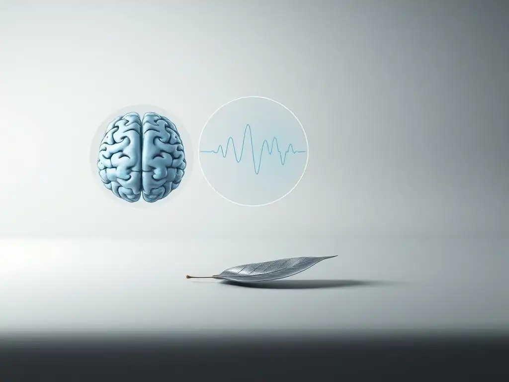
The true power in mental health imaging research strategies often lies not in a single technique, but in intelligently combining them. By integrating data from modalities that measure different aspects of brain structure, function, and chemistry, researchers can gain a far richer and more complete understanding of the biological underpinnings of mental illness.
Advantages of Multimodal Imaging in Mental Health Research
Multimodal imaging offers several key advantages over single-modality approaches. Firstly, it allows for the validation of findings across different levels of analysis. For example, if fMRI shows reduced activity in a brain region during a task, and structural MRI reveals volume loss in the same area, this provides converging evidence for dysfunction in that region. Secondly, it helps to disentangle complex relationships; combining fMRI and PET can reveal how altered neurotransmitter systems affect functional brain networks.
Furthermore, multimodal approaches enhance the potential for identifying robust biomarkers. A signature combining structural, functional, and neurochemical data points is likely to be more specific and sensitive for diagnosis or prediction than a single measure. This integrated view is essential for developing sophisticated mental health imaging research strategies that can capture the multifaceted nature of psychiatric disorders.
Enhanced Diagnostic Accuracy Through Integrated Data
Integrating data from multiple imaging modalities holds significant promise for improving the accuracy of psychiatric diagnosis, which currently relies heavily on subjective clinical assessments. For instance, combining structural MRI measures (like gray matter volume) with resting-state fMRI connectivity patterns and potentially PET measures of neurotransmitter systems could provide a more objective and comprehensive neurobiological profile of an individual.
Research is exploring how machine learning algorithms can classify patients based on these integrated multimodal datasets. Some studies have reported improved classification accuracy for differentiating between conditions like bipolar disorder and schizophrenia, or even between subtypes of depression, when using multimodal data compared to single-modality data. While still largely experimental, this approach is a key focus of advanced mental health imaging research strategies aimed at developing neuroimaging-based diagnostic aids.
Improved Understanding of Complex Brain-Behavior Relationships
Mental health conditions manifest as complex alterations in behavior, cognition, and emotion, arising from distributed dysfunction across multiple brain systems. Multimodal imaging allows researchers to link specific structural abnormalities to altered functional connectivity, and then relate these brain changes to observed symptoms or cognitive deficits. For example, combining DTI (white matter integrity) with fMRI (functional connectivity) can show how damage to communication pathways affects the synchronized activity between brain regions, which might then correlate with specific cognitive impairments seen in a disorder.
This integrated understanding is crucial for developing sophisticated models of psychopathology. It moves beyond simply identifying 'hot spots' of activity or volume change to understanding how entire brain networks are disrupted and how these disruptions give rise to the diverse symptoms experienced by individuals with mental illness. Such insights are invaluable for designing effective mental health imaging research strategies and therapeutic interventions.
Personalized Treatment Approaches Based on Individual Brain Signatures
Ultimately, one of the most exciting prospects of multimodal imaging is its potential to facilitate personalized medicine in mental health. By obtaining a detailed, multimodal neurobiological profile of an individual patient, clinicians might one day be able to predict their likely response to different types of treatment (e.g., specific medications, psychotherapy, neuromodulation techniques).
For example, a multimodal scan profile might reveal specific patterns of connectivity or neurotransmitter availability that predict a better response to a particular antidepressant or a specific type of cognitive behavioral therapy. While this is an area of active research, preliminary studies using multimodal data have shown promise in predicting treatment outcomes in conditions like depression and anxiety. Developing predictive biomarkers based on integrated brain imaging data is a major goal of future mental health imaging research strategies.
Examples of Multimodal Imaging Applications in Mental Health
Real-world applications of multimodal imaging are demonstrating its power in unraveling the complexities of mental disorders. These studies exemplify how combining techniques yields insights greater than the sum of their parts.
Combining fMRI and EEG to Study Cognitive Processes in Depression
Depression is characterized by altered emotional processing and cognitive biases. Combining fMRI and EEG allows researchers to investigate the spatial location and precise timing of brain activity during tasks related to emotional regulation or attention. For instance, fMRI might show reduced activity in prefrontal control regions and increased activity in the amygdala during emotional tasks, while simultaneous or concurrent EEG can capture the rapid sequence of neural events as the brain processes emotional stimuli, identifying deficits in early attentional capture or later cognitive reappraisal stages.
Studies using simultaneous fMRI-EEG in depressed patients have revealed specific temporal dynamics of aberrant emotional processing, such as early enhanced processing of negative stimuli followed by impaired later-stage regulation, localized to specific prefrontal-limbic circuits identified by fMRI. This multimodal approach provides a richer understanding of the dynamic interplay between brain regions and the timing of their activity, crucial for developing targeted interventions for cognitive and emotional symptoms in depression.
Integrating MRI and PET to Investigate Neuroinflammation in Schizophrenia
Schizophrenia is thought to involve neurodevelopmental issues and potentially ongoing neuroinflammatory processes. Combining structural MRI (to assess tissue volume/integrity), potentially DTI (white matter), and PET with tracers specific for neuroinflammation markers (like TSPO, which binds to activated microglia) allows researchers to investigate the relationship between structural brain changes, connectivity alterations, and inflammation in living patients.
Research integrating these modalities has provided evidence suggesting elevated neuroinflammation in certain brain regions in subgroups of patients with schizophrenia, particularly during acute phases of illness. These findings, combined with structural and functional data from the same individuals, help to build a more complete picture of the complex pathology in schizophrenia, potentially identifying inflammation as a target for novel therapies and refining mental health imaging research strategies for this disorder.
Using Multimodal Imaging to Predict Treatment Response in Anxiety Disorders
Predicting which patients with anxiety disorders will respond to specific treatments (e.g., cognitive behavioral therapy vs. medication) is a major clinical challenge. Multimodal imaging offers a promising avenue for developing predictive biomarkers. For example, combining resting-state fMRI connectivity patterns (e.g., amygdala connectivity), structural MRI measures (e.g., gray matter volume in fear-related regions), and potentially even EEG measures of threat reactivity can provide a comprehensive brain profile.
Studies have begun to explore whether specific patterns identified through such multimodal analyses can predict treatment outcomes. For instance, certain patterns of amygdala connectivity or prefrontal activity observed before treatment might be associated with a greater likelihood of responding to CBT. While more research is needed, these initial findings highlight the potential of integrated brain imaging data to personalize treatment selection in anxiety disorders, improving the efficiency and effectiveness of care.
Case Studies: Multimodal Imaging in Action
Examining specific research projects provides concrete examples of how multimodal imaging is applied to address complex questions in mental health.
Case Study 1: Multimodal Imaging to Differentiate Bipolar Disorder Subtypes
Differentiating between bipolar disorder subtypes (e.g., Bipolar I vs. Bipolar II) and from other conditions like major depressive disorder can be challenging based solely on clinical presentation. Researchers hypothesized that distinct neurobiological profiles might exist.
Methodology and Imaging Protocols
A research team recruited participants diagnosed with Bipolar I, Bipolar II, major depressive disorder, and healthy controls. Each participant underwent a battery of brain scans: a high-resolution structural MRI scan to assess gray matter volume and cortical thickness, a resting-state fMRI scan to measure functional connectivity within key mood regulation networks (like the default mode network and salience network), and a PET scan using a radiotracer targeting serotonin transporters (5-HTT), implicated in mood disorders. Data was collected using standardized protocols across all participants on the same scanners where possible to minimize variability.
The imaging data was then preprocessed and analyzed. Structural measures were extracted for regions of interest (e.g., prefrontal cortex, amygdala, hippocampus). Functional connectivity strength was calculated for relevant networks. 5-HTT binding potential was quantified in areas like the raphe nuclei and striatum. Advanced machine learning algorithms were then trained on the combined multimodal dataset to see if they could accurately classify individuals into their diagnostic groups based on their brain imaging profiles.
Key Findings and Clinical Implications
The study found that while single imaging modalities provided some differentiation, the combined multimodal dataset significantly improved classification accuracy, achieving approximately 80-85% accuracy in distinguishing between the three patient groups and controls. Specifically, the combination of reduced prefrontal gray matter volume (from MRI), altered functional connectivity within the salience network (from fMRI), and reduced 5-HTT binding in specific subcortical regions (from PET) formed a powerful signature that helped differentiate Bipolar I from Bipolar II and from major depression.
- Structural MRI contributed information about brain volume differences.
- Resting-state fMRI revealed distinct patterns of network connectivity.
- PET provided unique insights into serotonin system availability.
[ ] Discuss the implications of these findings for clinical practice.
[x] Explain how the combined data improved differentiation.
These findings suggest that distinct neurobiological signatures exist for different mood disorder diagnoses, supporting the use of multimodal mental health imaging research strategies to refine diagnostic categories. While not yet ready for clinical use, such research paves the way for future objective, imaging-assisted diagnostic tools that could help clinicians make more accurate diagnoses earlier, leading to more appropriate treatment selection for patients.
Case Study 2: Using fMRI and DTI to Understand Trauma's Impact on Brain Connectivity
Trauma exposure, particularly in early life, is a significant risk factor for developing various mental health conditions, including PTSD, depression, and anxiety. Researchers are keen to understand how trauma structurally and functionally alters brain networks.
Methodology and Imaging Protocols
In this study, researchers recruited adults with a history of childhood trauma and a diagnosis of PTSD, adults with childhood trauma but no PTSD, and healthy controls with no trauma history. Participants underwent structural MRI, Diffusion Tensor Imaging (DTI), and resting-state fMRI scans. The structural MRI was used to assess gray matter volume in regions like the hippocampus and amygdala, known to be sensitive to stress.
DTI was performed to evaluate the integrity and connectivity of major white matter tracts, particularly those connecting limbic areas (involved in emotion) and prefrontal cortex (involved in regulation). Resting-state fMRI measured functional connectivity within and between these same emotional and regulatory networks.
Data analysis involved quantifying regional gray matter volumes, calculating fractional anisotropy (FA) and mean diffusivity (MD) along key white matter tracts (measures of tract integrity), and assessing the strength of functional connections between predefined brain regions. Researchers then correlated these multimodal imaging measures with clinical assessments of trauma severity and PTSD symptom severity.
Key Findings and Clinical Implications
The study revealed a complex interplay between structural and functional alterations in the trauma-exposed groups, particularly pronounced in those with PTSD. The PTSD group showed reduced gray matter volume in the hippocampus (from structural MRI), reduced integrity (lower FA, higher MD) in white matter tracts connecting the amygdala and prefrontal cortex (from DTI), and altered functional connectivity in resting-state fMRI, specifically hyperconnectivity within the amygdala-centric fear network and hypoconnectivity between the amygdala and prefrontal regulatory regions.
- Structural MRI confirmed known volume changes in stress-sensitive regions.
- DTI provided evidence of disrupted communication pathways.
- fMRI showed the functional consequences of these structural/connectivity issues.
These findings, derived from a multimodal approach, provide strong evidence that trauma impacts both the structure of brain pathways and the functional communication between critical emotional and regulatory brain regions. The combination of DTI and fMRI was particularly powerful in showing how disrupted anatomical connections might underlie altered functional network dynamics observed in PTSD.
This integrated understanding is vital for developing targeted therapies, such as trauma-focused psychotherapies or neuromodulation techniques, aimed at restoring connectivity and function in these specific circuits. It also highlights the value of multimodal mental health imaging research strategies in elucidating the long-term neurobiological consequences of adverse experiences.
Challenges and Future Directions in Multimodal Mental Health Imaging
Despite its immense potential, multimodal imaging in mental health research faces significant challenges that need to be addressed to fully realize its promise.
Technical Challenges in Data Acquisition and Integration
Acquiring high-quality multimodal data is technically demanding. It requires participants to undergo multiple scans, potentially on different days and different scanners, which can introduce variability. Ensuring precise spatial and temporal alignment of data from different modalities (e.g., overlaying PET data onto MRI scans, or combining millisecond-resolution EEG with slower fMRI signals) requires sophisticated coregistration and fusion techniques. Artifacts (like head motion) can affect different modalities differently and require careful handling.
Integrating and analyzing the vast amounts of data generated by multiple imaging modalities presents a major computational challenge. Developing robust algorithms to combine diverse data types (structural volumes, functional time series, metabolic/receptor maps, electrical/magnetic signals) into a single, coherent analytical framework is an active area of research. Standardizing data acquisition protocols and analysis pipelines across different research sites is also crucial for enabling large-scale collaborative studies, which are essential for validating findings and developing generalizable mental health imaging research strategies.
Ethical Considerations in Using Brain Imaging Data
The increasing sophistication of brain imaging, particularly multimodal approaches, raises important ethical considerations. Brain scans produce highly sensitive personal data that could potentially reveal information about an individual's predisposition to mental illness, cognitive abilities, or even aspects of their personality. Ensuring the privacy and security of these large, complex datasets is paramount.
Furthermore, as imaging findings become more correlated with clinical conditions, there is a risk of misinterpretation or oversimplification, potentially leading to stigma or discrimination based on brain imaging profiles. Researchers and clinicians must be careful about how findings are communicated to participants and the public. Ethical guidelines for the use of neuroimaging data in research and clinical contexts are continually evolving, emphasizing informed consent, data anonymization, and responsible interpretation of results. These ethical considerations are integral to the responsible advancement of mental health imaging research strategies.
Future Trends: Artificial Intelligence and Advanced Image Analysis
The future of multimodal mental health imaging research strategies is inextricably linked with advancements in computational methods, particularly artificial intelligence (AI) and machine learning. These tools are becoming indispensable for handling the complexity and volume of multimodal data.
AI-Powered Image Analysis for Enhanced Detection
AI, especially deep learning techniques, is revolutionizing the analysis of brain images. These algorithms can automatically identify subtle patterns and features in imaging data that might be missed by traditional analysis methods, potentially improving the detection of early signs of mental illness or predicting disease progression. For example, deep learning models trained on large multimodal datasets (combining MRI, fMRI, PET) are being developed to identify complex spatial and temporal signatures associated with specific disorders.
AI can also assist in tasks like image segmentation (identifying specific brain regions), artifact removal, and data harmonization across different scanners or protocols. By automating and enhancing these steps, AI can make multimodal analysis more efficient and robust, accelerating the pace of mental health imaging research strategies.
The Role of Machine Learning in Multimodal Data Integration
Machine learning algorithms are particularly well-suited for integrating and finding meaningful patterns within diverse, high-dimensional multimodal datasets. Techniques like support vector machines, random forests, and neural networks can learn complex relationships between different types of imaging features (e.g., how structural volume in one area relates to functional connectivity in another and metabolic activity in a third) and clinical outcomes (e.g., diagnosis, symptom severity, treatment response).
Machine learning can be used for classification (e.g., diagnosing disorders), regression (e.g., predicting symptom severity), and clustering (e.g., identifying neurobiologically distinct patient subgroups). Developing sophisticated machine learning models capable of effectively leveraging the complementary information from multiple imaging modalities is a key area of focus for advancing mental health imaging research strategies towards personalized medicine and improved understanding of complex brain disorders.
Optimizing Mental Health Imaging Research Strategies for the Future
To maximize the impact of multimodal imaging on mental health care, several factors need optimization, ranging from study design to data sharing and clinical translation.
Best Practices for Multimodal Imaging Study Design
Designing effective multimodal imaging studies requires careful planning. Researchers must select modalities that are complementary and relevant to the specific research question (e.g., combining fMRI and EEG for temporal dynamics, or PET and MRI for structure-neurochemistry links). Standardized protocols for data acquisition across all modalities are essential to minimize unwanted variability. Sample sizes need to be sufficiently large to accommodate the complexity of the data and the use of advanced analytical techniques like machine learning.
Prospective designs that follow individuals over time, ideally capturing different stages of illness or treatment response, are particularly valuable for understanding the trajectory of brain changes. Integrating clinical, behavioral, and genetic data with imaging data in a multimodal framework provides an even richer dataset for analysis. Adhering to rigorous methodology and transparent reporting is crucial for the reproducibility and impact of these complex mental health imaging research strategies.
Collaboration and Data Sharing in Mental Health Research
Given the technical and computational demands of multimodal imaging and the need for large datasets to power advanced analyses, collaboration and data sharing are paramount. Individual research centers often lack the resources or patient numbers required for definitive multimodal studies. Initiatives promoting data sharing, such as the OpenfMRI, Human Connectome Project, and consortia focused on specific disorders (like the ENIGMA consortium), are critical for pooling resources and accelerating discovery.
Collaborative efforts allow researchers to harmonize data from different sites, validate findings in independent samples, and develop more robust analytical tools. Sharing data fosters transparency and allows the broader scientific community to build upon existing findings. This collaborative spirit is fundamental to advancing mental health imaging research strategies on a global scale.
The Impact of Multimodal Imaging on Mental Health Care
The ultimate goal of multimodal mental health imaging research strategies is to translate findings into improved mental health care. While still largely a research tool, multimodal imaging has the potential to impact clinical practice in several ways: by providing objective biomarkers for diagnosis and differential diagnosis, predicting treatment response to guide personalized therapy, monitoring disease progression, and potentially even identifying individuals at high risk before symptom onset.
Translating these complex findings from the lab to the clinic requires bridging the gap between researchers, clinicians, and technology developers. Developing user-friendly software for analyzing multimodal data in a clinical setting and establishing clinical guidelines for the interpretation of imaging findings are necessary steps. As the technology matures and evidence accumulates, multimodal imaging could become a routine part of comprehensive psychiatric assessment, transforming how mental health conditions are understood and treated.
Conclusion: Advancing Mental Health Understanding Through Integrated Brain Imaging
Multimodal imaging represents a significant leap forward in mental health imaging research strategies. By combining the strengths of different neuroimaging techniques, researchers can gain a more comprehensive and nuanced understanding of the complex structural, functional, and neurochemical alterations that underlie psychiatric disorders. This integrated approach provides a powerful lens through which to view the brain in both health and disease.
The Potential of Multimodal Imaging to Transform Mental Health Research
The potential of multimodal imaging to transform mental health research is vast. It moves the field beyond studying isolated brain features to investigating the dynamic interplay within and between brain networks. This holistic perspective is essential for unraveling the intricate pathology of conditions like schizophrenia, depression, anxiety disorders, and neurodevelopmental conditions.
By providing richer datasets, multimodal imaging fuels the development of advanced analytical techniques, including artificial intelligence and machine learning, which are critical for identifying subtle patterns and developing predictive models. This synergy between advanced imaging and computational power is driving the field towards the discovery of robust biomarkers for diagnosis, prognosis, and treatment prediction. Multimodal mental health imaging research strategies are poised to revolutionize our understanding of the brain basis of mental illness.
Frequently Asked Questions
What is multimodal imaging in the context of mental health research?
Multimodal imaging involves combining different types of brain scans, such as MRI and PET, to provide a more complete picture of brain structure and function. This allows researchers to gain a deeper understanding of mental health conditions.
Why is multimodal imaging considered a valuable research strategy?
It allows researchers to correlate structural abnormalities observed in MRI with functional changes detected by PET or EEG. This combined approach can reveal more nuanced relationships between brain activity and mental disorders.
How does multimodal imaging contribute to understanding specific mental health challenges?
By integrating data from various scans, researchers can identify specific neural circuits implicated in conditions like depression or schizophrenia. This can lead to more targeted treatment strategies based on individual brain characteristics.
What are some of the challenges associated with using multimodal imaging in mental health research?
The complexity of integrating data from different imaging modalities requires specialized expertise and advanced analytical techniques. Also, high costs of advanced imaging can limit the accessibility of multimodal studies.
How might mental health imaging research strategies, like multimodal imaging, impact future treatments?
By providing detailed insights into the biological underpinnings of mental illnesses, multimodal imaging can pave the way for personalized medicine approaches. This includes developing therapies that are tailored to an individual's unique brain profile.
Final Reflection: Unveiling the Mind's Landscape Through Multimodal Imaging
The convergence of advanced neuroimaging techniques offers an unprecedented opportunity to understand the complexities of mental illness, moving beyond simplistic models and towards a more nuanced, holistic view of the brain. This is not merely an academic pursuit; it holds the key to unlocking more effective diagnostic tools, personalized treatments, and ultimately, a brighter future for those struggling with mental health challenges.
The impact of multimodal imaging on our understanding of mental health is profound and far-reaching. It challenges the traditional, often reductionist, approaches to diagnosis and treatment, forcing us to consider the intricate interplay of brain regions and neurochemical systems. This shift in perspective is crucial, as it moves us away from simplistic categorizations and towards a more individualized understanding of mental illness. The brain is not a machine; it is a complex, dynamic system, and understanding its intricacies requires a multi-faceted approach.
This quote encapsulates the essence of why multimodal imaging is so vital. The integration of different imaging modalities allows researchers to build a richer, more comprehensive picture of the brain's structure and function, revealing subtle patterns and connections that would otherwise remain hidden.
Consider the potential. Imagine a future where personalized treatment plans are tailored to the unique neurobiological profile of each individual, maximizing efficacy and minimizing side effects. Imagine a future where early detection and intervention are the norm, preventing the debilitating effects of mental illness from taking hold. This future is not a fantasy; it is within our grasp.
You, the reader, have a role to play in this journey. By understanding the power of multimodal imaging and its potential to revolutionize mental health care, you can become a more informed advocate, a more engaged citizen, and a powerful force for positive change. Let us work together to translate the promise of this technology into tangible improvements in the lives of those who need it most.
Unlock Deeper Insights: Take the Next Step in Mental Health Understanding
Thanks for exploring the fascinating world of multimodal imaging with us! You've now gained valuable insights into how combining different scan types can revolutionize mental health research and diagnosis. Ready to delve further?
- Dive deeper into a specific modality: Choose one imaging technique discussed (fMRI, EEG, PET, etc.) and research its applications in more detail. This will help you understand the nuances of each method and its unique contribution to the field.
- Share this knowledge: Help spread awareness about the advancements in multimodal imaging by sharing this article with colleagues, friends, or family who might find it insightful. Let's ignite a conversation about mental health innovation!
- Consider the future: Think about how multimodal imaging might impact your own life or the lives of those you know. What breakthroughs do you anticipate in the coming years?
What are your thoughts on the future of mental health diagnosis with multimodal imaging techniques? Share your predictions below!