
Brain Imaging: A New Frontier in Mental Health Diagnosis and Understanding

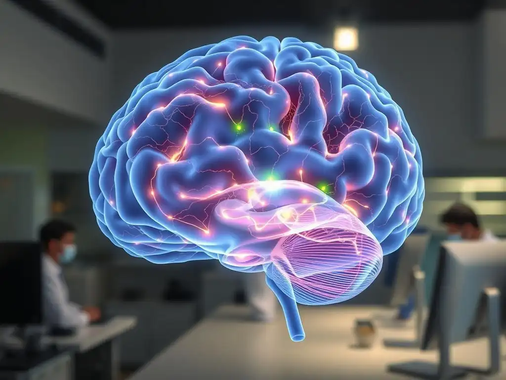
Welcome to Mind Care Guide, a sanctuary for your mental well-being. Ever wondered how we can truly see inside the mind, especially when facing mental health challenges? This is the promise of brain imaging in mental health diagnosis. Join us as we explore this fascinating frontier, revealing how cutting-edge technology is transforming our understanding and treatment of mental illness. What secrets lie hidden within these images, and how can they unlock a brighter future for mental healthcare? Let's delve in and uncover the possibilities...
Brain Imaging: Revolutionizing Mental Health Diagnosis and Treatment
- Introduction to Brain Imaging in Mental Health
- Understanding Brain Imaging Techniques
- The Role of Brain Imaging in Diagnosing Mental Health Disorders
- Brain Imaging for Understanding Mental Health Conditions
- Benefits of Brain Imaging in Mental Healthcare
- Ethical Considerations and Challenges in Neuroimaging
- Conclusion: The Future of Neuroimaging in Mental Health
-
Frequently Asked Questions
- What role does brain imaging play in mental health diagnosis?
- How accurate is brain imaging for diagnosing mental health disorders?
- What are the different types of brain imaging used in mental health?
- What are the potential benefits of using brain imaging in mental health treatment?
- Are there any risks or limitations associated with using brain imaging for mental health diagnosis?
- Final Reflection: Peering into the Mind: The Future of Mental Health Diagnosis
- Unlock Your Brain's Potential: Take the Next Step in Mental Wellness
Introduction to Brain Imaging in Mental Health
The Increasing Role of Neuroimaging in Mental Healthcare
Neuroimaging techniques have rapidly transitioned from purely research tools to playing an increasingly significant role in the complex landscape of mental healthcare. While not yet a standalone diagnostic test for most conditions, methods like MRI, fMRI, and PET scans are providing unprecedented insights into the structural, functional, and molecular underpinnings of the brain in individuals experiencing mental health challenges. This evolution is driven by decades of research demonstrating that many mental health conditions are associated with observable differences in brain structure, activity, and connectivity, offering a biological dimension to understanding these complex disorders.
The application of brain imaging for mental health diagnosis and understanding is helping to move the field towards a more biologically informed approach. For example, research using fMRI has identified differences in amygdala activity in individuals with anxiety disorders compared to healthy controls, often showing heightened responses to fearful stimuli.
Similarly, structural MRI studies frequently report volume differences in specific brain regions, such as the prefrontal cortex or hippocampus, in conditions like depression or schizophrenia. These findings, while often group-level averages and not yet definitive for individual diagnosis in most cases, are crucial for building a comprehensive picture of mental illness and exploring potential biomarkers.
Mind Care Guide's Perspective on Brain Imaging for Mental Wellness
At Mind Care Guide, we view brain imaging as a powerful, albeit evolving, tool in the pursuit of mental wellness. Our perspective is rooted in the understanding that mental health is intrinsically linked to brain health. We believe that neuroimaging techniques offer valuable avenues for research, enhancing our understanding of various conditions, and potentially paving the way for more precise and personalized interventions in the future. While acknowledging that brain imaging for mental health diagnosis is not a simple pass/fail test today, its potential to illuminate the biological underpinnings of distress is immense.
We advocate for the responsible and ethical use of brain imaging within mental healthcare contexts. This involves educating individuals about what these scans can and cannot reveal, ensuring data privacy, and interpreting findings within the broader context of clinical evaluation, personal history, and individual experiences. Mind Care Guide sees brain imaging as a complementary piece of the puzzle, contributing to a holistic understanding of mental health alongside psychotherapy, medication, lifestyle changes, and community support, ultimately aiming to empower individuals on their journey to well-being.
Understanding Brain Imaging Techniques
An Overview of Key Neuroimaging Modalities
A variety of techniques fall under the umbrella of brain imaging, each offering unique insights into different aspects of brain structure and function. Understanding these modalities is key to appreciating how brain imaging supports mental health diagnosis research and treatment development.
Magnetic Resonance Imaging (MRI) for Structural Analysis in Mental Health
Structural MRI provides detailed images of the brain's anatomy. It uses strong magnetic fields and radio waves to create images that differentiate between various tissues, such as gray matter, white matter, and cerebrospinal fluid. In mental health research, structural MRI is used to measure the volume and shape of specific brain regions.
Studies have shown, for example, that the average volume of the hippocampus may be reduced in individuals with chronic depression or PTSD compared to healthy controls. Analyzing cortical thickness, a measure of the outer layer of the brain, can also reveal differences associated with conditions like schizophrenia or bipolar disorder.
Functional Magnetic Resonance Imaging (fMRI) for Assessing Brain Activity
Functional MRI measures brain activity by detecting changes in blood flow. When a brain region is more active, it requires more oxygenated blood. fMRI detects the subtle magnetic differences between oxygenated and deoxygenated blood, providing a map of brain activity over time, often while a person performs a specific task or is in a resting state. This technique is invaluable for studying how different brain areas communicate (connectivity) and how specific circuits respond to emotional stimuli or cognitive challenges, offering insights into the functional disruptions seen in disorders like anxiety, depression, and OCD.
Positron Emission Tomography (PET) for Neurotransmitter Function
PET scanning uses small amounts of radioactive tracers that are injected into the bloodstream. These tracers bind to specific molecules in the brain, such as neurotransmitter receptors or metabolic markers like glucose. As the tracer decays, it emits positrons that are detected by the scanner, creating images that reflect the distribution and concentration of the target molecule. PET is particularly useful for studying neurotransmitter systems, like dopamine or serotonin pathways, which are implicated in conditions such as addiction, depression, and Parkinson's disease, providing a molecular window into brain function.
Electroencephalography (EEG) for Real-Time Brainwave Monitoring
EEG measures electrical activity in the brain using electrodes placed on the scalp. It detects the collective electrical signals from large groups of neurons firing simultaneously. EEG provides excellent temporal resolution, meaning it can capture brain activity changes in milliseconds, making it ideal for studying brain states (like sleep or alertness) and rapid responses to stimuli. While it offers less spatial resolution than MRI or PET, EEG is valuable for identifying patterns of brain activity associated with conditions like epilepsy, and research is exploring its potential for identifying markers related to attention deficits or certain mood disorders.
Diffusion Tensor Imaging (DTI) for Examining White Matter Tracts
DTI is a specialized MRI technique that measures the diffusion of water molecules in the brain. Since water diffuses more freely along the direction of nerve fibers (white matter tracts) than across them, DTI can be used to map the brain's white matter pathways and assess their integrity and connectivity. Disruptions in these communication lines between different brain regions are thought to play a role in various mental health conditions, including schizophrenia, autism spectrum disorder, and traumatic brain injury. DTI helps researchers understand how different parts of the brain are structurally connected.
How Brain Imaging Works: Basic Principles
Despite their different applications, most brain imaging techniques rely on detecting physical or chemical properties within the brain using external sensors. MRI and fMRI, for instance, leverage powerful magnetic fields and radiofrequency pulses to align and then manipulate the magnetic properties of water molecules in the body. The signals emitted as these molecules relax back to their original state are detected by the scanner and processed into detailed images. Structural MRI focuses on differences in tissue composition, while fMRI detects changes in blood oxygenation levels, which serve as a proxy for neural activity.
PET scanning, on the other hand, works by detecting gamma rays emitted from the radioactive tracer as it decays. The scanner maps where these emissions originate in the brain, showing the concentration of the tracer and, by extension, the activity or density of the molecules the tracer binds to.
EEG directly measures the electrical potentials generated by neuronal activity through electrodes on the scalp. While the underlying physics and technology vary significantly between modalities, the common goal is to translate complex biological signals within the brain into visual representations that can be analyzed to understand its structure, function, or chemical environment.
The Role of Brain Imaging in Diagnosing Mental Health Disorders
Advances in Diagnostic Accuracy Using Neuroimaging
The integration of brain imaging into mental health diagnosis is a significant area of research, aiming to improve the accuracy and objectivity of diagnosis, which traditionally relies heavily on self-report and clinical observation. While not yet definitive for individual cases across the board, neuroimaging is revealing specific patterns associated with various conditions, contributing to a more nuanced understanding and potentially aiding future diagnostic methods. The goal is not to replace clinical expertise but to provide complementary biological data.
Current research using brain imaging for mental health diagnosis focuses on identifying "neural signatures" – consistent structural, functional, or chemical patterns that differentiate between diagnostic groups or predict treatment response. For example, studies might look for specific patterns of reduced activity in certain brain regions during cognitive tasks or altered connectivity between brain networks. These findings, when robust and reproducible across different studies and populations, hold promise as potential biomarkers that could one day supplement clinical criteria, helping to refine diagnoses and personalize treatment approaches.
Identifying Neural Signatures of Major Depressive Disorder (MDD)
Neuroimaging studies in MDD frequently report abnormalities in regions involved in emotion regulation, reward processing, and cognitive control. Structural MRI research has often shown reduced volume in areas like the hippocampus and prefrontal cortex in individuals with MDD compared to healthy controls. Functional studies using fMRI commonly reveal altered activity and connectivity within circuits like the default mode network (DMN), salience network, and central executive network. For instance, increased connectivity within the DMN and reduced connectivity between the DMN and executive control networks have been observed in depressed individuals, potentially reflecting excessive self-focus and impaired cognitive control, respectively.
Brain Imaging in the Diagnosis of Generalized Anxiety Disorder (GAD)
In Generalized Anxiety Disorder, brain imaging research points towards dysregulation in circuits involved in processing fear and anxiety. fMRI studies often show heightened reactivity in the amygdala, a key region for processing threats, when individuals with GAD are exposed to emotional stimuli. Furthermore, altered connectivity between the amygdala and prefrontal regions, which are involved in regulating emotions, is frequently observed. This suggests a potential biological basis for the difficulty individuals with GAD experience in controlling their anxious thoughts and feelings, contributing to the ongoing exploration of brain imaging for mental health diagnosis in anxiety disorders.
Schizophrenia: Identifying Structural and Functional Abnormalities
Schizophrenia is one of the conditions where structural brain differences have been consistently reported in neuroimaging studies. These often include enlarged ventricles (fluid-filled spaces in the brain) and reduced gray matter volume in various regions, particularly in the temporal lobes, prefrontal cortex, and hippocampus, compared to healthy individuals. Functional imaging reveals disrupted connectivity between different brain regions and altered activity patterns during cognitive tasks, such as those involving working memory or executive function. PET studies have also shown abnormalities in dopamine receptor binding, which is a key focus in understanding the neurochemical basis of the disorder and developing treatments.
Autism Spectrum Disorder (ASD) and Connectivity Differences
Brain imaging in Autism Spectrum Disorder has largely focused on identifying differences in brain connectivity and development. While structural findings can be variable, many studies using fMRI and DTI highlight atypical patterns of neural connectivity.
This includes reports of both increased connectivity within certain localized brain regions and reduced long-range connectivity between different brain networks, particularly those involved in social processing and communication. These findings are helping researchers understand the diverse ways brain development might lead to the characteristic social and communication challenges experienced by individuals with ASD, enhancing the potential of brain imaging for mental health diagnosis research in this area.
ADHD Diagnosis: Insights into Frontal-Subcortical Circuits
Neuroimaging research in Attention-Deficit/Hyperactivity Disorder (ADHD) has primarily focused on the frontal-subcortical circuits, which play a critical role in attention, impulse control, and executive function. Structural MRI studies have sometimes shown slightly reduced volume in certain frontal lobe areas, the basal ganglia, and the cerebellum on average in groups of individuals with ADHD compared to controls.
Functional studies often reveal altered activity and connectivity within these circuits, particularly during tasks requiring sustained attention or inhibition. These findings support the understanding of ADHD as a disorder involving dysregulation in specific brain networks, contributing to the ongoing effort to use brain imaging for mental health diagnosis refinement.
Limitations and Challenges in Applying Brain Imaging for Diagnosis
Despite the exciting advances, applying brain imaging for mental health diagnosis on an individual level faces significant limitations. Most findings from neuroimaging studies are based on group averages, meaning that while differences may be statistically significant between a group of individuals with a disorder and a group of healthy controls, there is often substantial overlap between the groups. An individual's scan may not show the "typical" pattern for their diagnosis, or may show patterns associated with multiple conditions, making definitive diagnosis based solely on imaging unreliable for most disorders today.
Furthermore, brain structure and function are highly variable across individuals due to genetics, environment, experience, and even transient states like mood or fatigue. Factors such as medication use, age, gender, and the presence of co-occurring conditions can also influence scan results, complicating interpretation.
The sensitivity and specificity of current brain imaging techniques are generally not high enough to serve as standalone diagnostic tests for the majority of mental health conditions. Therefore, while brain imaging is a powerful research tool and holds promise for future diagnosis, it must currently be interpreted cautiously and within the context of a comprehensive clinical evaluation.
Brain Imaging for Understanding Mental Health Conditions
Research Applications: Unveiling the Neural Mechanisms
Beyond direct diagnosis, brain imaging is an indispensable tool for fundamental research aimed at understanding the underlying neural mechanisms of mental health conditions. By visualizing brain activity and structure, researchers can test hypotheses about how specific brain circuits contribute to symptoms, how disorders develop over time, and how different treatments exert their effects. This research is crucial for developing new and more effective interventions.
Neuroimaging allows scientists to observe the living brain in action, providing insights that were previously only possible through post-mortem studies or animal models. This has led to a deeper appreciation of the complexity of brain networks and how disruptions in connectivity or activity within these networks can manifest as diverse symptoms. For instance, studies have used fMRI to investigate the neural basis of hallucinations in schizophrenia or the altered reward processing observed in addiction, providing concrete biological targets for therapeutic development.
Exploring the Neurobiology of Trauma and Post-Traumatic Stress Disorder (PTSD)
Neuroimaging studies have greatly advanced our understanding of how trauma affects the brain and contributes to PTSD. Research frequently reports structural changes, such as reduced volume in the hippocampus and altered connectivity in fear-related circuits involving the amygdala and prefrontal cortex. fMRI studies often show exaggerated amygdala responses to trauma-related cues and diminished activity in prefrontal regions involved in emotional regulation when individuals with PTSD are processing traumatic memories. These findings provide biological correlates for symptoms like hyperarousal, avoidance, and intrusive memories, informing trauma-focused therapies.
Investigating the Neural Correlates of Effective Psychotherapy
Brain imaging is being used to explore how psychological treatments, such as cognitive behavioral therapy (CBT) or psychotherapy, change brain function. Studies have used fMRI to scan individuals before and after a course of therapy for conditions like depression or anxiety. Findings have shown that successful treatment is often associated with measurable changes in brain activity and connectivity, such as increased regulation of limbic regions by the prefrontal cortex or normalization of activity in reward pathways. This research provides biological evidence for the effectiveness of talk therapies and helps identify the neural mechanisms through which they work.
Understanding the Neural Pathways Involved in Addiction
PET and fMRI imaging have been pivotal in mapping the brain's reward system and understanding how it is hijacked by addictive substances and behaviors. PET studies using tracers for dopamine receptors have shown alterations in dopamine signaling in individuals with addiction, including reduced receptor availability in certain brain areas. fMRI studies reveal changes in connectivity and activity in circuits involving the prefrontal cortex, striatum, and other reward-related regions, demonstrating impaired impulse control and heightened responses to drug cues. This neurobiological understanding is critical for developing treatments that target these specific pathways.
The Effects of Pharmacological Interventions on Brain Function
Neuroimaging is widely used in psychopharmacology research to understand how psychiatric medications affect brain activity and chemistry. PET scans can measure how drugs bind to specific neurotransmitter receptors, confirming their mechanism of action in the living human brain. fMRI studies can assess changes in brain activity and connectivity patterns after medication administration, providing insights into how drugs like antidepressants or antipsychotics normalize or alter brain function. This research helps optimize dosages, predict treatment response, and develop new drug targets based on specific neurobiological pathways implicated in mental illness.
Future Directions in Neuroimaging Research
The future of neuroimaging in mental health research is incredibly promising, driven by advancements in technology, data analysis techniques, and computational power. There is a strong push towards integrating data from multiple imaging modalities (e.g., combining fMRI and EEG) to get a more complete picture of brain dynamics. The development of more sensitive scanners and novel tracers for PET will allow researchers to study a wider range of neurochemical systems and processes with greater precision.
Furthermore, researchers are increasingly using machine learning and artificial intelligence to analyze complex imaging datasets, aiming to identify subtle patterns that are not apparent through traditional statistical methods. This could potentially lead to the discovery of more reliable biomarkers for diagnosis, prognosis, and treatment prediction. Large-scale data sharing initiatives are also facilitating collaborative research, allowing scientists worldwide to analyze massive datasets and validate findings across diverse populations, accelerating the pace of discovery in understanding the biological basis of mental health conditions.
Benefits of Brain Imaging in Mental Healthcare
Enhancing Diagnostic Precision and Personalizing Treatment
While not yet a definitive diagnostic tool for most conditions, brain imaging holds significant potential to enhance diagnostic precision in the future by providing objective biological data that complements clinical assessment. As neural signatures become more clearly defined and detectable at the individual level, brain imaging for mental health diagnosis could help differentiate between conditions with overlapping symptoms or identify subtypes within a broad diagnosis, such as depression with melancholic features versus atypical depression.
This increased precision is foundational for moving towards personalized mental healthcare. Understanding the specific neural correlates of an individual's symptoms could allow clinicians to tailor treatment interventions more effectively. For instance, imaging might help determine whether a person is more likely to respond to a particular type of therapy or medication based on their unique brain profile, moving away from a trial-and-error approach that can delay recovery and cause distress.
Precision Medicine: Tailoring Interventions Based on Neuroimaging Profiles
The concept of precision medicine in mental health envisions using biological data, including neuroimaging, to guide treatment selection for individual patients. Research is exploring whether specific patterns observed in brain scans can predict how well someone will respond to different antidepressants, types of psychotherapy, or neuromodulation techniques like transcranial magnetic stimulation (TMS). For example, some studies suggest that connectivity patterns in certain prefrontal circuits might predict response to CBT for depression, while activity in other regions might predict response to antidepressant medication.
Predicting Treatment Outcomes Using Neuroimaging Biomarkers
Identifying neuroimaging biomarkers that predict treatment outcomes is a major focus. These biomarkers could be structural features, functional connectivity patterns, or levels of specific neurotransmitter receptors. The goal is to use baseline brain scans to forecast whether a patient is likely to benefit from a particular intervention. While still largely experimental, early findings are promising. For instance, certain resting-state fMRI connectivity patterns have been associated with remission rates in depression following specific treatments. Successful development of such biomarkers would allow clinicians to select the most effective treatment from the outset, improving patient outcomes and reducing healthcare costs.
Facilitating Early Identification and Intervention
Brain imaging research is also contributing to the potential for earlier identification of individuals at risk for developing certain mental health conditions. By studying populations at high genetic or environmental risk (e.g., individuals with a family history of schizophrenia or those exposed to significant early-life trauma), researchers are using imaging to identify subtle brain differences that may predate the onset of full-blown symptoms.
Identifying these early markers could open windows for preventative interventions or early-stage treatments that might be more effective than those administered later in the course of illness. While this research is complex and must be handled with extreme ethical care to avoid stigmatization, it holds the promise of shifting mental healthcare towards prevention and early intervention, potentially mitigating the long-term impact of serious mental health conditions.
Ethical Considerations and Challenges in Neuroimaging
Data Privacy, Security, and Informed Consent Procedures
The increasing use of brain imaging in research and potentially clinical settings raises significant ethical concerns regarding data privacy and security. Brain scan data is highly sensitive and potentially identifiable, containing information about an individual's brain structure and function. Ensuring that this data is collected, stored, and shared securely is paramount to protecting patient confidentiality and preventing misuse. Robust protocols for anonymization and data protection are essential.
Furthermore, obtaining truly informed consent for brain imaging studies or procedures is critical. Participants need to fully understand what the scan involves, how their data will be used, who will have access to it, and the potential implications of the findings, especially given the evolving and sometimes uncertain clinical relevance of imaging results. This requires clear communication and a commitment to transparency, ensuring individuals are empowered to make informed decisions about participating in brain imaging research or clinical applications.
Avoiding Over-Interpretation of Neuroimaging Findings
A significant challenge in neuroimaging is the risk of over-interpreting findings, both by researchers and the public. Brain imaging results are often complex, statistical in nature, and based on group averages. Presenting these findings as definitive individual-level diagnoses or as simple explanations for complex behaviors can be misleading and potentially harmful. For example, showing a brain scan with highlighted areas of activity can be compelling but doesn't fully capture the dynamic and networked nature of brain function or the influence of non-biological factors on mental health.
It is crucial for researchers, clinicians, and science communicators to exercise caution and humility when discussing brain imaging results. Emphasizing the correlational nature of many findings, the variability between individuals, and the current limitations of brain imaging for mental health diagnosis is essential. Avoiding deterministic language that suggests brain scans dictate a person's destiny or fully explain their experience is vital to prevent stigma and ensure responsible communication.
Mitigating Potential Biases in Neuroimaging Studies
Like all scientific research, neuroimaging studies can be susceptible to various forms of bias that can affect the interpretation of brain imaging for mental health diagnosis and understanding. These biases can include selection bias (e.g., recruiting only certain demographics), publication bias (e.g., preferentially publishing studies with statistically significant findings), and analytical bias (e.g., using specific data processing pipelines that might favor certain outcomes).
Researchers must employ rigorous methodologies, transparent reporting practices, and preregistration of studies to mitigate these biases. Efforts to increase sample diversity and replicability of findings across different research groups are also crucial for building a robust and reliable body of knowledge in neuroimaging. Acknowledging the potential for bias and actively working to minimize it is fundamental to advancing the ethical and accurate application of brain imaging in mental health.
Conclusion: The Future of Neuroimaging in Mental Health
The Potential of Neuroimaging to Transform Mental Healthcare
Brain imaging stands at the forefront of efforts to understand the biological basis of mental health conditions, holding immense potential to transform mental healthcare. While it is not yet a primary tool for mental health diagnosis in most clinical settings, its role in research is unparalleled. It provides a window into the living brain, allowing scientists to map the neural circuits involved in emotions, cognition, and behavior, and to identify how these circuits are altered in various disorders.
As technology continues to advance and our understanding deepens, brain imaging for mental health diagnosis may become more precise and clinically applicable at the individual level. This could lead to more accurate and timely diagnoses, personalized treatment plans based on an individual's unique neurobiological profile, and the development of entirely new therapies targeting specific brain pathways. The journey is complex and requires careful navigation of ethical considerations, but the promise of improved outcomes for individuals facing mental health challenges is a powerful driver.
Mind Care Guide's Commitment to Ethical and Responsible Neuroimaging
Mind Care Guide is committed to following and promoting the ethical and responsible integration of brain imaging into mental health discussions and potential future care. We believe in empowering individuals with accurate, evidence-based information about what neuroimaging can reveal, while also being transparent about its current limitations and the importance of clinical context. Our focus remains on holistic well-being, where biological insights from imaging are viewed as one piece of a larger puzzle that includes psychological, social, and environmental factors.
We are dedicated to providing resources and information that help demystify complex topics like brain imaging for mental health diagnosis, ensuring that our community feels informed and supported. As the field evolves, we will continue to advocate for patient privacy, informed consent, and the avoidance of reductionist or deterministic interpretations of neuroimaging findings, prioritizing the individual's experience and dignity above all else.
Continued Innovation and Refinement of Neuroimaging Techniques
The field of neuroimaging is characterized by continuous innovation and refinement. New scanning technologies are being developed that offer higher resolution, faster acquisition times, and greater sensitivity to specific biological processes. Advanced computational methods, including machine learning and artificial intelligence, are revolutionizing how complex imaging data is analyzed, enabling the identification of subtle patterns that were previously undetectable.
This ongoing progress means that the capabilities of brain imaging for mental health diagnosis and understanding are constantly expanding. Researchers are developing new paradigms to probe specific aspects of brain function relevant to mental illness, such as social cognition or reward sensitivity. As these techniques become more sophisticated and the findings more robust and replicable, the potential for neuroimaging to contribute meaningfully to both clinical practice and our fundamental understanding of the human mind will only continue to grow.
Frequently Asked Questions
What role does brain imaging play in mental health diagnosis?
Brain imaging techniques like MRI and PET scans can help visualize brain structure and function, potentially revealing abnormalities associated with mental health conditions. This can complement traditional diagnostic methods and offer insights into the underlying biological basis of mental illnesses.
How accurate is brain imaging for diagnosing mental health disorders?
While promising, brain imaging is not a standalone diagnostic tool. It's used in conjunction with clinical evaluations and patient history. Current accuracy varies depending on the specific disorder and imaging technique, requiring further research and refinement.
What are the different types of brain imaging used in mental health?
Common techniques include Magnetic Resonance Imaging (MRI) to visualize brain structure, Positron Emission Tomography (PET) to measure brain activity, and functional MRI (fMRI) to observe brain activity during specific tasks. Each provides unique information about the brain's health and function.
What are the potential benefits of using brain imaging in mental health treatment?
Brain imaging can help personalize treatment by identifying specific brain regions affected by a disorder. This could lead to more targeted therapies and improved outcomes, moving away from a one-size-fits-all approach.
Are there any risks or limitations associated with using brain imaging for mental health diagnosis?
Brain imaging can be expensive and time-consuming. There are also concerns about over-interpretation of results and potential for false positives or negatives. Some techniques, like PET scans, involve exposure to low levels of radiation.
Final Reflection: Peering into the Mind: The Future of Mental Health Diagnosis
The convergence of neuroscience and mental healthcare, spearheaded by advancements in brain imaging, is revolutionizing our understanding and treatment of mental illness. No longer are we solely reliant on subjective self-reporting; we are entering an era where objective biological markers can illuminate the complex neural pathways underlying conditions like depression, anxiety, and schizophrenia. This shift promises a future where diagnosis is more accurate, treatment is more personalized, and the stigma surrounding mental illness is significantly reduced.
The impact of brain imaging extends far beyond the clinical setting. It is reshaping our very understanding of consciousness, emotion, and the human condition. By visualizing the brain in action, we gain invaluable insights into the intricate interplay of neural networks and their relationship to behavior and experience. This understanding is crucial for fostering empathy and challenging the pervasive misconceptions surrounding mental health. The brain is the most complex organ in the known universe, and understanding its intricacies is key to unlocking the mysteries of the mind.
This quote underscores the ongoing journey of discovery and the immense potential of brain imaging to guide us.
Consider the implications of this research for your own life. Perhaps you know someone struggling with a mental health condition. Perhaps you yourself have experienced the challenges of navigating the complexities of mental wellbeing.
This article is not just a collection of scientific findings; it's an invitation to engage with a field that is rapidly transforming our ability to understand and address the mental health needs of individuals and communities. Let us embrace this new frontier, advocate for increased research funding, and work towards a future where brain imaging plays a pivotal role in creating a more compassionate and informed approach to mental healthcare.
Unlock Your Brain's Potential: Take the Next Step in Mental Wellness
Thanks for diving into the fascinating world of brain imaging and its role in mental health! We hope you gained a clearer understanding of how this technology is revolutionizing diagnosis and treatment. Now, it's time to put that knowledge into action.
- Explore brain imaging resources: Dive deeper into the advancements discussed by researching reputable organizations and institutions specializing in neuroimaging. This will help you stay informed about the latest breakthroughs.
- Share your insights: Start a conversation! Discuss this groundbreaking technology with friends, family, or online communities. Sharing knowledge empowers others and sparks crucial dialogue.
- Consider your own mental health journey: Reflect on how improved diagnostic tools like brain imaging could benefit you or someone you know. This is a powerful way to advocate for better mental healthcare.
What's one specific area of brain imaging technology you find most intriguing, and why?