
MEG: Pinpointing Brain Activity with Magnetic Precision in Mental Health

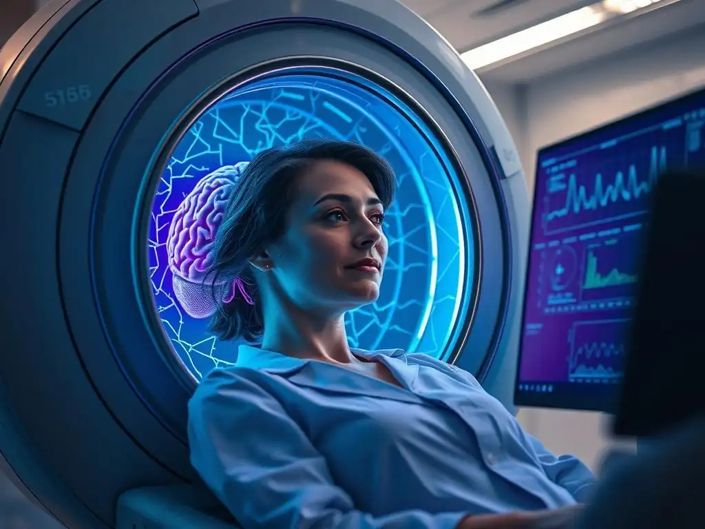
Welcome to Mind Care Guide, your trusted companion on the journey to mental well-being.
Ever wonder how scientists are unlocking the secrets of the brain to revolutionize mental health treatment? Our latest article delves into the fascinating world of MEG brain activity therapy, a cutting-edge technique offering unprecedented precision in understanding and addressing mental health challenges. Could this be the key to unlocking a brighter future for those struggling with mental health conditions? Explore the potential and the science behind it – your journey to understanding starts here.
MEG: Pinpointing Brain Activity with Magnetic Precision in Mental Health
- Introduction to Magnetoencephalography (MEG) in Mental Health
- How MEG Works: A Detailed Look at Magnetic Brain Imaging
- MEG Applications in Mental Health
- Advantages of MEG Over Other Brain Imaging Techniques
- Conditions Where MEG is Particularly Useful
- The Future of MEG in Mental Health Research and Treatment
- Limitations of MEG Technology
- Conclusion: MEG as a Powerful Tool for Understanding and Treating Mental Illness
-
Frequently Asked Questions
- What is MEG brain activity therapy and how does it relate to mental well-being?
- How does MEG differ from other brain imaging techniques like fMRI or EEG?
- What are some mental health challenges where MEG brain activity therapy might be helpful?
- How is a MEG brain activity scan performed, and is it safe?
- Where can I find more information and resources about MEG brain activity therapy for mental health?
- Final Reflection: MEG's Emerging Role in the Mental Health Revolution
- Unlock Your Brain's Potential: Take Charge of Your Mental Wellbeing
Introduction to Magnetoencephalography (MEG) in Mental Health
Understanding the Fundamentals of MEG Technology
Magnetoencephalography (MEG) is a non-invasive neuroimaging technique that measures the magnetic fields produced by electrical activity in the brain. Unlike electroencephalography (EEG), which measures electrical potentials on the scalp, MEG directly detects the tiny magnetic fields generated by neuronal currents, primarily those flowing tangentially to the scalp in the sulci.
These magnetic fields are extremely weak, typically ranging from a few femtoteslas (fT) to picoteslas (pT), which is billions of times weaker than the Earth's magnetic field. To capture these subtle signals, MEG systems require highly sensitive detectors, often Superconducting Quantum Interference Devices (SQUIDs), and must operate within a heavily magnetically shielded room to minimize environmental interference.
The core principle relies on the fact that electrical currents in neurons generate magnetic fields according to the laws of physics. Specifically, the synchronized activity of large populations of neurons, particularly pyramidal cells in the cerebral cortex, creates a net current dipole strong enough to produce detectable magnetic fields outside the head. MEG is particularly sensitive to currents flowing parallel to the scalp surface. Modern MEG systems typically employ hundreds of sensors distributed across the scalp, allowing for comprehensive coverage of the brain's magnetic activity and providing a detailed picture of where and when neural events occur.
The Role of MEG in Mapping Brain Activity
MEG excels at capturing the rapid fluctuations in brain activity that occur on a millisecond timescale. This high temporal resolution is crucial for understanding the dynamic nature of brain function and dysfunction in mental health conditions.
By precisely measuring the timing and location of magnetic signals, MEG allows researchers and clinicians to create detailed maps of brain activity, identifying which areas are active during specific cognitive processes, emotional states, or pathological events like seizures. This mapping capability extends to understanding functional connectivity – how different brain regions communicate with each other – which is often disrupted in various mental illnesses.
Beyond simply showing where activity happens, MEG provides insights into the type of activity by analyzing neural oscillations across different frequency bands (e.g., theta, alpha, beta, gamma). Alterations in these oscillatory patterns and their synchronization across brain regions are increasingly recognized as key features of conditions like schizophrenia, depression, and autism spectrum disorder. Analyzing these patterns with MEG can help pinpoint specific brain circuits involved, offering a deeper understanding of the neural underpinnings of these complex conditions. This detailed brain activity mapping is foundational for advancing research and potentially informing approaches like MEG brain activity therapy planning.
Why MEG is Crucial for Mental Health Research
MEG's unique combination of excellent temporal resolution and relatively good spatial resolution makes it an indispensable tool in mental health research. Many mental health conditions are characterized by subtle, rapid disruptions in neural timing and communication that other imaging modalities might miss. For instance, understanding the millisecond-by-millisecond processing of sensory information or the rapid shifts in attention is critical, and MEG is ideally suited for this. It allows researchers to observe the brain in action as it processes information, reacts to stimuli, or engages in cognitive tasks.
Furthermore, MEG provides a direct measure of neuronal activity, unlike techniques like fMRI which measure metabolic correlates (blood flow). This directness is valuable for understanding the fundamental electrical processes gone awry in mental disorders. By providing precise data on the location and timing of abnormal brain activity, MEG facilitates the identification of potential biomarkers for diagnosis, prognosis, and treatment response. This precision is vital for developing more targeted interventions and moving towards personalized mental healthcare, including exploring how MEG brain activity insights can inform therapeutic strategies.
How MEG Works: A Detailed Look at Magnetic Brain Imaging
The Principles of Magnetoencephalography: Detecting Magnetic Fields
The fundamental principle behind MEG is rooted in electromagnetism: any electrical current generates a magnetic field around it. In the brain, the primary source of these magnetic fields detectable outside the head is the flow of ionic currents within neurons, particularly the intracellular currents in the dendrites of pyramidal cells aligned perpendicularly to the cortical surface.
These currents produce weak magnetic fields that radiate outwards through the skull and scalp. Because magnetic fields are not distorted by tissues like the skull in the same way electrical fields are (which is a challenge for EEG), MEG offers better spatial localization of the source of neural activity compared to EEG.
To detect these minuscule magnetic fields, MEG systems employ highly sensitive magnetometers. The most common type are Superconducting Quantum Interference Devices (SQUIDs), which operate at extremely low temperatures (near absolute zero) maintained by liquid helium.
SQUIDs are exquisitely sensitive to magnetic flux changes and can detect fields as small as a few femtoteslas – a scale necessary to pick up brain signals amidst environmental noise. The array of SQUID sensors strategically placed around the head captures the spatial pattern of the magnetic field, which can then be used with sophisticated algorithms to estimate the location and strength of the underlying neural sources within the brain.
MEG Scanning Procedure: What to Expect
Undergoing an MEG scan is a non-invasive and generally comfortable procedure. The patient is typically asked to lie still on a bed or sit in a chair within a specially constructed magnetically shielded room. This room is essential to block out external magnetic noise from sources like electrical wiring, elevators, or nearby traffic, which would otherwise overwhelm the faint brain signals. Before entering the room, any metal objects (jewelry, belts, coins) must be removed as they can interfere with the magnetic field measurements.
Once positioned, the patient's head is placed within or under the helmet-shaped MEG sensor array. The sensors do not touch the head. The procedure itself usually involves minimal discomfort, primarily the need to remain still. Depending on the purpose of the scan, the patient may be asked to perform specific tasks (e.g., listening to sounds, looking at images, performing cognitive tests) or simply rest quietly. Data acquisition typically lasts between 30 minutes and an hour. The process is entirely passive for the patient, with no magnetic fields or currents applied to the brain.
Analyzing MEG Data: Interpreting Brain Activity Patterns
Analyzing MEG data is a complex process that requires specialized software and expertise. The raw data consists of continuous recordings from hundreds of sensors over time. The first step often involves filtering out noise and artifacts (like eye blinks or heartbeats). The core challenge is the "inverse problem": determining the location and strength of neural sources inside the brain from the magnetic fields measured outside the head. This problem does not have a unique solution, so advanced mathematical algorithms and models (like dipole fitting, minimum-norm estimates, or beamformers) are used to estimate the most probable sources.
Once the neural sources are localized, the data can be analyzed in various ways to understand brain function. This includes examining the power of oscillations in different frequency bands, analyzing the coherence or synchronization between different brain regions (functional connectivity), and tracking how brain activity evolves over time in response to stimuli or during tasks. These analyses yield insights into the patterns of brain activity that characterize normal cognitive processes or reveal abnormalities associated with mental health conditions, providing valuable information for understanding the neural basis of symptoms and potentially informing approaches like MEG brain activity therapy.
MEG Applications in Mental Health

Diagnosing Mental Health Conditions with MEG: Enhanced Precision
MEG offers the potential to enhance the precision of diagnosing certain mental health conditions by providing objective markers of brain dysfunction. While current psychiatric diagnosis primarily relies on clinical interviews and behavioral observations, MEG can reveal underlying neural circuit abnormalities. For example, studies have shown that specific patterns of neural oscillations or connectivity differences, as measured by MEG, may distinguish individuals with schizophrenia from healthy controls or from those with bipolar disorder. Research suggests that alterations in gamma oscillations, for instance, might be a signature of certain cognitive deficits in schizophrenia.
Furthermore, in conditions like epilepsy that often co-occur with mental health issues, MEG is a gold standard for localizing seizure onset zones, which can be critical for surgical planning and improving outcomes. For other conditions, like autism spectrum disorder (ASD), MEG studies have identified atypical connectivity patterns, such as reduced long-range coherence or altered local network interactions, which correlate with symptom severity.
While not yet a routine diagnostic tool for most mental illnesses, the ability of MEG to pinpoint precise timing and location of abnormal activity offers a promising avenue for developing more biologically informed diagnostic criteria and supporting clinical assessments.
MEG for Treatment Planning: Tailoring Interventions
The detailed brain activity maps provided by MEG can be invaluable for tailoring therapeutic interventions, moving towards a more personalized approach in mental healthcare. By identifying specific dysfunctional brain circuits or activity patterns in an individual, clinicians may be able to select the most appropriate treatment modality. For instance, if MEG reveals a specific area of aberrant activity or connectivity that is a target for neuromodulation techniques like Transcranial Magnetic Stimulation (TMS) or deep brain stimulation (DBS), the MEG data can help guide the precise placement and parameters of the stimulation, potentially increasing efficacy.
MEG can also provide insights relevant to pharmacological treatments or psychotherapy. Understanding the specific neural pathways affected in an individual might help predict their response to different medications or inform the focus of cognitive behavioral therapy. While still an active area of research, the concept of using MEG-derived insights into brain activity patterns to personalize treatment strategies is a core component of advancing towards precision psychiatry, where "MEG brain activity therapy" refers not to MEG itself as therapy, but to therapy guided by MEG data.
Monitoring Treatment Response with MEG: Objective Assessments
Monitoring how a patient's brain activity changes in response to treatment is crucial for assessing efficacy and making adjustments. MEG offers an objective way to track these changes at the neural level. For example, studies have used MEG to observe changes in neural oscillations or connectivity patterns in patients with depression undergoing antidepressant treatment or psychotherapy. Reductions in symptoms have sometimes been correlated with normalization of certain MEG-measured brain activity features.
For neuromodulation therapies, MEG can potentially track the immediate and longer-term effects of stimulation on targeted brain circuits. By performing MEG scans before and after a course of treatment, clinicians and researchers can gain objective evidence of whether the intervention is having the desired impact on the brain's electrical activity. This objective monitoring capability can help determine if a treatment is working, predict relapse, or guide modifications to the treatment plan, thereby enhancing the effectiveness of mental health interventions and providing valuable data for evaluating the success of approaches related to MEG brain activity therapy.
Advantages of MEG Over Other Brain Imaging Techniques
MEG vs. EEG: Comparing Temporal Resolution
Both MEG and EEG measure the electrical activity of the brain non-invasively, and both boast excellent temporal resolution, capturing neural events on the millisecond scale. This is significantly faster than techniques like fMRI or PET, which measure slower metabolic or hemodynamic responses. However, MEG offers a crucial advantage over EEG in terms of spatial localization accuracy. While EEG measures electrical potentials at the scalp, these signals are distorted as they pass through different tissues (brain, CSF, skull, scalp), making it challenging to precisely pinpoint the source of activity deep within the brain or even in the cortex.
MEG measures magnetic fields, which pass through tissues without significant distortion. This means that the magnetic field pattern measured outside the head more accurately reflects the location of the neural current source within the brain. While EEG is more sensitive to currents flowing radially (perpendicular to the scalp), MEG is more sensitive to currents flowing tangentially (parallel to the scalp), providing complementary information. For applications requiring precise timing and relatively accurate source localization, especially for cortical activity, MEG often holds an advantage over EEG, making it a powerful tool for studying the rapid dynamics underlying mental processes and disorders.
MEG vs. fMRI: Spatial Resolution and Direct Neural Activity
Functional Magnetic Resonance Imaging (fMRI) is widely used in neuroscience and mental health research due to its high spatial resolution, typically on the order of a few millimeters. It measures brain activity indirectly by detecting changes in blood flow and oxygenation (the BOLD signal), which are metabolic correlates of neural activity.
While fMRI can tell us where activity is occurring with good spatial detail, its temporal resolution is limited to the scale of seconds because the hemodynamic response is slow. This means fMRI cannot capture the rapid, millisecond-scale timing of neural events that are critical for many brain functions and dysfunctions.
MEG, in contrast, directly measures the magnetic fields generated by neural currents, providing a real-time window into brain activity with millisecond precision. While MEG's spatial resolution for deep brain structures is limited, its ability to localize cortical sources is often comparable to or better than fMRI, particularly when considering the challenges of relating the BOLD signal precisely to neural spiking activity.
The complementary strengths of MEG (temporal precision, direct neural measure) and fMRI (spatial precision, subcortical sensitivity) often make them powerful together, with researchers frequently combining data from both modalities for a more complete picture of brain function and dysfunction relevant to mental health and potential MEG brain activity therapy.
Non-Invasive Nature of MEG: A Patient-Friendly Approach
One of the significant advantages of MEG, shared with EEG and fMRI, is its completely non-invasive nature. The procedure does not involve injections, radiation (unlike PET or SPECT), or the application of magnetic fields or electrical currents to the brain. This makes it a safe and comfortable option for repeated studies, which is particularly important for longitudinal research tracking disease progression or monitoring the effects of treatment over time.
The non-invasive aspect also makes MEG suitable for studying vulnerable populations, such as children or individuals with certain medical conditions, who might not be eligible for techniques involving radiation or contrast agents. The ease of the procedure, requiring only that the participant remain still while their brain activity is passively recorded, contributes to its patient-friendly profile. This safety and comfort level facilitates broader participation in research studies aimed at understanding the neural basis of mental health conditions and evaluating interventions, including those related to optimizing brain activity patterns relevant to therapy.
Conditions Where MEG is Particularly Useful
MEG in Epilepsy Research: Identifying Seizure Foci
MEG is considered a gold standard tool in the presurgical evaluation of epilepsy, particularly for patients with focal epilepsy whose seizures originate from a specific brain region. By capturing the interictal (between seizures) and sometimes ictal (during seizures) magnetic spikes associated with abnormal neuronal firing, MEG can precisely localize the epileptogenic zone – the area of the brain responsible for generating seizures. This localization is critical because surgically removing this zone can significantly reduce or eliminate seizures in many patients.
Studies have shown that MEG localization information improves surgical outcomes. For example, research indicates that when the MEG-localized seizure focus is completely resected during surgery, patients have a significantly higher chance of becoming seizure-free compared to cases where the focus is not fully removed. While EEG also helps localize seizure activity, MEG's superior spatial localization, especially for cortical sources, often provides clearer and more precise information, making it an indispensable part of the comprehensive evaluation for complex epilepsy cases, including those where epilepsy co-occurs with other mental health challenges.
MEG for Autism Spectrum Disorder (ASD): Understanding Brain Connectivity
MEG has been extensively used to investigate the neural underpinnings of Autism Spectrum Disorder (ASD), a complex neurodevelopmental condition characterized by challenges in social interaction, communication, and restricted/repetitive behaviors. A key area of research in ASD focuses on atypical brain connectivity – how different brain regions communicate with each other. MEG's ability to measure neural oscillations and synchronization with high temporal resolution is ideal for studying these connectivity patterns, particularly functional connectivity.
MEG studies in ASD have reported various findings, including altered long-range connectivity (e.g., reduced synchronization between distant brain regions) and altered local connectivity (e.g., increased synchronization within specific regions), particularly in certain frequency bands like alpha, beta, and gamma. For example, some research suggests reduced coherence in the alpha band between frontal and posterior brain regions during social tasks.
These findings contribute to our understanding of the neural networks disrupted in ASD, potentially identifying biomarkers for diagnosis and providing insights that could inform targeted interventions or therapeutic strategies aimed at modulating brain activity patterns, sometimes discussed in the context of MEG brain activity therapy research.
MEG in Schizophrenia Research: Exploring Neural Oscillations
Schizophrenia is a severe mental illness characterized by disturbances in thought processes, perception, emotions, and behavior. Research suggests that abnormalities in neural oscillations and disrupted communication between brain regions play a significant role in the pathophysiology of schizophrenia symptoms, such as hallucinations, delusions, and cognitive deficits. MEG is a powerful tool for studying these oscillatory abnormalities due to its excellent temporal resolution.
MEG studies in schizophrenia have consistently reported alterations in gamma-band oscillations, which are thought to be involved in cognitive processes like perception, attention, and memory. For instance, reduced gamma-band power or impaired synchronization in response to auditory stimuli (like tones or clicks) has been observed in patients.
Abnormalities in other frequency bands, such as alpha and theta, and altered connectivity patterns have also been reported. These MEG findings provide crucial insights into the timing and coordination deficits in neural circuits in schizophrenia, contributing to the search for objective biomarkers and potential targets for novel treatments aimed at normalizing brain activity.
MEG in Depression and Anxiety Studies: Identifying Biomarkers
Depression and anxiety disorders are among the most common mental health conditions, characterized by persistent low mood, excessive worry, and various cognitive and somatic symptoms. While the exact neural mechanisms are still being unraveled, research points to dysfunction in brain circuits involved in emotion regulation, reward processing, and cognitive control. MEG is being used to identify specific patterns of brain activity and connectivity associated with these disorders.
MEG studies in depression have investigated patterns of resting-state activity, emotional processing, and cognitive control. Findings have included altered power in alpha or theta bands in certain brain regions, and atypical functional connectivity within networks like the default mode network, salience network, or executive control network.
For anxiety disorders, MEG research has explored neural responses to fearful stimuli and patterns of connectivity within fear and anxiety circuits. While findings are still emerging and heterogeneous, MEG offers the potential to identify neural signatures or biomarkers that could help differentiate subtypes of depression or anxiety, predict treatment response, or provide targets for interventions, contributing to the knowledge base for potential MEG brain activity therapy approaches.
The Future of MEG in Mental Health Research and Treatment

Advancements in MEG Technology: Improved Sensitivity and Resolution
The field of MEG technology is continuously evolving, with ongoing advancements promising even greater capabilities for mental health research and clinical applications. One significant area of development is the improvement of sensor technology beyond traditional SQUIDs. Optically Pumped Magnetometers (OPMs), for instance, are a new type of sensor that does not require cryogenic cooling with liquid helium, allowing for lighter, more flexible sensor arrays that can be placed closer to the scalp. This closer proximity can significantly increase the sensitivity to brain signals and potentially improve spatial resolution.
Furthermore, researchers are developing high-density MEG systems with an even larger number of sensors, providing more detailed spatial sampling of the magnetic fields. Improvements in hardware are coupled with advancements in software and analysis algorithms, leading to more robust source localization, more sophisticated connectivity analysis, and better methods for handling noise. These technological leaps are making MEG more powerful, potentially more accessible, and better equipped to uncover the subtle neural dynamics underlying complex mental health conditions.
Integrating MEG with Other Neuroimaging Modalities
Recognizing that each neuroimaging technique has its strengths and limitations, the future of brain research increasingly lies in integrating data from multiple modalities. Combining MEG's excellent temporal resolution and direct measure of neural activity with the high spatial resolution of fMRI or the metabolic information from PET can provide a more comprehensive picture of brain function and dysfunction. For example, using fMRI data to constrain the source localization of MEG signals can improve spatial accuracy.
Simultaneously acquiring MEG and EEG data is also becoming more common, leveraging the complementary sensitivity of the two techniques to different orientations of neural currents. Integrating MEG data with structural MRI provides anatomical context for the functional findings. This multimodal approach allows researchers to correlate rapid neural events (MEG) with metabolic changes (fMRI/PET) and structural abnormalities (MRI), offering a richer understanding of the complex interplay of factors contributing to mental illness. This integrated view is essential for developing truly effective, multi-faceted approaches to treatment and understanding how interventions impact brain activity across different levels.
MEG's Role in Precision Psychiatry
Precision psychiatry aims to move away from a one-size-fits-all approach to mental healthcare towards personalized diagnosis and treatment based on an individual's unique biological and clinical profile. MEG is poised to play a significant role in achieving this goal. By providing objective, individualized data on brain activity patterns, connectivity, and neural oscillations, MEG can help characterize the specific neural circuit dysfunction underlying a patient's symptoms.
This individualized neural profile, derived from MEG data, could potentially be used to: 1) refine diagnosis by identifying specific neural subtypes of conditions like depression or schizophrenia; 2) predict response to different treatments (e.g., predicting who will respond best to a particular medication or type of therapy); and 3) guide the application of targeted interventions like neuromodulation.
As our understanding of the brain-behavior relationship deepens, the high-resolution dynamic insights provided by MEG are expected to become increasingly central to tailoring mental health interventions and realizing the promise of personalized, effective care, including optimizing approaches related to MEG brain activity therapy.
Limitations of MEG Technology
Cost and Accessibility of MEG Scanners
Despite its powerful capabilities, one of the primary limitations of MEG technology is its high cost and relatively limited accessibility. A complete MEG system, including the sensors, cryogenic system (for SQUIDs), magnetically shielded room, and associated infrastructure, can cost several million US dollars. This substantial investment is a major barrier for many hospitals and research institutions, particularly outside of major academic centers.
Consequently, MEG scanners are not as widely available as MRI or even advanced EEG systems. This limited accessibility means that access to MEG for both research and clinical purposes is restricted to a relatively small number of specialized centers globally. This limits the number of studies that can be conducted, the diversity of populations that can be easily studied, and the routine clinical application of MEG findings, such as using detailed MEG brain activity data to guide therapy planning on a widespread basis.
Sensitivity to Environmental Noise
Although MEG systems are housed in specially designed magnetically shielded rooms, they remain extremely sensitive to residual magnetic noise from the environment. Even within a shielded room, sources like nearby electrical equipment, power lines, or even the movement of ferrous objects outside the building can potentially interfere with the detection of the brain's faint magnetic signals.
This sensitivity requires meticulous site selection and construction for MEG facilities and can sometimes complicate data acquisition, particularly in urban environments. While advanced filtering and noise cancellation techniques are employed during data analysis, minimizing environmental interference at the source remains crucial for obtaining high-quality MEG data. Overcoming this sensitivity challenge is an ongoing area of focus in MEG technology development, particularly with new sensor types like OPMs that have different shielding requirements.
Challenges in Data Analysis and Interpretation
Analyzing MEG data is a complex and computationally intensive process, requiring significant expertise and specialized software. The "inverse problem" – estimating the location and strength of neural sources from the measured magnetic fields – does not have a unique mathematical solution and relies on assumptions and models. Choosing the appropriate analysis methods and correctly interpreting the results can be challenging, and different analysis pipelines can sometimes yield slightly different findings.
Furthermore, linking complex patterns of neural activity and connectivity observed with MEG to specific symptoms or cognitive deficits in mental health conditions requires sophisticated statistical approaches and a deep understanding of neuroscience. The interpretation of oscillatory power or connectivity differences, for instance, needs to be done carefully within the context of specific tasks or resting states.
The complexity of analysis and interpretation means that significant training and expertise are needed to effectively utilize MEG data in research and clinical settings, which can also be a barrier to its broader application, including the development of robust methods for informing MEG brain activity therapy.
Conclusion: MEG as a Powerful Tool for Understanding and Treating Mental Illness
The Impact of MEG on Personalized Mental Healthcare
Magnetoencephalography (MEG) stands out as a powerful non-invasive tool that provides a unique window into the dynamic electrical activity of the human brain with unparalleled temporal precision. Its ability to directly measure neural currents and localize their sources, particularly in the cortex, offers insights into the rapid timing and intricate connectivity of brain circuits that are often disrupted in mental health conditions. While challenges related to cost, accessibility, and data analysis exist, ongoing technological advancements and analytical innovations are steadily increasing its potential and reach.
The impact of MEG on the future of mental healthcare is significant, particularly in driving the move towards personalized approaches. By providing objective, high-resolution data on an individual's specific patterns of brain activity and connectivity, MEG offers the possibility of moving beyond symptom-based diagnosis to a more biologically informed understanding of mental illness. This foundational knowledge is crucial for identifying specific neural targets for intervention, predicting individual responses to different treatments, and ultimately tailoring therapies to the unique needs of each patient. The vision of personalized medicine in mental health is heavily reliant on such precise neurobiological data.
MEG's Contribution to Understanding Neural Mechanisms in Mental Disorders
MEG has made, and continues to make, substantial contributions to our understanding of the neural mechanisms underlying a wide range of mental disorders, from epilepsy and autism spectrum disorder to schizophrenia, depression, and anxiety. By allowing researchers to observe the brain in action on a millisecond timescale, MEG has helped reveal subtle timing deficits, aberrant oscillatory patterns, and disrupted network connectivity that are characteristic of these conditions. For example, insights into altered gamma oscillations in schizophrenia or atypical connectivity in ASD have profoundly shaped our understanding of these complex disorders.
Beyond research, MEG's clinical utility, particularly in epilepsy, demonstrates its practical value in guiding interventions based on precise brain activity localization. As research progresses, the insights gained from MEG studies are expected to increasingly inform the development of novel therapeutic strategies aimed at normalizing dysfunctional brain activity. The data derived from MEG about brain activity patterns are foundational for developing targeted interventions and evaluating their effectiveness, paving the way for future approaches and research into what might be considered MEG brain activity therapy – therapy guided and optimized by precise brain activity measurements.
Frequently Asked Questions
What is MEG brain activity therapy and how does it relate to mental well-being?
MEG, or Magnetoencephalography, is a non-invasive neuroimaging technique that measures magnetic fields produced by electrical activity in the brain. It's used in mental health to pinpoint abnormal brain activity associated with various challenges and potentially guide targeted therapies.
How does MEG differ from other brain imaging techniques like fMRI or EEG?
While fMRI measures blood flow and EEG measures electrical activity with electrodes on the scalp, MEG directly measures magnetic fields, offering higher temporal resolution than fMRI and better spatial resolution than EEG, leading to more precise insights.
What are some mental health challenges where MEG brain activity therapy might be helpful?
MEG can be useful in understanding and potentially treating conditions like epilepsy, depression, anxiety disorders, and schizophrenia, by identifying specific areas of the brain that are functioning abnormally.
How is a MEG brain activity scan performed, and is it safe?
A MEG scan involves sitting or lying inside a large, helmet-shaped device. It's a completely non-invasive and painless procedure because it only measures magnetic fields, posing no risk of radiation or other harm.
Where can I find more information and resources about MEG brain activity therapy for mental health?
You can explore reputable medical websites, research institutions, and resources like the Mind Care Guide for comprehensive information on MEG. Consulting with a qualified mental health professional is also highly recommended.
Final Reflection: MEG's Emerging Role in the Mental Health Revolution
The non-invasive precision of magnetoencephalography (MEG) is no longer a futuristic dream; it's rapidly becoming a vital tool in understanding and treating mental health conditions. This technology offers a level of detail in brain activity mapping previously unimaginable, paving the way for more accurate diagnoses and personalized therapeutic interventions. The implications for the future of mental healthcare are profound and incredibly exciting.
The journey of understanding the human brain, particularly in the context of mental illness, has been long and arduous. MEG's ability to capture the intricate dance of neural activity in real-time offers a transformative shift in our approach. The brain is the most complex organ in the body, and understanding its intricacies is crucial to advancing mental health care.
This quote underscores the importance of continued research and development in this field, and MEG stands as a beacon of progress in that endeavor. The potential to move beyond broad diagnostic categories and towards truly individualized treatments based on precise brain activity patterns is a powerful motivator for continued exploration.
Consider the possibilities: Imagine a future where personalized brainwave feedback, guided by real-time MEG data, helps individuals manage anxiety or depression more effectively. Imagine the potential for earlier and more accurate diagnoses of neurological disorders, leading to earlier interventions and improved outcomes. This isn't science fiction; it's the promise of MEG. You, the reader, are now part of this unfolding story. Take a moment to reflect on the potential of MEG and its impact on the future of mental health. What role can you play in furthering this vital research and its application?
Unlock Your Brain's Potential: Take Charge of Your Mental Wellbeing
Thanks for diving deep into the fascinating world of MEG and its potential for mental health! You've now gained valuable insights into this groundbreaking technology. Let's put that knowledge to work. Here’s what you can do next:
- Share this article with someone who might benefit from understanding MEG's role in mental healthcare. Imagine the impact of spreading awareness!
- Explore resources about brain health and mental wellness in your area. Discover what local support networks and initiatives are available.
- Learn more about MEG research: Look for studies and articles on this incredible technology and how it's shaping the future of mental health treatment.
What surprising application of MEG do you think will have the biggest impact on mental healthcare in the next decade?