
ADHD and the Brain: Imaging Insights into Attention, Impulsivity, and Hyperactivity

Welcome to Mind Care Guide, a space dedicated to nurturing your mental well-being. Ever wondered what's really happening inside the brain of someone with ADHD? Prepare to delve into fascinating ADHD brain imaging insights that reveal the neural underpinnings of attention, impulsivity, and hyperactivity. What if understanding the science could unlock new paths to support and self-discovery? Let's explore this together...
ADHD and the Brain: Imaging Insights into Attention, Impulsivity, and Hyperactivity
- Introduction: Unveiling ADHD Through Brain Imaging
- Neuroscience of ADHD: Key Brain Regions and Networks Involved
-
Brain Imaging Techniques for Studying ADHD: A Comparative Overview
- Structural MRI: Examining Brain Volume and Morphology in ADHD
- Functional MRI (fMRI): Investigating Brain Activity and Connectivity in ADHD
- Diffusion Tensor Imaging (DTI): Mapping White Matter Tracts in ADHD
- Positron Emission Tomography (PET): Neurotransmitter Systems in ADHD
- Electroencephalography (EEG): Brainwave Patterns and ADHD
- ADHD Brain Imaging Insights: Specific Findings and Research Highlights
- The Future of ADHD Brain Imaging: Personalized Medicine and Treatment Monitoring
- Limitations and Challenges of Brain Imaging in ADHD
- Conclusion: The Power of Brain Imaging in Understanding and Managing ADHD
-
Frequently Asked Questions
- What is the role of brain imaging in understanding ADHD?
- How can ADHD brain imaging insights help in diagnosis?
- What specific brain regions are often examined in ADHD brain imaging studies?
- Can brain imaging insights contribute to the development of new ADHD treatments?
- Are ADHD brain imaging findings consistent across all individuals with ADHD?
- Final Reflection: Unveiling the ADHD Brain: A Journey of Understanding and Hope
- Unlock Your Brain's Potential: Take Charge of ADHD
Introduction: Unveiling ADHD Through Brain Imaging
Understanding ADHD: Symptoms and Diagnostic Criteria
Attention-Deficit/Hyperactivity Disorder (ADHD) is a complex neurodevelopmental condition characterized by persistent patterns of inattention and/or hyperactivity-impulsivity that interfere with functioning or development. Diagnosis relies on behavioral criteria outlined in diagnostic manuals like the DSM-5. Symptoms typically manifest before age 12 and are present in two or more settings, such as home and school. Inattention symptoms can include difficulty focusing, being easily distracted, forgetting daily tasks, and poor organization. Hyperactivity symptoms involve excessive movement, fidgeting, and restlessness, while impulsivity may present as difficulty waiting turns, interrupting others, and making hasty decisions without considering consequences.
These behavioral manifestations are believed to stem from differences in brain structure and function. While behavioral observations remain the cornerstone of diagnosis, research using advanced imaging techniques is providing increasingly detailed ADHD brain imaging insights, helping us understand the underlying neural basis of these symptoms. This neurobiological perspective complements the clinical diagnosis by exploring the "why" behind the observable behaviors.
The Role of Brain Imaging in ADHD Research and Diagnosis
Brain imaging has revolutionized our understanding of psychiatric and neurodevelopmental conditions, including ADHD. Techniques such as Magnetic Resonance Imaging (MRI), Positron Emission Tomography (PET), and Electroencephalography (EEG) allow researchers to non-invasively peer into the living brain and examine its structure, function, and connectivity. This has been crucial in moving beyond purely behavioral descriptions to identifying potential biological markers associated with ADHD.
While brain imaging is not currently used as a standalone diagnostic tool for ADHD in routine clinical practice, it plays a vital role in research. Studies using these techniques provide valuable ADHD brain imaging insights into the neural circuits that may be atypical in individuals with the disorder. This research helps validate behavioral observations with biological evidence, informs theories about the causes of ADHD, and holds potential for identifying subtypes or predicting treatment responses in the future.
Neuroscience of ADHD: Key Brain Regions and Networks Involved
Prefrontal Cortex: Executive Function and Attention Deficits in ADHD
The prefrontal cortex (PFC), particularly the dorsal and ventral regions, is a critical area for executive functions, including attention, working memory, planning, and impulse control. Numerous ADHD brain imaging insights point to structural and functional differences in the PFC of individuals with ADHD.
Studies using structural MRI have often reported slightly smaller volumes in certain PFC subregions, such as the ventrolateral prefrontal cortex and anterior cingulate cortex, in children and adolescents with ADHD compared to typically developing peers. While these volume differences are subtle on an individual level and not universal, they are consistent findings in group-level analyses across many studies.
Functional imaging studies, particularly fMRI, reveal altered activity patterns in the PFC during tasks requiring sustained attention or inhibitory control. For example, during go/no-go tasks (where participants must respond quickly to a 'go' signal but withhold response to a 'no-go' signal), individuals with ADHD often show reduced activation in prefrontal areas associated with inhibition compared to control groups. This diminished activity is thought to contribute directly to the difficulties with impulse control and sustained attention that are hallmarks of the disorder.
Basal Ganglia: Motor Control and Impulsivity in ADHD
The basal ganglia, a group of subcortical structures including the striatum (caudate nucleus and putamen), globus pallidus, and substantia nigra, are involved in motor control, habit formation, and reward processing. ADHD brain imaging insights from structural MRI studies have frequently noted smaller volumes in the caudate nucleus and putamen in individuals with ADHD, particularly in childhood. While these differences may normalize or change in adulthood, they represent a common finding in pediatric ADHD neuroimaging research.
Functional imaging further suggests that the basal ganglia exhibit altered activity and connectivity in individuals with ADHD, contributing to both motor restlessness (hyperactivity) and difficulty inhibiting responses (impulsivity). PET studies have explored the dopamine system within the basal ganglia, finding reduced dopamine transporter (DAT) availability in the striatum in some individuals with ADHD. Given dopamine's role in reward, motivation, and motor control, this finding provides a potential neurochemical link to symptoms like reduced motivation for non-preferred tasks and difficulties with regulating movement and inhibiting impulsive actions.
Default Mode Network (DMN): Its Role in Inattention and Mind-Wandering in ADHD
The Default Mode Network (DMN) is a large-scale brain network that is typically active when a person is not focused on the external world – during mind-wandering, introspection, or thinking about the past or future. It usually deactivates when an individual engages in a task requiring external attention. ADHD brain imaging insights from resting-state fMRI have highlighted atypical patterns in the DMN in individuals with ADHD. Specifically, studies often show reduced deactivation of the DMN during attention-demanding tasks, meaning the network remains overly active when it should be quiet.
Furthermore, researchers have observed altered connectivity within the DMN and between the DMN and other networks, such as the executive control network and the dorsal attention network. In individuals with ADHD, there is often weaker anticorrelation between the DMN and task-positive networks. This means the DMN doesn't "switch off" effectively when the brain needs to focus on a task. This persistent or intrusive DMN activity is strongly associated with symptoms of inattention, distractibility, and excessive mind-wandering, providing a compelling neural explanation for these core difficulties in ADHD.
Cerebellum: Emerging Evidence of its Involvement in ADHD
While traditionally associated primarily with motor coordination, the cerebellum is increasingly recognized for its role in cognitive and affective processes, including attention, working memory, and timing. Emerging ADHD brain imaging insights suggest the cerebellum is also involved in the neurobiology of ADHD. Structural MRI studies have sometimes reported smaller cerebellar volumes, particularly in specific lobules, in individuals with ADHD compared to control groups. These findings, while perhaps less consistently reported than those in the PFC or basal ganglia, are gaining traction in the literature.
Functional imaging studies have also demonstrated altered cerebellar activity and connectivity in individuals with ADHD, particularly during tasks involving timing, attention shifting, and executive functions. The cerebellum is highly interconnected with the prefrontal cortex and basal ganglia, forming circuits that are crucial for coordinated thought and action. Dysfunction within these cerebello-cortical and cerebello-basal ganglia loops is hypothesized to contribute to the timing deficits, motor restlessness, and difficulties with smooth cognitive transitions often observed in ADHD.
Brain Imaging Techniques for Studying ADHD: A Comparative Overview
Structural MRI: Examining Brain Volume and Morphology in ADHD
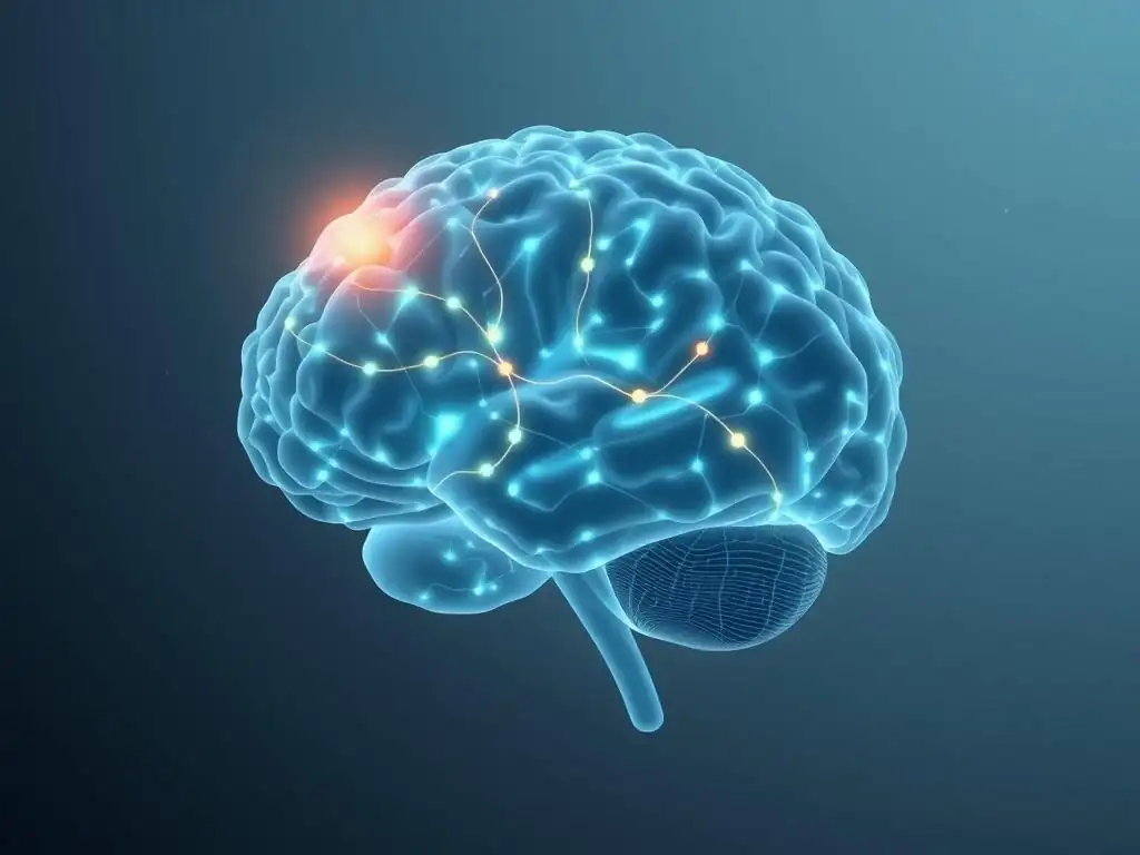
Structural Magnetic Resonance Imaging (MRI) is a foundational technique in obtaining ADHD brain imaging insights. It uses strong magnetic fields and radio waves to create detailed images of brain anatomy, allowing researchers to measure the size (volume) and shape (morphology) of different brain regions. This technique has been instrumental in identifying macroscopic structural differences between individuals with ADHD and typically developing individuals.
Gray Matter Volume Differences in ADHD Brains
Numerous structural MRI studies have focused on gray matter volume, which represents brain tissue primarily composed of neuronal cell bodies. As mentioned earlier, common findings in pediatric ADHD include subtly reduced total brain volume and smaller volumes in specific regions like the prefrontal cortex, basal ganglia (caudate, putamen), and cerebellum.
For instance, meta-analyses pooling data from many studies often report average volume reductions of about 3-5% in key subcortical structures like the caudate nucleus in children with ADHD. While these differences are averages and significant overlap exists between groups, they provide evidence of atypical brain development or structure associated with the disorder.
White Matter Integrity and Connectivity in ADHD
Structural MRI, particularly when combined with advanced techniques like Diffusion Tensor Imaging (DTI), also allows examination of white matter. White matter consists of myelinated axons, the "cables" that connect different brain regions. Differences in white matter integrity, often measured by indices like fractional anisotropy (FA), can indicate variations in the organization, myelination, or density of these connections.
ADHD brain imaging insights from white matter studies have frequently shown altered integrity in tracts connecting frontal regions with striatal, parietal, and occipital areas. For example, studies have reported reduced FA in tracts within the corpus callosum or fronto-striatal pathways in individuals with ADHD, suggesting potential disruptions in communication efficiency between these critical brain regions involved in attention and executive control.
Functional MRI (fMRI): Investigating Brain Activity and Connectivity in ADHD
Functional MRI (fMRI) measures brain activity by detecting changes in blood flow, based on the principle that active brain regions consume more oxygen and receive increased blood supply (the BOLD signal). This technique provides dynamic ADHD brain imaging insights into how different brain areas function individually and interact within networks during specific tasks or at rest.
Task-Based fMRI: Studying Neural Responses to Cognitive Tasks in ADHD
Task-based fMRI involves scanning the brain while participants perform cognitive tasks, such as those requiring attention, working memory, or inhibitory control. This allows researchers to identify which brain regions are more or less active in individuals with ADHD during specific challenges.
As noted previously, typical findings include reduced activation in prefrontal and parietal regions during tasks requiring sustained attention or response inhibition, and altered activation in basal ganglia during reward or timing tasks. For example, during a working memory task, individuals with ADHD might show less robust recruitment of dorsolateral prefrontal cortex compared to controls, correlating with poorer performance on the task.
Resting-State fMRI: Examining Brain Connectivity Patterns in ADHD
Resting-state fMRI captures brain activity when participants are simply resting quietly in the scanner, not performing any explicit task. This approach is used to study functional connectivity – how correlated the activity is between different brain regions, which reflects how strongly they are communicating within large-scale networks. Resting-state fMRI has yielded crucial ADHD brain imaging insights into network dysregulation.
Key findings include altered connectivity within the DMN, executive control network, and salience network, as well as disrupted interactions between these networks. For instance, weaker connectivity within frontal-parietal attention networks or atypical connectivity patterns within the cingulo-opercular network (involved in maintaining task goals) are often observed in individuals with ADHD.
Diffusion Tensor Imaging (DTI): Mapping White Matter Tracts in ADHD
Diffusion Tensor Imaging (DTI) is a specialized MRI technique that measures the diffusion of water molecules in brain tissue. Because water diffusion is restricted by the structure of white matter fibers, DTI can be used to infer the orientation and integrity of these axonal bundles. DTI provides detailed ADHD brain imaging insights into the brain's wiring diagram.
DTI metrics like fractional anisotropy (FA) and mean diffusivity (MD) are often used to characterize white matter. As mentioned earlier, studies in ADHD frequently report lower FA and higher MD in specific white matter tracts, particularly those connecting frontal regions to subcortical structures and posterior brain areas.
For example, studies have shown reduced FA in the superior longitudinal fasciculus, a major tract connecting frontal and parietal lobes, which is crucial for attention and language processing. These findings suggest potential inefficiencies or alterations in the structural connections that support rapid and coordinated information processing necessary for effective attention and cognitive control.
Positron Emission Tomography (PET): Neurotransmitter Systems in ADHD
Positron Emission Tomography (PET) is a nuclear imaging technique that uses small amounts of radioactive tracers to visualize and measure metabolic processes or the distribution of specific molecules in the brain, such as neurotransmitter receptors or transporters. PET imaging has been particularly valuable in providing ADHD brain imaging insights into the neurochemical underpinnings of the disorder, focusing largely on the dopamine and norepinephrine systems.
Dopamine Transporter Density and Activity in ADHD
The dopamine system is strongly implicated in ADHD due to its roles in reward, motivation, attention, and motor control. PET studies using tracers that bind to dopamine transporters (DATs) have yielded important, albeit sometimes inconsistent, ADHD brain imaging insights. Some early studies reported increased DAT density in the striatum of individuals with ADHD compared to controls, suggesting a potential mechanism for reduced dopamine signaling (more transporters clearing dopamine from the synapse).
However, subsequent studies and meta-analyses have had more variable findings, with some showing no difference or even reduced DAT availability in certain subgroups or age ranges. Despite the variability, the focus on dopamine transporters highlights the sustained interest in dopaminergic dysfunction as a core feature of ADHD neurobiology.
Norepinephrine and ADHD: PET Imaging Findings
The norepinephrine system is also critical for attention, arousal, and executive function, and is another key target for ADHD medications. PET imaging of norepinephrine transporters or receptors is more technically challenging than dopamine imaging, leading to fewer studies in this area. However, the available ADHD brain imaging insights from PET studies focusing on norepinephrine suggest potential alterations in this system as well.
Some research has investigated the alpha-2A adrenergic receptor, which is involved in prefrontal cortex function and targeted by some ADHD medications. Findings, though preliminary, support the idea that altered norepinephrine signaling, particularly in frontal networks, contributes to attentional and executive function deficits in ADHD.
Electroencephalography (EEG): Brainwave Patterns and ADHD
Electroencephalography (EEG) measures electrical activity in the brain using electrodes placed on the scalp. It records brainwaves, which reflect the summed activity of large populations of neurons. EEG offers excellent temporal resolution, capturing brain activity changes on the millisecond scale, providing a different type of ADHD brain imaging insights compared to the spatial resolution of MRI or PET.
Quantitative EEG (qEEG) and ADHD Diagnosis
Quantitative EEG (qEEG) involves analyzing the raw EEG data to extract numerical measures of brainwave activity, such as the power or amplitude of different frequency bands (e.g., theta, alpha, beta). Some qEEG studies in ADHD have reported differences in characteristic brainwave patterns compared to controls, such as a higher theta/beta ratio (more slow-wave theta activity relative to fast-wave beta activity), particularly in frontal regions.
This pattern is sometimes interpreted as reflecting reduced cortical arousal or inefficient processing. While qEEG findings are not currently definitive enough for standalone clinical diagnosis, research continues to explore specific qEEG markers that might correlate with ADHD symptoms or help differentiate subtypes, contributing to the growing body of ADHD brain imaging insights.
ADHD Brain Imaging Insights: Specific Findings and Research Highlights
Attention Deficits and Brain Activity: Imaging Studies
Imaging studies consistently link attention deficits in ADHD to atypical activity and connectivity in brain networks responsible for focused attention and cognitive control. Task-based fMRI studies, for example, repeatedly show reduced activation in the dorsal attention network (involving parietal and frontal regions) and the executive control network (centered in the dorsolateral prefrontal cortex and posterior parietal cortex) during tasks requiring sustained or selective attention.
Conversely, resting-state fMRI reveals increased connectivity within the default mode network and reduced anticorrelation between the DMN and task-positive networks. These ADHD brain imaging insights suggest that the brain in ADHD struggles to efficiently engage the networks needed for external focus while simultaneously failing to suppress internal distractions originating from the DMN, directly explaining the difficulties with concentration and susceptibility to mind-wandering.
Impulsivity and Brain Function: Imaging Evidence
Impulsivity, a core symptom of ADHD, is strongly associated with dysfunction in brain regions involved in inhibitory control and reward processing. ADHD brain imaging insights from fMRI studies using tasks like the stop-signal task (requiring participants to inhibit a planned response) often demonstrate reduced activity in key inhibitory control areas, including the right inferior frontal gyrus and the supplementary motor area, in individuals with ADHD.
Furthermore, studies examining reward processing, often involving the striatum and ventral prefrontal cortex, have shown altered responses to anticipated or received rewards, sometimes characterized by blunted responses or steeper 'delay discounting' (preferring smaller immediate rewards over larger delayed ones). These findings provide clear neural correlates for the difficulty individuals with ADHD face in inhibiting prepotent responses and delaying gratification, contributing significantly to our understanding of impulsive behaviors.
Hyperactivity and Neural Correlates: Imaging Research
While hyperactivity can be the most outwardly visible symptom, its neural correlates are also revealed through imaging. Hyperactivity involves excessive motor activity and restlessness, often linked to dysregulation in motor control circuits involving the basal ganglia and cerebellum. Structural ADHD brain imaging insights have noted volume differences in these areas, while functional studies show altered activity during motor tasks or even at rest.
For instance, fMRI studies have sometimes shown atypical connectivity within motor networks or between motor areas and the prefrontal cortex, regions involved in regulating movement. PET studies pointing to differences in the dopamine system within the basal ganglia also offer a plausible neurochemical basis for motor dysregulation. Although research on the specific neural basis of hyperactivity is perhaps less extensive than that on attention or impulsivity, the available imaging data consistently points towards involvement of subcortical and cerebello-cortical circuits.
Neuroimaging Findings in Different ADHD Subtypes
ADHD is categorized into predominantly inattentive presentation (ADHD-I), predominantly hyperactive-impulsive presentation (ADHD-HI), and combined presentation (ADHD-C). ADHD brain imaging insights have sought to determine if distinct neural profiles underlie these subtypes, though findings are not always consistent, partly due to diagnostic overlap and developmental changes.
Predominantly Inattentive Type: Imaging Characteristics
Individuals with the predominantly inattentive type (ADHD-I) primarily exhibit symptoms of inattention. Neuroimaging studies of this subtype sometimes report findings that are more pronounced in regions associated with attentional networks, perhaps showing greater alterations in the connectivity or deactivation of the Default Mode Network compared to other subtypes.
Some studies have also noted structural differences that may be more prominent in parietal or temporal regions involved in attention processing. While there is overlap with findings in other subtypes, the focus on attentional difficulties in ADHD-I is often reflected in ADHD brain imaging insights highlighting dysfunction in networks like the DMN and dorsal attention network.
Predominantly Hyperactive-Impulsive Type: Imaging Characteristics
The predominantly hyperactive-impulsive type (ADHD-HI) is characterized mainly by symptoms of hyperactivity and impulsivity. ADHD brain imaging insights for this subtype have sometimes emphasized differences in subcortical structures, particularly the basal ganglia, and brain regions involved in inhibitory control. Some studies suggest that structural differences in the caudate nucleus might be more pronounced in this group, or that functional alterations during inhibitory tasks are more evident. However, due to the developmental trajectory of ADHD (many children with ADHD-HI transition to ADHD-C or ADHD-I in adolescence/adulthood), disentangling subtype-specific imaging findings can be challenging.
Combined Type: Imaging Characteristics
The combined type (ADHD-C) is the most common presentation and involves significant symptoms of both inattention and hyperactivity-impulsivity. ADHD brain imaging insights in this group often reveal a pattern of findings that encompasses alterations seen in both the inattentive and hyperactive-impulsive subtypes. This typically includes structural and functional differences in prefrontal cortex, basal ganglia, cerebellum, and widespread alterations in large-scale brain networks such as the DMN, executive control network, and salience network. Because ADHD-C presents with the full spectrum of symptoms, its neuroimaging profile often reflects the most comprehensive set of atypical brain characteristics observed across ADHD research.
The Future of ADHD Brain Imaging: Personalized Medicine and Treatment Monitoring
Using Brain Imaging to Predict Treatment Response in ADHD
A major goal for ADHD brain imaging insights is to move beyond group-level differences to individual prediction, specifically predicting how a person with ADHD will respond to different treatments, such as stimulant medication or behavioral therapy. Research is exploring whether specific brain imaging markers – structural volumes, functional connectivity patterns, or neurochemical profiles – can predict response. For instance, some preliminary studies suggest that certain patterns of prefrontal or striatal activity seen in fMRI might correlate with a better response to stimulant medication.
While still in the research phase, the potential is significant. Imagine a scenario where brain imaging could help clinicians select the most effective treatment path before trying different options, saving time and reducing frustration for individuals and families. This move towards using ADHD brain imaging insights for prediction is a key step towards personalized medicine in psychiatry.
Neurofeedback and Brain Training for ADHD: Imaging-Guided Approaches
Neurofeedback is a type of biofeedback that uses real-time displays of brain activity (often from EEG or fMRI) to teach individuals how to self-regulate their brain function. For ADHD, neurofeedback often targets specific brainwave patterns (like the theta/beta ratio in EEG) or activity in specific brain regions or networks (like the PFC or DMN in fMRI). ADHD brain imaging insights from studies identifying atypical activity patterns provide the rationale for these interventions.
While the efficacy of neurofeedback for ADHD is still debated and requires more robust research, imaging techniques are crucial for both guiding the development of these therapies and evaluating their effects. fMRI-based neurofeedback, for example, directly trains individuals to modulate activity in regions like the anterior cingulate cortex or networks like the DMN, based on the ADHD brain imaging insights showing their dysregulation. Imaging can potentially help identify which individuals are most likely to benefit and track the neural changes associated with successful training.
Developing Personalized Treatment Strategies Based on Brain Imaging Biomarkers
Ultimately, the long-term vision for ADHD brain imaging insights is to contribute to the development of truly personalized treatment strategies. This involves identifying specific neurobiological profiles or "biomarkers" from imaging data that correspond to different underlying causes or symptom clusters within ADHD. For example, an individual showing pronounced DMN dysregulation might benefit most from interventions targeting mind-wandering or attention switching, while someone with stronger evidence of striatal dopamine system differences might respond better to dopaminergic medications.
Developing personalized strategies requires large-scale, multimodal imaging studies that integrate different types of ADHD brain imaging insights (structural, functional, chemical) with genetic, behavioral, and clinical data. This complex approach aims to move beyond the current symptom-based diagnosis to a more biologically informed understanding that can guide tailored therapeutic interventions for each individual's unique brain profile.
Limitations and Challenges of Brain Imaging in ADHD
Sample Size and Heterogeneity in ADHD Research
Despite significant progress, brain imaging research in ADHD faces challenges. One major limitation is sample size. While some large-scale initiatives exist (like the ADHD-200 Consortium or ENIGMA), many individual studies have relatively small sample sizes, which can limit the generalizability of findings and make it difficult to detect subtle effects or replicate results.
Furthermore, ADHD is highly heterogeneous; individuals with the same diagnosis can have different symptom profiles, co-occurring conditions (like anxiety, depression, or learning disabilities), and varying developmental trajectories. This heterogeneity makes it challenging to identify consistent ADHD brain imaging insights across diverse groups and can obscure underlying biological differences.
Methodological Considerations in Brain Imaging Studies
Brain imaging studies involve complex data acquisition and analysis pipelines, and methodological variations can significantly impact results. Differences in scanner hardware, acquisition parameters, preprocessing steps, and statistical analysis methods can lead to inconsistencies across studies. For example, different fMRI tasks or resting-state analysis techniques can yield varying functional connectivity results. Ensuring standardization and using rigorous methods are crucial but challenging across different research sites. Furthermore, factors like participant motion within the scanner, which can be particularly problematic in individuals with ADHD, can introduce artifacts and affect data quality, potentially biasing ADHD brain imaging insights.
Ethical Implications of Brain Imaging in ADHD
The increasing ability to visualize and interpret brain function in conditions like ADHD also raises important ethical considerations. One concern is the potential for misinterpreting group-level research findings as deterministic explanations for individual behavior. Presenting ADHD brain imaging insights should be done carefully to avoid biological reductionism or suggesting that brain differences excuse behavior without considering the interplay of biological, environmental, and social factors.
There are also ethical considerations around using imaging in clinical decision-making before it is fully validated, the privacy of imaging data, and the potential for stigma associated with having a "different" brain. It is crucial that research and clinical translation of ADHD brain imaging insights proceed with careful consideration of these ethical dimensions.
Conclusion: The Power of Brain Imaging in Understanding and Managing ADHD
Summarizing Key ADHD Brain Imaging Insights
Brain imaging has profoundly advanced our understanding of Attention-Deficit/Hyperactivity Disorder. Through techniques like structural and functional MRI, DTI, PET, and EEG, researchers have gained critical ADHD brain imaging insights into the neural underpinnings of attention deficits, impulsivity, and hyperactivity. Key findings consistently point to atypical structure, function, and connectivity in brain regions and networks including the prefrontal cortex, basal ganglia, cerebellum, and the default mode network. These differences are associated with core ADHD symptoms, providing biological evidence for the neurodevelopmental nature of the disorder.
The Importance of Multimodal Approaches in ADHD Research and Clinical Practice
No single imaging technique provides a complete picture of the complex brain differences in ADHD. The most powerful ADHD brain imaging insights come from multimodal approaches that combine information from different techniques (e.g., structural volumes with functional connectivity or neurochemistry). Integrating imaging data with genetic studies, cognitive assessments, and clinical profiles is essential for building a comprehensive model of ADHD. As research progresses, integrating these diverse data sources holds the greatest promise for identifying robust biomarkers, understanding the disorder's heterogeneity, and ultimately translating findings into clinical practice.
Call to Action: Further Research and Clinical Translation of Brain Imaging Findings in ADHD
While current ADHD brain imaging insights have significantly deepened our knowledge, much work remains. Further research is needed to replicate findings, explore developmental trajectories across the lifespan, investigate treatment mechanisms, and understand the interaction between brain biology and environmental factors.
The ultimate goal is the successful clinical translation of these findings – moving from research discoveries to tools that can aid in diagnosis, predict treatment response, and guide personalized interventions. Continued investment in rigorous, large-scale imaging research, coupled with careful ethical consideration, is essential to harness the full potential of brain imaging in improving the lives of individuals with ADHD.
Frequently Asked Questions
What is the role of brain imaging in understanding ADHD?
Brain imaging techniques, such as MRI and PET scans, allow researchers to visualize brain structure and activity, providing insights into the neurological differences associated with ADHD. These scans help us understand how ADHD affects brain function.
How can ADHD brain imaging insights help in diagnosis?
While not a primary diagnostic tool, brain imaging can support the diagnosis of ADHD by revealing patterns of brain activity and structure that are commonly observed in individuals with the condition. This assists in identifying potential neurological markers.
What specific brain regions are often examined in ADHD brain imaging studies?
Commonly studied regions include the prefrontal cortex (responsible for executive functions), the basal ganglia (involved in motor control and reward), and the cerebellum (critical for motor coordination).
Can brain imaging insights contribute to the development of new ADHD treatments?
Yes, by identifying specific neurological differences associated with ADHD, brain imaging can help researchers develop targeted treatments, such as medications or therapies that address these specific brain functions.
Are ADHD brain imaging findings consistent across all individuals with ADHD?
No, there is variability in brain structure and function among individuals with ADHD. Imaging findings can reveal trends, but it's crucial to remember that ADHD is a complex and heterogeneous condition.
Final Reflection: Unveiling the ADHD Brain: A Journey of Understanding and Hope
The insights gleaned from brain imaging studies are revolutionizing our understanding of ADHD, moving beyond simplistic labels and revealing the complex neurological underpinnings of this condition. This is not merely an academic pursuit; it holds the key to unlocking more effective treatments and fostering greater empathy and acceptance for those living with ADHD.
The journey of understanding ADHD, as illuminated by these imaging studies, underscores the crucial need for a shift in perspective. We are moving away from a deficit model, focusing solely on what's "wrong," towards a model that appreciates the unique neural architecture of the ADHD brain. The brain is not a machine to be fixed, but a garden to be cultivated.
This quote highlights the potential for growth and adaptation inherent within the ADHD brain, a potential often overlooked in traditional approaches. The ongoing research emphasizes the plasticity of the brain, offering hope for personalized interventions and improved outcomes.
Consider the implications of these findings. How can we use this newfound knowledge to create more inclusive environments, both at home and in the workplace? How can we leverage the strengths often associated with ADHD – creativity, innovation, and hyperfocus – while addressing the challenges? You have the power to advocate for a more nuanced understanding of ADHD, to challenge stigmas, and to support individuals in their journey towards self-acceptance and success. Let the insights shared in this article inspire you to learn more, to empathize deeply, and to contribute to a future where neurodiversity is celebrated and supported.
Unlock Your Brain's Potential: Take Charge of ADHD
Thanks for exploring the fascinating world of ADHD and brain imaging with us! We hope you gained valuable insights into this often-misunderstood condition. Now, it's time to put that knowledge into action.
- Prioritize one small, achievable step today: Whether it's researching local support groups, trying a new organizational technique, or simply scheduling a check-in with your doctor, even a small step forward makes a big difference.
- Share this knowledge: Pass along this article to someone who might benefit from understanding ADHD better. You could make a real impact on their life!
- Explore mindfulness techniques: Many find mindfulness practices helpful in managing ADHD symptoms. Look into guided meditations or simple breathing exercises to see if they could be beneficial for you.
What one small change will you make this week to better understand and manage your attention, impulsivity, and hyperactivity?