
PTSD Through the Lens: Imaging Trauma's Impact on the Brain and Memory


Welcome to Mind Care Guide, a safe space to explore the complexities of mental well-being.
Have you ever wondered how trauma leaves its indelible mark, not just on our minds, but on the very structure of our brains? In our featured article, "PTSD Through the Lens," we delve into the fascinating world of PTSD brain imaging memory impact, revealing groundbreaking insights into how trauma reshapes neural pathways and affects memory processing. Join us as we uncover the science behind the struggle, offering a deeper understanding and potential pathways to healing. Intrigued? Explore the neuroscience of mental health and discover more within.
PTSD Through the Lens: Imaging Trauma's Impact on the Brain and Memory
- Introduction: Understanding PTSD and the Power of Brain Imaging
- The Neuroscience of Trauma: PTSD and the Brain
- Brain Imaging Techniques for PTSD Research and Diagnosis
- PTSD Brain Imaging Memory Impact: A Closer Look
- Clinical Applications of PTSD Brain Imaging
- The Future of PTSD Brain Imaging Research
- Conclusion: The Promise of Brain Imaging for Understanding and Treating PTSD
-
Frequently Asked Questions
- What is the primary focus of using brain imaging in PTSD research?
- How does PTSD affect memory, and can brain imaging reveal this impact?
- What specific brain regions are typically examined in PTSD brain imaging studies?
- Can brain imaging be used to diagnose PTSD?
- What are the potential benefits of understanding the "PTSD brain imaging memory impact" connection for treatment?
- Final Reflection: Unveiling the Silent Scars: PTSD, the Brain, and the Power of Understanding
- Unveil Your Brain's Resilience: Steps to Understanding PTSD
Introduction: Understanding PTSD and the Power of Brain Imaging
The Growing Need for Objective PTSD Assessment
Post-traumatic stress disorder (PTSD) is a complex mental health condition that can develop after someone experiences or witnesses a traumatic event. While clinical interviews and self-report questionnaires are the standard methods for diagnosing PTSD, these approaches can sometimes be subjective or influenced by factors like memory recall bias or reluctance to disclose symptoms.
The subjective nature of these assessments highlights a significant need for more objective methods to confirm diagnosis, understand symptom severity, and potentially predict treatment outcomes. Developing objective markers, such as those derived from brain imaging, could lead to more accurate and timely diagnoses, ensuring individuals receive the appropriate support and interventions sooner.
The limitations of purely subjective assessment become particularly apparent in cases where trauma survivors struggle to articulate their experiences or where symptoms overlap with other conditions. For instance, dissociative symptoms in PTSD can make it difficult for individuals to provide a linear narrative of their trauma or current struggles.
Objective measures offer the potential to cut through some of this complexity, providing a clearer picture of the underlying biological correlates of the disorder. This is crucial not only for clinical diagnosis but also for research aimed at uncovering the fundamental mechanisms driving PTSD, including the PTSD brain imaging memory impact.
How Brain Imaging Enhances PTSD Understanding
Brain imaging techniques offer an unprecedented window into the living human brain, allowing researchers and clinicians to visualize its structure, function, and neurochemistry. By applying these powerful tools to the study of PTSD, we can gain insights into how trauma physically and functionally alters the brain. This goes beyond simply observing symptoms; it allows us to explore the neural circuits and regions that are disrupted, providing a biological basis for the experienced difficulties, such as intrusive memories, hyperarousal, and emotional dysregulation.
Utilizing methods like magnetic resonance imaging (MRI) and positron emission tomography (PET), scientists can identify structural changes, observe patterns of neural activity during specific tasks or at rest, and even investigate the distribution of neurotransmitters. This enhanced understanding of the PTSD brain imaging memory impact and broader neural correlates is vital for refining diagnostic criteria, developing more targeted therapies, and ultimately reducing the burden of this disorder. It moves us closer to a precision medicine approach, where treatments can be tailored based on an individual's specific brain profile.
The Neuroscience of Trauma: PTSD and the Brain
PTSD: A Definition and Overview of Symptoms
Post-traumatic stress disorder (PTSD) is a psychiatric disorder that can occur in people who have experienced or witnessed a traumatic event such as a natural disaster, a serious accident, a terrorist act, war/combat, or rape or other violent personal assault.
The diagnostic criteria, as outlined in the Diagnostic and Statistical Manual of Mental Disorders (DSM-5), include a range of symptoms clustered into four main categories: re-experiencing symptoms (e.g., flashbacks, nightmares, intrusive thoughts), avoidance symptoms (e.g., avoiding places, people, or activities that remind one of the trauma), negative alterations in cognition and mood (e.g., difficulty remembering aspects of the trauma, negative beliefs about oneself or the world, persistent negative emotional state), and alterations in arousal and reactivity (e.g., irritability, difficulty concentrating, hypervigilance, exaggerated startle response).
These symptoms must persist for more than a month and cause clinically significant distress or impairment in social, occupational, or other important areas of functioning. It's important to note that not everyone who experiences trauma develops PTSD. The development of the disorder is influenced by a complex interplay of factors, including the nature of the trauma, individual resilience, genetic predispositions, and environmental support. Understanding the neural underpinnings, often explored through PTSD brain imaging memory impact studies, is key to developing more effective interventions for this often debilitating condition.
The Role of the Amygdala in PTSD and Fear Processing
The amygdala, a small, almond-shaped region deep within the temporal lobe, plays a critical role in processing emotions, particularly fear and threat detection. In individuals with PTSD, the amygdala is frequently found to be hyperactive, meaning it shows an exaggerated response to stimuli, even those that are not directly threatening but may be associated with the trauma.
This overactivity contributes to the heightened state of fear, anxiety, and hypervigilance characteristic of the disorder. Research using functional MRI (fMRI) has consistently demonstrated increased amygdala activation in response to trauma-related cues in individuals with PTSD compared to healthy controls or trauma-exposed individuals without PTSD.
For example, studies have shown that when presented with images or sounds reminiscent of their trauma, individuals with PTSD exhibit significantly stronger amygdala responses. This persistent state of hyper-responsivity can make it difficult for individuals to feel safe and can trigger intense fear or panic reactions in everyday situations.
Understanding this amygdala dysregulation through brain imaging is crucial for developing treatments that aim to modulate these fear responses, such as exposure therapy or pharmacological interventions targeting fear circuitry. The amplified fear signals processed by the amygdala also likely contribute to the distorted processing of traumatic memories, impacting the overall PTSD brain imaging memory impact.
Hippocampal Dysfunction and Memory Impairment in PTSD
The hippocampus, another key structure in the brain's limbic system, is primarily involved in the formation, organization, and storage of new memories, especially declarative memories (memories of facts and events). In PTSD, the hippocampus is often found to be smaller in volume and functionally impaired. This dysfunction is thought to contribute to the characteristic memory problems experienced by individuals with PTSD, such as difficulty recalling details of the trauma itself (dissociative amnesia) while simultaneously suffering from intrusive, fragmented flashbacks.
Structural MRI studies have reported reduced hippocampal volume in individuals with PTSD, with reductions sometimes estimated to be in the range of 5% to 10% compared to non-traumatized controls, although findings can vary. This volume reduction may precede or be a consequence of the trauma and the resulting chronic stress.
Functionally, the hippocampus in PTSD may struggle to properly contextualize traumatic memories, leading to them being experienced as vivid, present-moment threats rather than past events. This complex interplay between hippocampal structure and function profoundly affects the PTSD brain imaging memory impact, contributing to the intrusive re-experiencing symptoms that are a hallmark of the disorder.
Prefrontal Cortex and Emotional Regulation Deficits in PTSD
The prefrontal cortex (PFC), particularly the medial prefrontal cortex (mPFC) and the anterior cingulate cortex (ACC), plays a critical role in executive functions, including emotional regulation, decision-making, and inhibiting inappropriate responses. In healthy individuals, the PFC exerts top-down control over the amygdala, helping to dampen fear responses when a threat is no longer present. In PTSD, this regulatory circuit is often disrupted. Brain imaging studies frequently show reduced activity in the mPFC and ACC, suggesting a diminished capacity of the PFC to inhibit the hyperactive amygdala.
This weakened top-down control contributes significantly to the emotional dysregulation, impulsivity, and difficulty modulating fear and anxiety seen in PTSD. For example, fMRI studies have shown reduced activation in the mPFC when individuals with PTSD attempt to regulate their emotions in response to negative stimuli. This deficit in the brain's "executive control" system, combined with amygdala hyperactivity and hippocampal dysfunction, creates a challenging neural environment that perpetuates PTSD symptoms and contributes to the complex PTSD brain imaging memory impact, where memories are poorly regulated and highly emotionally charged.
Brain Imaging Techniques for PTSD Research and Diagnosis
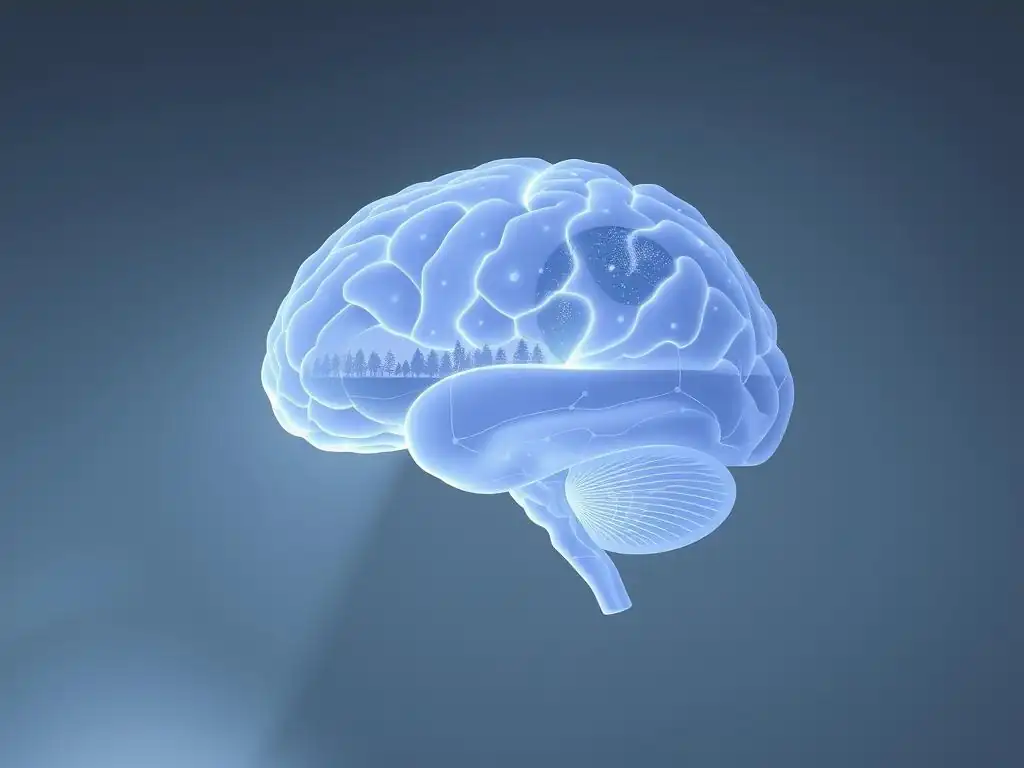
Structural MRI: Identifying Anatomical Differences in PTSD Brains
Structural Magnetic Resonance Imaging (MRI) is a widely used non-invasive technique that provides detailed images of the brain's anatomy. By measuring the signals emitted by water molecules in different tissues when placed in a strong magnetic field, MRI can differentiate between gray matter (neuronal cell bodies), white matter (nerve fibers), and cerebrospinal fluid. In PTSD research, structural MRI is primarily used to investigate differences in the size, shape, and integrity of specific brain regions compared to healthy control groups. This allows researchers to identify potential anatomical correlates of the disorder.
Studies using structural MRI have consistently pointed to alterations in key areas involved in processing fear, memory, and emotion. As mentioned earlier, reduced hippocampal volume is a frequently reported finding, though the consistency and magnitude of this finding can vary across studies depending on factors like trauma type, age, and comorbidity. While structural changes identified by MRI are not typically used for individual diagnosis yet, they provide valuable insights into the neurobiological underpinnings of PTSD and highlight potential targets for intervention. This anatomical information is foundational to understanding the PTSD brain imaging memory impact.
Exploring Hippocampal Volume Reduction in PTSD with MRI
Numerous structural MRI studies have investigated the hippocampus in individuals with PTSD. While findings are not perfectly uniform, a large body of evidence suggests that individuals with PTSD often exhibit smaller hippocampal volumes compared to control groups. Meta-analyses, combining data from multiple studies, have supported this finding, estimating average volume reductions that can range from around 5% to 10%. For instance, one prominent meta-analysis involving hundreds of participants found a significant effect size indicating reduced hippocampal volume in PTSD.
The interpretation of this finding is complex; it's debated whether the smaller hippocampus is a pre-existing vulnerability factor that makes someone more susceptible to developing PTSD after trauma, or if it's a consequence of the trauma and the associated chronic stress exposure (which is known to be neurotoxic to the hippocampus). Longitudinal studies tracking individuals after trauma exposure are helping to shed light on this question. Regardless of causality, reduced hippocampal volume is strongly associated with memory impairments observed in PTSD, underscoring its relevance to the PTSD brain imaging memory impact.
Investigating Amygdala Hyperactivity Using Structural MRI
While functional MRI is the primary tool for assessing amygdala activity, structural MRI can provide some insights into potential anatomical correlates related to amygdala function in PTSD. Some studies have reported subtle structural differences in the amygdala in individuals with PTSD, such as altered gray matter density or volume, although these findings are less consistent than those for hippocampal volume. For example, some research has suggested increased amygdala volume in certain PTSD subtypes or populations, while others have found no significant difference or even decreased volume.
It's important to distinguish that structural MRI reveals the size and shape of the amygdala, not its neural activity level. However, anatomical variations could potentially be linked to functional changes. For instance, altered connectivity between the amygdala and other brain regions, which can sometimes be inferred from structural patterns or more directly measured with techniques like DTI, could contribute to amygdala hyperactivity. Therefore, while structural MRI doesn't directly measure hyperactivity, it provides crucial anatomical context for understanding the functional abnormalities observed in this key fear processing region and its role in the PTSD brain imaging memory impact.
Functional MRI (fMRI): Revealing Brain Activity Patterns in PTSD
Functional Magnetic Resonance Imaging (fMRI) measures brain activity by detecting changes in blood flow. When a brain region is more active, it consumes more oxygen, and blood flow to that area increases (the BOLD signal - Blood-Oxygen-Level Dependent). By observing these changes while individuals perform tasks (like recalling memories or viewing emotional stimuli) or while they are at rest, fMRI can map which brain areas are more or less active and how different areas communicate with each other. fMRI is a cornerstone technique for understanding the neural circuits underlying PTSD symptoms.
In PTSD research, fMRI is extensively used to investigate the brain's response to trauma-related cues, emotional regulation challenges, and resting-state brain activity. These studies have consistently revealed patterns of abnormal activity and connectivity within the fear circuitry (amygdala, mPFC), memory systems (hippocampus), and executive control networks (PFC, ACC). The dynamic nature of fMRI allows researchers to see how the brain behaves in real-time, offering profound insights into the functional basis of the disorder and the mechanisms behind the PTSD brain imaging memory impact.
fMRI Studies of Fear Conditioning and Extinction in PTSD
Fear conditioning is a learning process where a neutral stimulus becomes associated with a fearful outcome, leading to a conditioned fear response. Extinction is the process of learning that the neutral stimulus is no longer associated with the fearful outcome, leading to a reduction in the fear response. These processes are mediated by brain regions like the amygdala, hippocampus, and prefrontal cortex. fMRI studies examining fear conditioning and extinction in PTSD have provided critical insights into the disorder's pathophysiology.
Research consistently shows that individuals with PTSD exhibit altered fear extinction learning. They may show exaggerated or prolonged amygdala activation during the extinction phase, indicating difficulty learning that a previously threatening cue is now safe. They also often show reduced activity in the ventromedial prefrontal cortex (vmPFC), a region crucial for inhibiting conditioned fear responses.
For example, studies might show control participants exhibiting a significant decrease in amygdala activity and increase in vmPFC activity over extinction trials, while PTSD participants show blunted or absent changes. This impaired fear extinction is considered a core deficit in PTSD, contributing to persistent fear responses and intrusive re-experiencing, directly impacting the PTSD brain imaging memory impact related to trauma cues.
Resting-State fMRI and Network Connectivity in PTSD
Resting-state fMRI (rs-fMRI) measures brain activity while an individual is awake but not performing a specific task. It focuses on spontaneous fluctuations in the BOLD signal, which are highly correlated between functionally connected brain regions, forming large-scale brain networks. Rs-fMRI allows researchers to investigate the intrinsic connectivity patterns of the brain in PTSD, providing insights into how different brain regions communicate with each other when the individual is simply at rest.
Studies using rs-fMRI in PTSD have revealed widespread alterations in network connectivity. A common finding is increased connectivity within the salience network (which includes the amygdala and insula, involved in detecting important stimuli) and decreased connectivity within the default mode network (DMN, involved in self-referential thought and memory) and the executive control network.
For example, research might show increased connectivity between the amygdala and other limbic areas, potentially reflecting a state of heightened internal threat monitoring even at rest. Altered connectivity between the hippocampus and other DMN regions may also contribute to the disorganized and intrusive nature of traumatic memories, highlighting the PTSD brain imaging memory impact on intrinsic brain function.
Positron Emission Tomography (PET): Examining Neurochemical Imbalances in PTSD
Positron Emission Tomography (PET) is an imaging technique that uses small amounts of radioactive tracers to visualize and measure metabolic processes or the distribution of specific molecules in the brain, such as neurotransmitter receptors or transporters. Unlike MRI, which primarily focuses on structure and blood flow (as a proxy for activity), PET provides direct information about the brain's neurochemistry. This is particularly valuable for understanding the role of specific neurotransmitter systems, like serotonin, dopamine, and norepinephrine, which are implicated in the stress response and mood regulation, and are often dysregulated in PTSD.
By using tracers that bind to specific receptors or transporters, PET allows researchers to quantify their density and availability in different brain regions. This provides a different, yet complementary, perspective to structural and functional imaging, offering insights into the molecular basis of PTSD symptoms. PET studies contribute significantly to understanding the complex biological landscape of the disorder and the potential targets for pharmacological interventions, including those aimed at modulating the PTSD brain imaging memory impact at a neurochemical level.
PET Imaging of Serotonin Transporters in PTSD
The serotonin system plays a crucial role in regulating mood, anxiety, and stress responses. The serotonin transporter (SERT) is responsible for clearing serotonin from the synaptic cleft, thus regulating the strength and duration of serotonin signaling. Studies using PET with tracers that bind to SERT have investigated whether there are alterations in SERT availability in the brains of individuals with PTSD.
Findings from these PET studies have been somewhat mixed, but some research suggests altered SERT binding in certain brain regions in PTSD. For instance, some studies have reported reduced SERT availability in areas like the midbrain or amygdala, while others have found no significant differences or even increases in certain regions.
These variations may depend on factors like trauma type, comorbidity with depression, and medication status. However, evidence of altered serotonin system function, as indicated by PET, supports the use of Selective Serotonin Reuptake Inhibitors (SSRIs), which block SERT, as a common pharmacological treatment for PTSD, potentially modulating mood and anxiety symptoms and indirectly influencing the PTSD brain imaging memory impact.
Dopamine and PTSD: Insights from PET Scans
The dopamine system is involved in reward, motivation, and salience processing, but also plays a role in the stress response and fear memory consolidation. Dysregulation of the dopamine system has been implicated in various psychiatric disorders, including PTSD. PET imaging with tracers that bind to dopamine receptors (like D2/D3 receptors) or the dopamine transporter (DAT) allows researchers to investigate dopamine system function in PTSD.
PET studies of the dopamine system in PTSD have also yielded varied results. Some research has suggested alterations in dopamine receptor availability or dopamine release in areas like the striatum or prefrontal cortex. For example, some studies have reported reduced D2/D3 receptor availability in certain brain regions, which could impact reward processing and motivation, symptoms often affected in PTSD.
Other research has explored dopamine release in response to stress or trauma cues. These findings suggest that dopamine dysregulation may contribute to symptoms such as anhedonia, irritability, and altered responses to trauma-related stimuli, further complicating the understanding of the PTSD brain imaging memory impact.
Diffusion Tensor Imaging (DTI): Mapping White Matter Integrity in PTSD
Diffusion Tensor Imaging (DTI) is a specialized MRI technique that measures the diffusion of water molecules within biological tissues. In the brain, water diffusion is anisotropic (directional) along white matter tracts because the myelin sheaths and axons act as barriers. DTI exploits this property to infer the orientation and integrity of white matter fiber bundles, which are crucial for connecting different brain regions and enabling communication between them. DTI provides insights into the structural connectivity of the brain.
In PTSD research, DTI is used to investigate whether trauma and the development of PTSD are associated with alterations in the integrity of white matter tracts connecting key brain regions involved in emotion regulation, memory, and fear processing. Measures derived from DTI, such as fractional anisotropy (FA) and mean diffusivity (MD), can indicate changes in the organization, myelination, or density of white matter fibers. Disruption of these connections could impair communication between critical brain areas, contributing to the observed functional deficits in PTSD, including those related to the PTSD brain imaging memory impact.
DTI Studies of White Matter Tracts Affected by Trauma
DTI studies in PTSD have focused on white matter tracts that connect regions known to be dysfunctional in the disorder, such as the uncinate fasciculus (connecting the amygdala and hippocampus to the prefrontal cortex), the cingulum bundle (part of the limbic system), and tracts within the corpus callosum. Findings have often reported reduced fractional anisotropy (FA) or increased mean diffusivity (MD) in these tracts in individuals with PTSD, suggesting reduced white matter integrity.
For example, multiple studies have found reduced FA in the uncinate fasciculus in PTSD patients. This finding is particularly relevant as this tract is a major pathway for communication between the amygdala and the prefrontal cortex, critical for regulating fear responses. Impaired integrity in this pathway could contribute to the weakened top-down control over amygdala hyperactivity seen in PTSD. Similarly, alterations in the cingulum bundle could affect emotional processing and cognitive control. These DTI findings highlight how structural disconnection, revealed through imaging, underpins the functional impairments and contributes to the complex PTSD brain imaging memory impact.
PTSD Brain Imaging Memory Impact: A Closer Look
How Trauma Alters Memory Encoding and Retrieval
Traumatic experiences have a profound and often disruptive impact on memory. Unlike typical memories, which are usually integrated into a coherent narrative of one's life, traumatic memories in PTSD can be fragmented, disorganized, and intensely emotional. The extreme stress and neurobiological changes occurring during and after trauma appear to alter the processes of memory encoding (forming new memories) and retrieval (accessing stored memories). High levels of stress hormones like cortisol and norepinephrine during trauma can enhance the encoding of sensory and emotional aspects of the event, while impairing the encoding of contextual details (like time, place, and sequence).
This leads to memories that are strong in sensory and emotional intensity but weak in narrative structure. When these memories are retrieved, they often lack context, feeling like they are happening in the present moment (flashbacks) rather than being recalled as past events. The difficulty in retrieving a complete, coherent narrative of the trauma is also common. This selective impairment and enhancement of different memory components is a hallmark of the PTSD brain imaging memory impact, reflecting the dysregulation of brain systems involved in memory processing under extreme stress.
The Impact of PTSD on Declarative Memory Systems
Declarative memory, which includes episodic memory (memory for specific events) and semantic memory (memory for facts and concepts), relies heavily on the hippocampus and its connections to cortical areas. In PTSD, the dysfunction and volume reduction often observed in the hippocampus significantly impact declarative memory systems. Individuals with PTSD frequently report difficulties with recalling specific details of the traumatic event itself, as well as broader impairments in forming and retrieving non-trauma-related declarative memories.
For example, someone with PTSD might struggle to remember aspects of their daily life, recent conversations, or appointments, alongside their difficulty accessing a complete narrative of their trauma. This suggests that the impact of PTSD extends beyond trauma-specific memories to affect general declarative memory function. The compromised ability of the hippocampus to properly encode and consolidate new information, and to retrieve existing memories, is a key aspect of the PTSD brain imaging memory impact, contributing to cognitive difficulties experienced by those with the disorder.
The Role of Brain Imaging in Correlating Memory Deficits with Brain Changes
Brain imaging techniques are invaluable for understanding the relationship between the observed memory deficits in PTSD and the underlying neurobiological changes. By combining cognitive assessments of memory performance with structural and functional brain imaging data, researchers can identify specific brain abnormalities that correlate with different types of memory impairment. This allows for a more precise understanding of the neural basis of the PTSD brain imaging memory impact.
For instance, studies might correlate reduced hippocampal volume (measured with structural MRI) with poorer performance on verbal or visual memory tasks. Similarly, fMRI studies can examine brain activity patterns during memory encoding or retrieval tasks and relate these patterns to memory accuracy and the subjective experience of memory (e.g., intrusive vs. voluntary recall). PET imaging can add a neurochemical layer by investigating how neurotransmitter system alterations in memory-related regions correlate with memory performance. This multi-modal imaging approach provides compelling evidence linking specific brain changes to the complex memory disturbances seen in PTSD.
Studies Linking Hippocampal Activity and Memory Performance in PTSD
Numerous fMRI studies have specifically investigated the relationship between hippocampal activity and memory performance in individuals with PTSD. These studies often involve participants performing memory tasks while in the scanner, such as trying to encode new information, recall previously learned material, or differentiate between trauma-related and neutral stimuli.
Findings from these studies consistently show altered hippocampal activity patterns in PTSD during memory tasks. For example, some studies have reported hypoactivity (reduced activity) in the hippocampus during the encoding or retrieval of neutral or positive information, potentially explaining the difficulties with general declarative memory. Conversely, during the processing of trauma-related stimuli, hippocampal activity might be altered in a way that contributes to the fragmentation and lack of contextualization characteristic of intrusive memories.
For example, a study might show a negative correlation between hippocampal activity during trauma recall and the ability to place the memory in the past. These direct links between hippocampal function, as visualized by fMRI, and memory performance provide strong evidence for the neural basis of the PTSD brain imaging memory impact.
Clinical Applications of PTSD Brain Imaging
Improving PTSD Diagnosis and Treatment Planning
While not yet a standard diagnostic tool, brain imaging holds significant promise for improving the diagnosis and treatment planning for PTSD. The objective neurobiological data provided by imaging could potentially complement clinical assessments, leading to more accurate and timely diagnoses, particularly in complex cases or when distinguishing PTSD from other conditions with overlapping symptoms. Identifying specific brain signatures or biomarkers associated with PTSD could help reduce misdiagnosis and ensure individuals receive appropriate care sooner.
Furthermore, imaging could aid in treatment planning. For example, identifying specific patterns of amygdala hyperactivity or PFC hypoactivity might suggest that a particular type of therapy focusing on emotional regulation or fear extinction could be more effective for that individual. Similarly, understanding the extent of hippocampal dysfunction could inform the approach to therapies involving memory processing. This personalized approach, guided by insights from PTSD brain imaging memory impact and other neural correlates, could lead to more efficient and effective treatment strategies tailored to the individual's unique brain profile.
Predicting Treatment Response Using Brain Imaging Biomarkers
One of the most exciting potential clinical applications of PTSD brain imaging is the prediction of treatment response. Currently, predicting whether an individual will benefit from a specific therapy (like psychotherapy or medication) is challenging, often involving a trial-and-error process. Brain imaging offers the possibility of identifying neurobiological markers that predict how well someone will respond to a particular intervention before treatment even begins.
Research is actively exploring potential imaging biomarkers for predicting response to therapies like trauma-focused psychotherapy (e.g., Prolonged Exposure, Cognitive Processing Therapy) and pharmacological treatments (e.g., SSRIs). For instance, baseline levels of amygdala reactivity, vmPFC activity, or connectivity patterns within fear and executive control networks, as measured by fMRI, have shown promise in predicting response to psychotherapy.
Similarly, PET measures of neurotransmitter system function might predict response to medication. Identifying these predictors, including those related to the PTSD brain imaging memory impact, could allow clinicians to select the most appropriate treatment from the outset, saving time, reducing patient distress, and improving outcomes.
Monitoring Treatment Effectiveness Through Longitudinal Brain Imaging Studies
Longitudinal brain imaging studies, where individuals are scanned before, during, and after treatment, are crucial for understanding how therapies work and for monitoring their effectiveness at a neurobiological level. By comparing brain scans over time, researchers can observe whether successful treatment is associated with normalization of previously observed brain abnormalities. This provides objective evidence of treatment-induced brain changes.
For example, studies have used fMRI to show that effective trauma-focused psychotherapy is associated with decreased amygdala reactivity and increased activity in the vmPFC when processing trauma cues, reflecting improved emotional regulation and fear extinction. Structural MRI studies have sometimes shown increases in hippocampal volume following successful treatment, although this finding is less consistent. PET studies might reveal changes in neurotransmitter receptor availability.
These imaging findings provide objective validation that therapies are having their intended effect on the brain circuits underlying PTSD symptoms, including modifying the PTSD brain imaging memory impact. This ability to visualize treatment-related changes can also help refine therapeutic techniques and understand why some individuals respond better than others.
The Future of PTSD Brain Imaging Research
Advanced Imaging Techniques and Machine Learning Applications
The field of brain imaging is continuously evolving, with new techniques and analytical methods emerging. Advanced MRI techniques, such as ultra-high field MRI (7T and beyond) offer even greater spatial resolution, allowing visualization of finer brain structures. Advanced diffusion imaging models can provide more detailed information about white matter microstructure. Combining different imaging modalities (e.g., simultaneous fMRI-PET) can offer richer, multi-dimensional data about brain function and neurochemistry.
Furthermore, the application of machine learning and artificial intelligence (AI) to brain imaging data is revolutionizing the field. Machine learning algorithms can analyze complex patterns across vast amounts of imaging data to identify subtle biomarkers that might not be apparent through traditional statistical methods. These approaches hold immense potential for improving the accuracy of diagnosis, predicting treatment response, and even identifying individuals at risk for developing PTSD after trauma exposure. Machine learning applied to PTSD brain imaging memory impact data could potentially classify different subtypes of memory dysfunction or predict which individuals are most likely to experience debilitating flashbacks.
Personalized Medicine Approaches to PTSD Treatment
The ultimate goal of much of this research is to move towards a personalized medicine approach for PTSD treatment. Currently, treatment guidelines often recommend a stepped-care approach, starting with therapies that are generally effective but without specific guidance on which therapy is best for a particular individual. Brain imaging data, combined with genetic information, clinical history, and other factors, could potentially provide the basis for selecting the most effective treatment strategy for each person.
Imagine a future where a brain scan could help determine whether an individual's PTSD is characterized primarily by amygdala hyper-reactivity requiring emotion regulation focus, or by severe hippocampal dysfunction suggesting a need for memory processing support, or a specific neurochemical imbalance indicating likely response to a particular medication. This level of personalized insight, informed by the comprehensive understanding of the PTSD brain imaging memory impact and other neural correlates, could transform PTSD care, leading to faster recovery and better long-term outcomes.
Ethical Considerations in PTSD Brain Imaging
As brain imaging technology becomes more powerful and its potential clinical applications grow, it's crucial to consider the ethical implications. Issues surrounding data privacy and security are paramount, as brain scans contain highly sensitive personal information. Ensuring that imaging data is collected, stored, and used responsibly is essential.
Furthermore, there are ethical considerations related to the interpretation and use of imaging findings. It's important to avoid deterministic views of the brain, recognizing that imaging findings represent biological correlates, not destiny. Using imaging data to predict risk or treatment response must be done cautiously and transparently, avoiding stigmatization or discrimination. Ensuring equitable access to advanced imaging technologies and the personalized treatments they might enable is another critical ethical challenge. Addressing these ethical considerations is vital as brain imaging continues to advance our understanding of PTSD and its PTSD brain imaging memory impact.
Conclusion: The Promise of Brain Imaging for Understanding and Treating PTSD
Summarizing Key Findings and Future Directions
Brain imaging techniques have significantly advanced our understanding of the neurobiological basis of PTSD. Studies using structural MRI have revealed anatomical differences, such as reduced hippocampal volume and potential amygdala alterations. Functional MRI has provided insights into abnormal activity and connectivity patterns within fear, memory, and executive control networks, highlighting amygdala hyperactivity, mPFC hypoactivity, and disrupted network communication.
PET imaging has shed light on potential neurochemical imbalances, particularly within the serotonin and dopamine systems. DTI has shown evidence of impaired white matter integrity, suggesting structural disconnections between key brain regions. These findings collectively paint a picture of a brain significantly altered by trauma, with profound implications for emotional processing, fear regulation, and, notably, the PTSD brain imaging memory impact.
The future of PTSD brain imaging research is bright, with ongoing advancements in imaging technology and analytical methods, including machine learning. These developments promise to provide even more detailed insights into the disorder's complexities. Future directions include identifying robust imaging biomarkers for diagnosis and treatment prediction, developing personalized treatment approaches based on individual brain profiles, and conducting large-scale, multi-site studies to ensure findings are generalizable. Continued research is also needed to clarify causal relationships between trauma, brain changes, and symptom development, and to understand the long-term impact of treatment on brain function.
Brain Imaging: A Powerful Tool for Mind Care Guide Readers
For readers of Mind Care Guide, understanding the role of brain imaging in PTSD research offers valuable perspective. It underscores that PTSD is not simply a psychological reaction but a condition with tangible neurobiological underpinnings. Seeing how trauma impacts specific brain regions involved in fear and memory, as revealed by imaging, can help validate the experiences of those living with the disorder and reduce self-blame. It highlights the fact that symptoms like intrusive flashbacks or hypervigilance are rooted in altered brain function, including the significant PTSD brain imaging memory impact.
While brain imaging isn't yet a routine part of clinical care for most individuals with PTSD, its potential is immense. It represents a powerful tool in the ongoing effort to develop more effective diagnostic methods, predict who will respond best to which treatment, and ultimately personalize care. As research progresses, the insights gained from imaging will continue to inform the development of new therapies and refine existing ones, offering hope for better outcomes and improved quality of life for those affected by trauma. Stay informed about these advancements, as they represent the cutting edge of understanding and treating PTSD.
Frequently Asked Questions
What is the primary focus of using brain imaging in PTSD research?
Brain imaging in PTSD research primarily aims to visualize and understand how trauma impacts brain structure and function. This helps identify neurological markers associated with the disorder.
How does PTSD affect memory, and can brain imaging reveal this impact?
PTSD can severely impair memory, leading to flashbacks and difficulty with recall. Brain imaging shows changes in regions like the hippocampus and amygdala, which are crucial for memory processing.
What specific brain regions are typically examined in PTSD brain imaging studies?
Commonly examined regions include the amygdala (emotional processing), hippocampus (memory consolidation), and prefrontal cortex (executive function). These areas are often dysregulated in individuals with PTSD.
Can brain imaging be used to diagnose PTSD?
While brain imaging can reveal physiological differences in individuals with PTSD, it is not currently used as a sole diagnostic tool. Diagnosis relies on clinical evaluation and symptom assessment.
What are the potential benefits of understanding the "PTSD brain imaging memory impact" connection for treatment?
Understanding this connection can lead to more targeted and effective treatments. It can aid in developing therapies aimed at rehabilitating affected brain regions and improving memory function.
Final Reflection: Unveiling the Silent Scars: PTSD, the Brain, and the Power of Understanding
The intricate relationship between PTSD, brain imaging, and memory is not merely an academic pursuit; it's a crucial step towards understanding and effectively treating a pervasive condition that affects millions. The ability to visualize the neurological impact of trauma offers a powerful tool for both diagnosis and the development of targeted therapeutic interventions, moving beyond anecdotal evidence to a more precise and compassionate approach to care.
The journey through this exploration of PTSD's impact on the brain and memory has underscored the profound and lasting consequences of trauma on the human experience. It's a stark reminder of the invisible wounds that can shape lives, often silently and subtly. The advancements in brain imaging techniques, while still evolving, offer a glimpse into the complex mechanisms at play, allowing us to better understand the challenges faced by those living with PTSD. The mind is not a vessel to be filled, but a fire to be kindled.
– Plutarch. This quote resonates deeply, highlighting the need to nurture and heal the "fire" within those struggling with the debilitating effects of trauma.
Consider the implications of this research. What does it mean for future treatment approaches? How can we, as individuals and as a society, better support those affected by PTSD? You have the power to contribute to a more informed and compassionate world. By sharing this knowledge, engaging in open conversations about trauma, and advocating for improved mental healthcare access, you can help break down the stigma surrounding PTSD and create a more supportive environment for healing and recovery. Let this exploration be a catalyst for empathy, understanding, and a commitment to fostering resilience in the face of adversity.
Unveil Your Brain's Resilience: Steps to Understanding PTSD
Thanks for taking the time to explore the intricate connection between PTSD, brain imaging, and memory. We hope you gained valuable insights into this often misunderstood condition. Now, let's move forward with actionable steps:
- Deepen your understanding: Research support groups or therapy options specific to PTSD in your area. This is your primary step towards finding the right path to healing and support.
- Share this knowledge: Send this article to someone you know who might benefit from understanding PTSD better. A simple act of sharing can make a big difference.
- Consider exploring: Mindfulness techniques, such as meditation or deep breathing exercises, can help manage symptoms. Look into resources to find what works for you.
What personal step will you take today to better understand or support someone dealing with the impact of trauma?