
Pediatric Brain Imaging: Studying Mental Health Development in Children and Adolescents

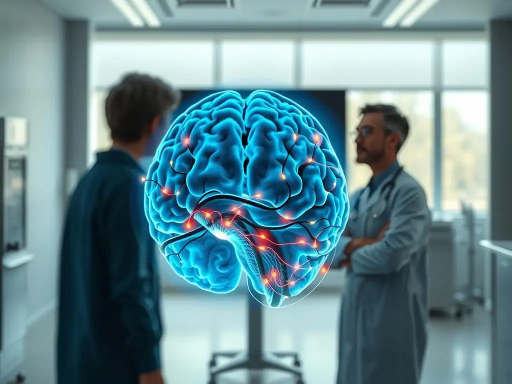
Welcome to Mind Care Guide, a space dedicated to nurturing your mental well-being. Ever wondered how the developing brain impacts childhood mental health? In this article, we delve into the fascinating world of childhood mental health brain imaging, offering a glimpse into the groundbreaking research shaping our understanding. Discover how these advanced techniques are helping us unlock the secrets behind mental health challenges in children and adolescents, and what this means for early intervention and future care. Ready to explore the intricate connection between the brain and the mind?
Pediatric Brain Imaging: Studying Mental Health Development
- Introduction to Pediatric Mental Health Brain Imaging
- Understanding Brain Development
- Overview of Brain Imaging Techniques
- Applications of Brain Imaging in Mental Health Research
- Ethical Considerations in Brain Imaging Research
- Advancements and Future Directions in Brain Imaging
- Resources for Mental Health Awareness
- Conclusion: The Future of Brain Imaging in Understanding and Improving Mental Health
-
Frequently Asked Questions
- What is pediatric brain imaging and how does it relate to childhood mental health?
- Why is brain imaging important for understanding childhood mental health challenges?
- What are some common brain imaging techniques used in the study of childhood mental health brain imaging?
- How can the findings from childhood mental health brain imaging studies be used to improve mental well-being?
- Are there any risks associated with brain imaging in children, and how are they minimized?
- Final Reflection: The Evolving Landscape of Childhood Mental Health
- Unlock Your Child's Potential: Next Steps for Brain Health
Introduction to Pediatric Mental Health Brain Imaging
The Importance of Brain Imaging in Understanding Mental Health
Understanding the complexities of mental health conditions in children and adolescents requires looking beyond observable behaviors. While clinical assessments and behavioral observations are crucial, they don't reveal the underlying biological processes occurring within the developing brain. This is where childhood mental health brain imaging plays a transformative role.
By providing non-invasive windows into brain structure and function, these techniques allow researchers and clinicians to explore the neural basis of mental health disorders, potentially identifying differences or patterns associated with conditions like anxiety, depression, ADHD, and autism spectrum disorder (ASD). This deeper biological understanding is vital for developing more targeted and effective interventions tailored to the specific needs of young people.
Research utilizing pediatric brain imaging for mental health has shown significant progress in recent decades. For instance, studies have indicated that youth with certain anxiety disorders may exhibit heightened activity in brain regions involved in processing fear, such as the amygdala, compared to typically developing peers.
Similarly, investigations into attention-deficit/hyperactivity disorder using brain imaging in child and adolescent mental health have pointed towards differences in connectivity within neural networks responsible for executive function and attention. These findings, while often correlational and part of ongoing research, provide critical insights into how the brain develops and functions differently in individuals experiencing mental health challenges, paving the way for potential early identification and intervention strategies.
Focus on Neuroscience of Mental Health
The field of neuroscience of mental health specifically investigates the biological underpinnings of psychiatric conditions, seeking to understand how alterations in brain structure, function, and connectivity contribute to symptoms and experiences. Within this field, childhood mental health brain imaging is indispensable. The developing brain is a dynamic system, undergoing rapid changes throughout childhood and adolescence.
These critical periods of development are also times when many mental health conditions first emerge. Studying the brain during these formative years allows researchers to not only identify potential biological markers of risk or resilience but also to understand how environmental factors, experiences, and interventions might influence neural trajectories.
By focusing on the neuroscience of mental health through brain imaging in child and adolescent mental health, researchers can explore questions like: Are there specific patterns of brain development that increase vulnerability to depression? How does trauma experienced in early childhood impact the maturation of stress-response circuits? Can brain imaging predict which young people are most likely to respond to a particular therapy?
While definitive answers are still being sought, the insights gained from this research are steadily advancing our understanding of mental health from a biological perspective. This neuroscientific lens complements psychological and social approaches, providing a more comprehensive view of the factors contributing to mental well-being and illness in youth.
Understanding Brain Development
Typical Brain Maturation Processes
Brain development is a complex, lifelong process, but childhood and adolescence are particularly critical periods characterized by rapid changes. One key process is synaptogenesis, the formation of new synapses (connections between neurons), followed by synaptic pruning, where less used connections are eliminated, making the brain more efficient. Another crucial process is myelination, the insulation of nerve fibers with a fatty substance called myelin, which speeds up neural communication. These processes occur at different rates across various brain regions, with sensory and motor areas maturing earlier than higher-order cognitive areas like the prefrontal cortex.
During adolescence, significant structural and functional changes continue, particularly in the prefrontal cortex (involved in planning, decision-making, and impulse control) and limbic system (involved in emotion and reward). The limbic system tends to mature earlier than the prefrontal cortex, which some theories suggest contributes to the characteristic risk-taking and heightened emotional reactivity often observed during the teenage years. Understanding these typical developmental trajectories is essential for childhood mental health brain imaging research, as it provides a baseline against which to compare the brains of young people experiencing mental health difficulties and identify potential deviations from normative development.
Key Brain Regions Involved in Mental Health
Several brain regions and networks are consistently implicated in mental health and are key targets for childhood mental health brain imaging studies. The amygdala, part of the limbic system, is central to processing emotions, particularly fear and threat, and is often found to show altered activity in anxiety disorders and trauma-related conditions.
The hippocampus, also within the limbic system, is crucial for memory formation and is affected by stress and depression. The prefrontal cortex (PFC), especially the dorsolateral and ventromedial areas, plays a vital role in executive functions, emotion regulation, and social cognition; disruptions here are linked to conditions like ADHD, depression, and conduct disorders.
Beyond individual regions, research using brain imaging in child and adolescent mental health increasingly focuses on neural networks – interconnected regions working together. The default mode network (DMN), active during rest and self-referential thought, and the salience network, which detects important stimuli and switches between other networks, are frequently studied. Altered connectivity or activity within these networks has been observed in various conditions, including ASD, depression, and schizophrenia. Understanding how these regions and networks develop and interact in childhood provides critical insights for pediatric brain imaging for mental health.
How Brain Imaging Techniques Help Visualize Brain Development
Brain imaging techniques offer non-invasive ways to visualize and measure aspects of brain development in living children and adolescents. Structural MRI can track changes in gray matter volume (neuron cell bodies) and white matter volume (myelinated axons) across development, showing how different brain regions grow or shrink over time. For example, studies using structural MRI have mapped the pruning of gray matter in the cortex from childhood through adolescence.
Functional MRI (fMRI) allows researchers to observe brain activity patterns and functional connectivity (how different brain regions communicate) during tasks or at rest. This helps visualize how neural networks mature and become more integrated or specialized over development.
Diffusion Tensor Imaging (DTI) specifically maps white matter tracts, revealing the integrity and organization of the brain's connections, which are undergoing significant myelination and refinement throughout childhood and adolescence. By applying these various techniques, childhood mental health brain imaging research can build a comprehensive picture of typical brain maturation and identify how developmental trajectories might differ in youth with mental health challenges.
Overview of Brain Imaging Techniques
Magnetic Resonance Imaging (MRI)
Magnetic Resonance Imaging (MRI) is a widely used non-invasive technique in childhood mental health brain imaging research. It uses strong magnetic fields and radio waves to create detailed images of the brain's structure and function. MRI is safe for children as it does not use ionizing radiation, unlike X-rays or CT scans. However, it requires participants to lie still inside a scanner, which can be challenging for some young children. Specialized protocols and child-friendly environments are often used to help make the scanning experience more comfortable and successful for pediatric populations.
MRI provides high-resolution images, making it excellent for visualizing anatomical details. Different types of MRI sequences can highlight different tissue properties, allowing researchers to differentiate between gray matter, white matter, and cerebrospinal fluid. The versatility of MRI makes it a cornerstone of studies investigating the neurobiological basis of mental health conditions in youth, from examining overall brain size to looking at specific regions and their connections.
Structural MRI: Examining Brain Anatomy
Structural MRI provides static images that detail the physical characteristics of the brain, including its shape, size, and tissue composition. Researchers use structural MRI to measure the volume or thickness of different brain regions, such as the cortex, hippocampus, or amygdala. They can also assess measures like cortical surface area or gyrification (the folding of the cortex). By comparing these structural measures between groups of children – for example, those with and without a particular mental health condition – researchers can identify potential anatomical differences.
Studies utilizing structural MRI in childhood mental health brain imaging have, for instance, reported differences in amygdala volume in youth with anxiety disorders or variations in cortical thickness in certain areas in children with ADHD or ASD compared to control groups. While structural differences don't tell the whole story of brain function, they can indicate potential developmental variations that might be associated with increased vulnerability to certain mental health challenges. These studies often involve large datasets to account for normal variations in brain anatomy across development.
Functional MRI (fMRI): Assessing Brain Activity
Functional MRI (fMRI) measures brain activity by detecting changes in blood flow. When a brain region is active, it requires more oxygenated blood, which has different magnetic properties than deoxygenated blood. fMRI detects these changes in blood oxygenation levels (the BOLD signal). By measuring the BOLD signal while participants perform tasks (like emotional processing, cognitive challenges, or social interactions) or simply rest, researchers can identify which brain regions are active and how different regions communicate with each other (functional connectivity).
In the context of childhood mental health brain imaging, fMRI is invaluable for understanding how neural circuits function in youth with conditions like depression or anxiety. For example, fMRI studies have shown altered connectivity between the amygdala and prefrontal cortex in anxious adolescents during emotional regulation tasks. Research on ADHD using fMRI has revealed differences in activity within attention and executive function networks. Resting-state fMRI, which measures brain activity while the participant is not performing a specific task, is also widely used to study intrinsic functional networks and their connectivity patterns in young populations with various mental health conditions.
Electroencephalography (EEG)
Electroencephalography (EEG) is a non-invasive technique that measures electrical activity in the brain using electrodes placed on the scalp. Unlike MRI, which provides excellent spatial resolution, EEG excels at temporal resolution, meaning it can capture brain activity changes very quickly, within milliseconds. This makes it ideal for studying dynamic brain processes related to attention, perception, and cognitive processing. EEG is relatively inexpensive and less sensitive to movement than fMRI, making it particularly useful for studies with infants and young children.
EEG recordings show brain waves, which are patterns of electrical activity characterized by different frequencies (e.g., alpha, beta, theta, delta waves). Researchers can analyze these wave patterns during different states (like sleep or wakefulness) or in response to specific stimuli.
Event-Related Potentials (ERPs), derived from EEG data, measure the brain's electrical response to specific events or stimuli, providing insights into sensory processing, attention, and cognitive functions. Childhood mental health brain imaging using EEG has been employed to study conditions like epilepsy (where abnormal electrical activity is central), as well as to investigate attention deficits in ADHD or altered responses to social stimuli in ASD.
Magnetoencephalography (MEG): A Non-Invasive Method for Studying Brain Function
Magnetoencephalography (MEG) is another neuroimaging technique that measures brain activity. Similar to EEG, MEG captures neural activity directly by detecting the tiny magnetic fields produced by electrical currents in neurons. Like EEG, MEG offers excellent temporal resolution, allowing researchers to pinpoint when brain activity occurs with high precision. However, MEG has better spatial resolution than EEG for activity originating from deeper brain structures, although it is less sensitive to activity from deep structures compared to fMRI. MEG is also completely non-invasive and silent, which can be advantageous for pediatric studies compared to the noisy environment of an MRI scanner.
MEG is often used in childhood mental health brain imaging research to study the timing and location of neural responses during cognitive or sensory tasks. For example, MEG has been used to investigate auditory processing differences in children with dyslexia or ASD, or to examine the timing of attention-related brain responses in ADHD. While MEG is less widely available than MRI or EEG due to its cost and specialized equipment, it offers unique capabilities for understanding the rapid sequence of events in neural processing that may be disrupted in various developmental and mental health conditions.
Diffusion Tensor Imaging (DTI): Mapping White Matter Tracts
Diffusion Tensor Imaging (DTI) is a specialized MRI technique that measures the diffusion of water molecules in the brain. Water diffuses more freely along the direction of white matter fibers (axons) than across them because the myelin sheath and cell membranes act as barriers. By measuring the direction and magnitude of water diffusion, DTI can infer the orientation and integrity of white matter tracts, the bundles of myelinated axons that connect different brain regions. This allows researchers to map the structural connectivity of the brain.
In childhood mental health brain imaging, DTI is crucial for understanding how the brain's wiring develops and how differences in white matter tracts might relate to mental health challenges. For instance, studies using DTI have identified alterations in white matter pathways connecting regions involved in social processing in youth with ASD.
Research on ADHD has pointed to differences in tracts within networks related to attention and impulse control. DTI provides insights into the structural backbone of neural networks, complementing functional measures by showing how the physical connections between brain regions might contribute to typical or atypical brain function and development.
Applications of Brain Imaging in Mental Health Research
Identifying Neural Correlates of Anxiety Disorders
Childhood anxiety disorders are among the most common mental health conditions in youth, affecting an estimated 7% of children aged 3-17. Brain imaging studies have been instrumental in identifying neural patterns associated with pediatric anxiety. Research using fMRI often shows heightened reactivity or altered connectivity of the amygdala, a key region for processing fear and threat, in anxious children and adolescents when presented with emotional stimuli. For example, one study might show greater amygdala activation in anxious youth compared to controls when viewing fearful faces.
Furthermore, studies using fMRI and DTI have investigated the circuits involved in emotion regulation, particularly the connections between the amygdala and the prefrontal cortex. Findings often suggest weaker or altered connectivity in these pathways in anxious youth, which could contribute to difficulties in regulating fear and worry. While no single brain scan can diagnose anxiety, these findings from childhood mental health brain imaging contribute to understanding the biological vulnerabilities associated with the condition and can inform the development of neurobiologically informed therapies.
Brain Imaging Studies of ADHD
Attention-Deficit/Hyperactivity Disorder (ADHD) is characterized by persistent patterns of inattention and/or hyperactivity-impulsivity, affecting about 9% of children aged 3-17. Brain imaging in child and adolescent mental health has provided significant insights into the neural basis of ADHD. Structural MRI studies have, on average, found slightly smaller volumes in certain brain regions like the prefrontal cortex, caudate nucleus, and cerebellum in children with ADHD compared to their peers, although there is considerable overlap between groups. These differences are often subtle and not diagnostic for individuals.
Functional MRI studies have highlighted differences in activity and connectivity within brain networks involved in attention, executive function, and reward processing. For instance, studies often show reduced activity in prefrontal regions during tasks requiring sustained attention or inhibitory control. Resting-state fMRI research has also pointed to altered connectivity within the default mode network and between the DMN and other networks in youth with ADHD. These findings from childhood mental health brain imaging help researchers understand the neural circuits that may contribute to the core symptoms of inattention, impulsivity, and hyperactivity observed in ADHD.
Investigating the Neurological Basis of Autism Spectrum Disorder (ASD)
Autism Spectrum Disorder (ASD) is a complex developmental condition characterized by challenges with social interaction and communication, and restricted or repetitive behaviors or interests. Brain imaging in child and adolescent mental health has revealed significant heterogeneity in the brains of individuals with ASD, reflecting the spectrum nature of the disorder. Early structural MRI studies sometimes reported larger brain volumes in young children with ASD, particularly in certain regions, followed by potentially slower growth in later childhood compared to typical development.
Functional and connectivity studies using fMRI and DTI are crucial in ASD research. Many studies have reported differences in functional connectivity, often showing reduced long-range connectivity (connections between distant brain regions) and increased local connectivity (connections within nearby regions) in certain networks, although findings can vary. For example, altered connectivity within social brain networks or language processing regions is frequently observed.
DTI studies have also identified differences in white matter tracts connecting these areas. These findings from childhood mental health brain imaging contribute to understanding the diverse neural atypicalities associated with ASD and how they might relate to the characteristic behavioral traits.
Exploring Brain Changes in Depression
Pediatric depression is a serious mood disorder that can affect children and adolescents, with prevalence estimates for major depressive disorder reaching around 2-3% in children and 5-8% in adolescents. Brain imaging in child and adolescent mental health has begun to shed light on the neural circuits involved in youth depression. Structural MRI studies have sometimes noted smaller hippocampal volumes or altered cortical thickness in certain areas in depressed youth, though findings are not always consistent.
Functional MRI studies frequently identify altered activity and connectivity within brain networks related to emotion processing, reward, and cognitive control. For instance, research might show reduced activity in reward circuits (like the striatum) when anticipating positive outcomes, or heightened activity in regions involved in processing negative emotions. Altered connectivity within the default mode network and between the DMN and other regions is also commonly observed. These findings from childhood mental health brain imaging suggest that depression in youth is associated with disruptions in neural circuits that regulate mood, motivation, and self-referential thinking.
Understanding the Impact of Trauma on Development
Experiences of trauma and adversity in childhood can have profound and lasting impacts on brain development and mental health. Brain imaging in child and adolescent mental health is being used to investigate these effects. Studies in youth exposed to trauma have sometimes reported smaller volumes in regions like the hippocampus and amygdala, which are highly sensitive to stress hormones. However, findings can be complex and depend on the type, timing, and severity of the trauma.
Functional MRI studies have shown altered reactivity in stress and fear circuits, such as the amygdala, and differences in how these regions connect with areas involved in emotion regulation, like the prefrontal cortex. For example, youth with a history of trauma might show exaggerated amygdala responses to threat cues or reduced activation in PFC regions during emotion regulation tasks.
DTI studies have also explored potential alterations in white matter tracts connecting these regions. This childhood mental health brain imaging research underscores how adverse experiences can sculpt the developing brain, increasing vulnerability to conditions like PTSD, anxiety, and depression later in life.
Ethical Considerations in Brain Imaging Research
Informed Consent and Assent
Conducting brain imaging research with children and adolescents presents unique ethical challenges, particularly regarding consent. Since children are considered a vulnerable population, direct consent must be obtained from a parent or legal guardian. However, it is also crucial to obtain assent from the child participants themselves, especially from older children and adolescents who have the capacity to understand the study's procedures, risks, and benefits. Assent means the child actively agrees to participate after being informed in age-appropriate language.
The process of obtaining informed consent and assent for childhood mental health brain imaging must be thorough and transparent. Researchers must explain the procedures, including what it's like to be in the scanner, the time commitment, and any potential discomforts (like noise or needing to stay still). They must clearly state that participation is voluntary and that the child or parent can withdraw at any time without penalty. Ensuring both the parent and child feel comfortable and fully understand their involvement is paramount.
Data Privacy and Confidentiality
Brain imaging data is inherently sensitive and potentially identifiable, raising significant concerns about privacy and confidentiality, especially when studying vulnerable populations like children with mental health conditions. Researchers must implement robust measures to protect participant data. This typically involves anonymizing or de-identifying the data as soon as possible, replacing names and other identifying information with unique codes. Data should be stored securely on protected servers with restricted access.
When sharing data, particularly in large-scale collaborative studies common in childhood mental health brain imaging, strict protocols must be followed to ensure data remains de-identified. Researchers must comply with relevant privacy regulations, such as HIPAA in the United States. Participants and their families should be fully informed about how their data will be collected, stored, used, and potentially shared, ensuring they understand the steps taken to protect their privacy.
Minimizing Risk and Maximizing Benefit
All research involving human participants, especially children, must carefully weigh potential risks against potential benefits. In pediatric brain imaging for mental health, the physical risks of techniques like MRI, EEG, and MEG are generally considered minimal, as they are non-invasive and do not involve radiation. However, there can be psychological risks, such as anxiety about being in a scanner, discomfort from noise, or distress if a child finds a task difficult. Researchers must take steps to minimize these risks, such as using mock scanners for practice, providing ear protection, and having trained staff present to support the child.
The potential benefits of childhood mental health brain imaging research are primarily societal: advancing scientific understanding of mental health development, which can lead to better diagnostic tools, prevention strategies, and treatments in the future. While individual participants may not receive direct clinical benefit from a research scan, they contribute to knowledge that can ultimately help other children. Ethical research design requires that the potential gains in knowledge justify the time commitment and minimal risks undertaken by the young participants and their families.
Advancements and Future Directions in Brain Imaging
Developing Novel Brain Imaging Techniques
The field of brain imaging is continuously evolving, with researchers developing novel techniques and refining existing ones to improve spatial and temporal resolution, reduce scanning time, and enhance comfort, particularly for pediatric populations. For example, advancements in MRI hardware and pulse sequences are leading to faster scanning, better signal-to-noise ratio, and the ability to acquire multiple types of data simultaneously. New analysis methods, such as advanced machine learning algorithms, are also pushing the boundaries of what can be learned from complex brain imaging datasets.
Beyond refinements to current methods, entirely new approaches are being explored. Portable or wearable brain imaging technologies, such as diffuse optical tomography (DOT) or newer generations of MEG systems, are being developed that might eventually allow for brain activity measurement in more naturalistic settings outside the confines of a traditional scanner. These advancements hold promise for overcoming some of the limitations of current childhood mental health brain imaging, potentially making studies more accessible and comfortable for young participants and allowing for the investigation of brain function during real-world tasks.
Integrating Brain Imaging with Other Measures of Mental Health
The brain is just one piece of the puzzle in understanding complex mental health conditions. A significant future direction in childhood mental health brain imaging is the integration of imaging data with other levels of information. This includes genetic data (e.g., common variants or rare mutations), epigenetic markers (changes in gene expression), behavioral assessments (from parents, teachers, and the child), cognitive testing, physiological measures (like heart rate or cortisol levels), and environmental factors (such as family dynamics or exposure to stress).
By combining brain imaging data with these diverse measures, researchers can build more comprehensive models of mental health development. For example, a study might investigate how genetic risk factors interact with early environmental stress to influence the development of anxiety-related brain circuits, which in turn affects a child's behavioral response to fear. This multimodal approach in brain imaging in child and adolescent mental health aims to identify complex interactions between biological, psychological, and environmental factors that contribute to mental well-being and vulnerability.
Personalized Approaches to Mental Healthcare Using Brain Imaging
Ultimately, a major goal of childhood mental health brain imaging research is to contribute to more personalized and effective mental healthcare for children and adolescents. While currently primarily a research tool, there is significant interest in whether brain imaging might one day help predict which young people are at highest risk for developing a condition, how a condition might progress, or which specific treatment (e.g., a particular therapy or medication) is most likely to be effective for an individual child.
For example, ongoing research is exploring whether patterns of brain activity or connectivity observed in fMRI scans might predict response to cognitive behavioral therapy (CBT) for anxiety or depression in youth. Other studies are investigating if DTI measures of white matter integrity could predict outcomes in children with ADHD receiving stimulant medication.
While these applications are still largely experimental and not yet used in routine clinical practice, the hope is that future advancements in childhood mental health brain imaging will provide clinicians with additional tools to tailor interventions to the unique brain and needs of each child, moving towards precision psychiatry for youth.
Resources for Mental Health Awareness
Support for Families and Caregivers
Navigating mental health challenges in children and adolescents can be difficult for families and caregivers. Fortunately, numerous resources are available to provide support, information, and guidance. These include national and local mental health organizations focused on children and families, which offer helplines, support groups, educational materials, and directories of mental health professionals. Pediatricians are also a crucial first point of contact and can provide referrals to specialists.
Many organizations provide specific information about various conditions like anxiety, depression, ADHD, and ASD, including guidance on recognizing symptoms, seeking help, and supporting a child through treatment. Online resources, while requiring careful vetting for credibility, can also offer valuable information and connect families with support networks. Seeking support is a sign of strength, and connecting with others who understand or accessing professional guidance can make a significant difference in managing childhood mental health challenges.
Accessing Information on Brain Imaging Studies and Mental Health
For those interested in learning more about childhood mental health brain imaging research specifically, academic institutions and research centers often have websites detailing their ongoing studies and findings. Scientific journals publish research results, though these can be highly technical. Reputable mental health organizations and science communication websites sometimes publish summaries or articles explaining brain imaging findings in more accessible language.
It is important to access information from reliable sources, such as university research pages, government health institutes (like the National Institute of Mental Health in the U.S.), or established mental health charities. These sources can provide accurate information about the role of brain imaging in advancing our understanding of mental health development in children and adolescents, while also managing expectations about the current clinical applicability of these techniques.
Conclusion: The Future of Brain Imaging in Understanding and Improving Mental Health
Childhood mental health brain imaging has emerged as a powerful tool for exploring the intricate relationship between brain development and mental well-being in children and adolescents. Techniques like MRI, fMRI, EEG, MEG, and DTI provide unprecedented insights into the structural, functional, and connectivity differences associated with various mental health conditions in youth. While still primarily a research endeavor, these studies are fundamentally changing how we understand the biological underpinnings of conditions like anxiety, depression, ADHD, and ASD during critical developmental periods.
As the field continues to advance with novel techniques, multimodal approaches, and sophisticated data analysis, the potential for childhood mental health brain imaging to inform clinical practice grows. While not yet a diagnostic tool for individual children, the knowledge gained is contributing to the development of more targeted interventions and holds promise for personalized approaches to mental healthcare in the future. Ethical considerations surrounding pediatric research, including informed consent, data privacy, and participant well-being, remain paramount as researchers continue to unlock the secrets of the developing brain to improve the lives of children and adolescents facing mental health challenges.
Frequently Asked Questions
What is pediatric brain imaging and how does it relate to childhood mental health?
Pediatric brain imaging uses techniques like MRI to study the brain structure and function of children and adolescents. This can help researchers and clinicians understand the biological basis of mental health development and disorders.
Why is brain imaging important for understanding childhood mental health challenges?
Brain imaging provides a non-invasive way to visualize the developing brain. This allows researchers to identify potential abnormalities or differences in brain activity that may contribute to mental health issues in children.
What are some common brain imaging techniques used in the study of childhood mental health brain imaging?
Common techniques include Magnetic Resonance Imaging (MRI), which provides detailed structural and functional images. Other methods include Electroencephalography (EEG) for measuring brain electrical activity and Positron Emission Tomography (PET) for assessing brain metabolism.
How can the findings from childhood mental health brain imaging studies be used to improve mental well-being?
By identifying neural correlates of mental disorders, brain imaging can help develop targeted interventions. This can lead to more personalized and effective treatments for children and adolescents struggling with mental health challenges.
Are there any risks associated with brain imaging in children, and how are they minimized?
While MRI is generally safe, some concerns exist regarding sedation for younger children and potential exposure to radiation with certain techniques. Protocols are in place to minimize these risks and prioritize the child's safety.
Final Reflection: The Evolving Landscape of Childhood Mental Health
The non-invasive study of the developing brain through imaging techniques offers unprecedented insight into the complex origins of childhood mental health disorders. This understanding is not merely academic; it holds the key to earlier diagnosis, more effective interventions, and ultimately, a brighter future for millions of children struggling silently. The implications are profound, extending far beyond the research lab and into the lives of families and communities worldwide.
The journey of understanding the intricate relationship between brain structure, function, and mental well-being in children is far from over, but the progress made is undeniably inspiring. The ability to visualize the developing brain, to witness the subtle shifts associated with mental health challenges, has revolutionized our approach to care. The brain is the most complex organ in the body, and understanding its development is crucial to understanding mental health.
This simple statement underscores the monumental task before us, but also the immense potential for positive change that lies within our grasp. The ongoing research detailed in this article serves as a testament to the dedication and innovation driving this crucial field.
Consider the implications of this research for your own life. Perhaps you are a parent, educator, or simply a concerned citizen. Regardless of your background, understanding the science behind childhood mental health empowers you to advocate for better resources, to recognize early warning signs, and to foster a more compassionate and supportive environment for young people.
Let the images of the developing brain, presented in this article, serve as a reminder of the incredible potential within each child, and the vital importance of supporting their mental well-being. Engage with the ongoing conversation, learn more about the latest advancements, and become part of the solution. The future of childhood mental health depends on it.
Unlock Your Child's Potential: Next Steps for Brain Health
Thanks for exploring the fascinating world of pediatric brain imaging and its role in understanding mental health development! You've gained valuable insights into how we can better support young minds. Now, let's take action.
- Dive deeper: Explore reputable resources online, like the National Institute of Mental Health (NIMH) website, to learn more about child and adolescent mental health research and available support services. This is your primary action to continue learning.
- Share your knowledge: Discuss this article with a friend, family member, or healthcare professional to spark conversation about the importance of early intervention and support systems for children.
- Consider attending a local mental health awareness event or workshop to learn practical strategies for supporting children's well-being.
What specific steps will you take today to prioritize the mental health of the children in your life?