
AI and the Brain: How Machine Learning Enhances Mental Health Imaging Analysis and Interpretation

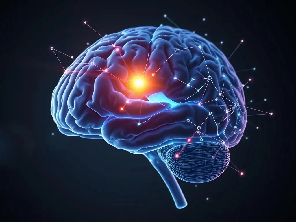
Welcome to Mind Care Guide, your trusted source for navigating the complexities of mental well-being. Feeling overwhelmed by the possibilities of modern neuroscience? Imagine a future where AI can unlock the secrets hidden within brain scans, leading to earlier and more accurate diagnoses. This week, we delve into the fascinating world of AI mental health imaging analysis, exploring how machine learning is revolutionizing the way we understand and treat mental health conditions. Intrigued? Discover how this cutting-edge technology is paving the way for a brighter future for mental healthcare.
AI and the Brain: Revolutionizing Mental Health Imaging Analysis
- Introduction to AI in Mental Health Imaging
- AI and Machine Learning Fundamentals in Neuroimaging
- AI-Powered Techniques for Neuroimaging Analysis
-
Applications of AI in Analyzing Specific Mental Health Conditions
- AI in Depression Diagnosis and Treatment Monitoring Using fMRI
- Machine Learning for Early Detection of Mild Cognitive Impairment and Alzheimer's Disease Using MRI
- AI-Enhanced Imaging for Understanding Schizophrenia's Neural Correlates Using PET
- Using AI to Analyze Amygdala Activity in Anxiety Disorders Using fMRI
- Benefits of AI-Enhanced Neuroimaging Analysis
- Ethical Considerations and Challenges in AI-Driven Neuroimaging
- Future Directions in AI for Mental Health Neuroimaging
- Conclusion: Transforming Mental Health Through AI in Neuroimaging
-
Frequently Asked Questions
- How is AI mental health imaging analysis being used to improve diagnoses?
- What types of mental health conditions can benefit from AI mental health imaging analysis?
- What are the potential benefits of using AI mental health imaging analysis for treatment planning?
- What are the ethical considerations surrounding the use of AI mental health imaging analysis?
- How accessible is AI mental health imaging analysis currently, and what are the future prospects?
- Final Reflection: The Dawn of AI-Powered Mental Healthcare
- Unlock the Power of AI in Mental Health: Take the Next Step
Introduction to AI in Mental Health Imaging
The Role of Neuroimaging in Mental Health Research and Clinical Practice
Neuroimaging techniques like Magnetic Resonance Imaging (MRI), functional MRI (fMRI), Positron Emission Tomography (PET), and Electroencephalography (EEG) have become indispensable tools in understanding the complex landscape of mental health. These methods allow researchers and clinicians to peer into the living brain, observing its structure, function, and metabolic activity. Structural MRI provides detailed images of brain anatomy, revealing potential differences in gray matter volume or cortical thickness associated with various conditions.
fMRI measures brain activity by detecting changes in blood flow, offering insights into neural circuits and connectivity patterns relevant to mood, cognition, and behavior. PET scans can visualize specific neurotransmitter systems or metabolic processes, crucial for understanding disorders like depression or addiction.
In clinical practice, neuroimaging can sometimes aid in differential diagnosis, rule out organic causes for psychiatric symptoms (like tumors or strokes), and potentially inform treatment selection. For instance, identifying specific patterns of connectivity via fMRI might help predict response to certain therapies. While not yet a primary diagnostic tool for most psychiatric disorders due to their complexity and heterogeneity, the data derived from these scans are vital for advancing our fundamental understanding of how mental health conditions manifest in the brain.
Researchers analyze these images to identify biomarkers – measurable indicators of a biological state – that could eventually lead to more objective diagnostic criteria and personalized treatment approaches. The sheer volume and complexity of the data generated, however, pose significant challenges for traditional analysis methods.
Limitations of Traditional Neuroimaging Analysis Methods
Analyzing neuroimaging data traditionally involves labor-intensive, manual, or semi-automated processes that can be time-consuming and susceptible to human variability. Identifying subtle structural anomalies or complex functional connectivity patterns across vast datasets requires expert knowledge and significant computational resources. Manual delineation of brain regions, for example, can vary between different analysts, introducing inter-rater variability that might affect the reliability of findings. Furthermore, traditional statistical methods often focus on group averages or predefined regions of interest, potentially overlooking subtle, yet significant, patterns or variations present in individual brains.
The sheer scale of modern neuroimaging studies exacerbates these limitations. A single fMRI scan can generate millions of data points per subject, and studies often involve hundreds or even thousands of participants. Sifting through this immense data to identify meaningful biomarkers or understand complex neural interactions using conventional methods is a monumental task.
This difficulty in processing and interpreting high-dimensional data limits the speed at which new discoveries can be translated into clinical applications and highlights the need for more efficient and robust analytical tools. This is where the potential for advanced AI mental health imaging analysis becomes clear, offering ways to handle this data deluge.
AI's Impact on Enhancing Neuroimaging Interpretation
Artificial intelligence, particularly machine learning and deep learning, is rapidly transforming the field of neuroimaging analysis by providing powerful tools to overcome the limitations of traditional methods. AI algorithms can process vast amounts of complex brain imaging data much faster and more consistently than humans, reducing analysis time from hours to minutes or even seconds for certain tasks. This acceleration is crucial for both research, enabling quicker validation of hypotheses, and potentially for future clinical applications where timely results are important.
Moreover, AI models can identify subtle, complex patterns and relationships within neuroimaging data that might be invisible to the human eye or traditional statistical approaches. By learning directly from the data, these algorithms can uncover novel biomarkers, improve the accuracy of automated brain region segmentation, and enhance the prediction of clinical outcomes based on brain characteristics.
The application of AI in brain imaging analysis is not only improving the efficiency and reliability of existing methods but also opening up entirely new avenues for research, leading to a deeper and more nuanced understanding of the neural underpinnings of mental health conditions. This burgeoning area of AI mental health imaging analysis holds significant promise for the future.
AI and Machine Learning Fundamentals in Neuroimaging
Differentiating AI, Machine Learning, and Deep Learning in Neuroimaging
Understanding the relationship between AI, machine learning, and deep learning is fundamental to appreciating their application in neuroimaging analysis. Artificial intelligence (AI) is the broadest concept, referring to the simulation of human intelligence in machines programmed to think and learn. It encompasses any technique that enables computers to mimic human cognitive functions, such as problem-solving, learning, and decision-making, when applied to tasks like analyzing brain scans.
Machine learning (ML) is a subset of AI that provides systems the ability to automatically learn and improve from experience without being explicitly programmed. In the context of neuroimaging, ML algorithms are trained on large datasets of brain scans to identify patterns, make predictions, or classify images.
Deep learning (DL), in turn, is a specialized subfield of machine learning that uses artificial neural networks with multiple layers (hence "deep") to learn complex patterns from data. These deep architectures, such as convolutional neural networks (CNNs) for image analysis, are particularly well-suited for processing high-dimensional data like brain images, automatically extracting relevant features without manual intervention, which is highly valuable for sophisticated AI mental health imaging analysis.
Common Machine Learning Algorithms for Mental Health Neuroimaging
Various machine learning algorithms are employed in the analysis of neuroimaging data for mental health research. Support Vector Machines (SVMs) are frequently used for classification tasks, such as distinguishing between individuals with a specific mental health condition and healthy controls based on structural or functional brain features.
SVMs work by finding the optimal hyperplane that separates different classes in a high-dimensional feature space. Another popular method is Random Forests, an ensemble learning technique that builds multiple decision trees and combines their outputs to improve accuracy and robustness, often used for both classification and regression tasks, like predicting symptom severity from brain data.
Other algorithms include Logistic Regression for binary classification, K-Nearest Neighbors (KNN) for classifying data points based on the majority class of their neighbors, and clustering algorithms like K-Means for identifying subgroups within patient populations based on neuroimaging patterns. Principal Component Analysis (PCA) and Independent Component Analysis (ICA) are common unsupervised learning techniques used for dimensionality reduction and identifying independent components of brain activity in fMRI data. These diverse ML techniques provide a powerful toolkit for researchers applying AI mental health imaging analysis to complex brain data.
Deep Learning Architectures for Advanced Neuroimaging Analysis
Deep learning architectures have revolutionized neuroimaging analysis, particularly for tasks involving complex pattern recognition and feature extraction directly from raw or minimally preprocessed images. Convolutional Neural Networks (CNNs) are the most prominent deep learning models in neuroimaging, excelling at image-based tasks like segmentation, classification, and registration. CNNs use convolutional layers to automatically learn hierarchical features from images, from basic edges to complex structural patterns, making them ideal for analyzing the spatial relationships within brain scans. For instance, 3D CNNs are used to process volumetric MRI data directly, identifying subtle atrophy patterns associated with conditions like Alzheimer's.
Recurrent Neural Networks (RNNs), and their variants like Long Short-Term Memory (LSTM) networks, are useful for analyzing sequential data, which can include time-series fMRI data or longitudinal studies tracking brain changes over time. Autoencoders and Generative Adversarial Networks (GANs) are employed for tasks like data augmentation, anomaly detection, and generating synthetic images for training purposes, helping to overcome limitations posed by small datasets. These advanced deep learning models enable more sophisticated AI mental health imaging analysis, allowing for deeper insights into the complex neural correlates of mental disorders.
AI-Powered Techniques for Neuroimaging Analysis
Automated Segmentation of Brain Regions Using AI
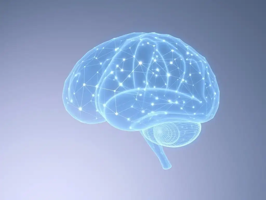
Automated segmentation of brain regions is a fundamental step in neuroimaging analysis, crucial for measuring volumes, cortical thickness, or analyzing activity within specific areas. Traditionally, this involved manual tracing or using semi-automated atlas-based methods that can be time-consuming and prone to errors or variability. AI, particularly deep learning models like Convolutional Neural Networks (CNNs), has dramatically improved the accuracy and speed of this process. Models trained on large datasets of expert-segmented brain images can automatically and consistently delineate dozens or even hundreds of distinct brain structures, including complex and variable regions like the hippocampus, amygdala, or specific cortical areas.
For example, researchers using 3D U-Net architectures, a type of CNN, have achieved highly accurate segmentation of subcortical structures in T1-weighted MRI scans. These models can process a full brain volume in minutes, whereas manual segmentation might take hours for just a few structures. This automation reduces inter-rater variability to near zero and enables large-scale analysis of brain morphology across massive datasets, facilitating the identification of subtle structural differences linked to mental health conditions that might have been missed by less precise methods. This enhanced capability is a core component of modern AI mental health imaging analysis.
Biomarker Discovery in Neuroimaging with Machine Learning
Machine learning algorithms are powerful tools for discovering potential neuroimaging biomarkers for mental health conditions. Unlike traditional methods that often test pre-defined hypotheses about specific regions or connections, ML can explore complex patterns across the entire brain or within functional networks to identify features that best differentiate between groups (e.g., patients vs. controls) or predict clinical outcomes (e.g., treatment response, disease progression). Algorithms like Support Vector Machines, Random Forests, and deep learning models are trained on neuroimaging data along with clinical information to find these discriminative patterns.
For instance, studies have used ML to identify functional connectivity patterns derived from fMRI data that can potentially serve as biomarkers for major depressive disorder or schizophrenia. An ML model might learn that a specific pattern of reduced connectivity between the prefrontal cortex and amygdala is highly correlated with depressive symptoms.
Similarly, algorithms analyzing structural MRI data can identify subtle combinations of volume or shape changes across multiple brain regions indicative of early neurodegenerative processes associated with cognitive decline. While still largely a research tool, this data-driven approach facilitated by AI mental health imaging analysis holds significant promise for developing objective and predictive markers for mental health challenges.
AI-Driven Pattern Recognition in Brain Activity
AI is revolutionizing the analysis of complex brain activity patterns captured by techniques like fMRI and EEG. These methods generate massive amounts of time-series data reflecting dynamic neural interactions. Traditional analysis often focuses on simple metrics like average activity in a region or pairwise connectivity. AI, particularly deep learning and advanced machine learning techniques, can identify more complex, non-linear patterns within this dynamic data, such as distributed network interactions, transient brain states, or subtle timing differences in neural responses.
For example, researchers are using deep learning models to classify different cognitive states or predict behavior based on real-time fMRI data by recognizing subtle, distributed patterns of activity across multiple brain networks. Similarly, AI algorithms analyzing EEG data can identify characteristic patterns of brain waves associated with conditions like epilepsy or sleep disorders with high accuracy.
Beyond classification, AI can also model the underlying generative processes of brain activity, potentially leading to a deeper understanding of how neural circuits malfunction in mental illness. This advanced pattern recognition is a frontier in AI mental health imaging analysis, providing unprecedented insights into the dynamic brain.
Applications of AI in Analyzing Specific Mental Health Conditions
AI in Depression Diagnosis and Treatment Monitoring Using fMRI
Major Depressive Disorder (MDD) is a complex condition with heterogeneous symptoms and potential underlying neural correlates. fMRI studies have revealed that depression is associated with altered connectivity within and between various brain networks, including the default mode network (DMN), executive control network (ECN), and salience network. Traditional analysis struggles to capture the full complexity of these network dysfunctions at an individual level. AI, specifically machine learning and deep learning algorithms, is being applied to fMRI data to identify patterns of functional connectivity that can potentially aid in the diagnosis of depression and predict treatment response.
Research studies, often involving datasets of hundreds or thousands of participants, have trained ML models to differentiate individuals with MDD from healthy controls based on fMRI connectivity features, reporting accuracies often exceeding 70-80% in research settings. These models might identify specific patterns like hyperconnectivity within the DMN or hypoconnectivity between the ECN and DMN as key discriminators. Furthermore, AI is being explored to predict which patients are likely to respond to specific treatments, such as psychotherapy or antidepressant medication, by analyzing baseline fMRI scans.
For instance, certain pre-treatment connectivity profiles identified by an ML model might indicate a higher likelihood of remission with a particular therapy. While not yet a clinical diagnostic tool, this application of AI mental health imaging analysis is actively being researched to improve precision in depression care.
Machine Learning for Early Detection of Mild Cognitive Impairment and Alzheimer's Disease Using MRI
Mild Cognitive Impairment (MCI) and Alzheimer's Disease (AD) are neurodegenerative conditions characterized by progressive cognitive decline and specific structural changes in the brain, such as atrophy in the hippocampus and other medial temporal lobe structures. Early and accurate detection is crucial for intervention and management. Structural MRI is a standard tool for assessing brain atrophy, but visual inspection and traditional quantitative methods can be challenging for identifying subtle changes, especially in the early stages of MCI. Machine learning algorithms are proving highly effective in analyzing structural MRI data to identify these subtle patterns indicative of early neurodegeneration.
ML models, including SVMs and deep learning CNNs, are trained on large datasets of MRI scans from healthy individuals, patients with MCI, and patients with AD. These algorithms learn to recognize complex volumetric and shape changes across multiple brain regions that are characteristic of these conditions. Studies have demonstrated that ML models can achieve high accuracy (often above 85-90% in research cohorts) in differentiating AD from healthy aging and also in predicting which individuals with MCI are likely to progress to AD within a few years.
For example, an AI model might detect a specific pattern of subtle atrophy not just in the hippocampus but also in related cortical areas that strongly correlates with future cognitive decline. This use of machine learning for AI mental health imaging analysis is a promising area for improving early diagnostic capabilities for dementia.
AI-Enhanced Imaging for Understanding Schizophrenia's Neural Correlates Using PET
Schizophrenia is a severe mental illness associated with complex alterations in brain structure, function, and neurochemistry. Positron Emission Tomography (PET) is a neuroimaging technique particularly useful for studying neurochemical processes, such as dopamine receptor binding or glucose metabolism, which are thought to be implicated in schizophrenia pathophysiology. Analyzing PET data traditionally involves quantifying tracer uptake in predefined regions, which may not capture the full spatial complexity or subtle variations across individuals. AI-enhanced imaging analysis is being applied to PET data to gain a deeper understanding of schizophrenia's neural correlates.
AI algorithms, including machine learning classifiers and deep learning models, can analyze PET images to identify complex patterns of metabolic activity or receptor availability that differentiate individuals with schizophrenia from controls or relate to specific symptom dimensions. For example, an AI model might analyze dopamine D2/D3 receptor binding patterns across the striatum and extrastriatal regions, identifying subtle deviations that are highly predictive of a schizophrenia diagnosis.
Furthermore, combining PET data with other modalities like MRI or clinical data using multi-modal AI approaches can provide a more comprehensive picture of the neural underpinnings of the disorder. This application of AI mental health imaging analysis using PET is contributing to a more nuanced understanding of the neurochemical basis of schizophrenia.
Using AI to Analyze Amygdala Activity in Anxiety Disorders Using fMRI
Anxiety disorders are characterized by excessive fear and worry, often linked to dysregulation in brain regions involved in processing emotion, particularly the amygdala. fMRI studies frequently examine amygdala activity and its connectivity with other brain areas during the presentation of emotional stimuli or during resting states. Traditional analysis might focus on average amygdala activation levels, but AI can provide a more detailed and nuanced understanding of its complex activity patterns and interactions within neural circuits in individuals with anxiety disorders.
AI algorithms, such as machine learning classifiers or deep learning models, can analyze the dynamic patterns of amygdala activity and its functional connectivity with regions like the prefrontal cortex or hippocampus from fMRI data. These models can identify subtle, distributed patterns of activity that are highly correlated with anxiety severity or specific anxiety disorder subtypes. For instance, an AI model might detect a specific pattern of hyper-responsivity in the amygdala coupled with reduced connectivity to regulatory prefrontal regions that is characteristic of social anxiety disorder.
Furthermore, AI can analyze how amygdala activity changes during therapeutic interventions, potentially serving as a biomarker for treatment effectiveness. This application of AI mental health imaging analysis is enhancing our ability to probe the neural basis of fear and anxiety.
Benefits of AI-Enhanced Neuroimaging Analysis
Increased Diagnostic Accuracy and Reliability
One of the most significant benefits of applying AI to neuroimaging analysis is the potential for increased diagnostic accuracy and reliability for mental health conditions. Traditional methods, relying heavily on visual inspection and manual measurements, can be subjective and prone to inter-rater variability. AI algorithms, once trained, apply consistent criteria across all scans, reducing this variability and improving the objectivity of the analysis. Furthermore, AI models can identify subtle patterns across multiple brain regions that might be missed by the human eye, leading to potentially more precise differentiation between conditions or between patients and healthy individuals.
In research settings, AI models have demonstrated impressive performance in classification tasks. For example, studies using deep learning to analyze structural MRI scans for Alzheimer's disease have reported diagnostic accuracies exceeding 90% in specific datasets. While translating these results to clinical practice requires rigorous validation on diverse populations, the inherent ability of AI to process complex, high-dimensional imaging data consistently and identify subtle biomarkers holds immense promise for improving the accuracy and reliability of neuroimaging interpretation in mental healthcare. This enhanced accuracy in AI mental health imaging analysis could eventually lead to earlier and more precise diagnoses.
Accelerated Neuroimaging Data Analysis
The sheer volume of data generated by modern neuroimaging scanners presents a major bottleneck in research and potential clinical workflows. Analyzing a single high-resolution structural MRI or a lengthy fMRI session using traditional manual or semi-automated methods can take hours. This time-consuming process limits the throughput of studies and can delay the availability of results. AI-powered techniques dramatically accelerate this process.
Automated segmentation algorithms based on deep learning, for instance, can segment a full brain volume into dozens or hundreds of regions in minutes, compared to hours for manual methods. Similarly, processing and analyzing large fMRI datasets for functional connectivity using AI can be significantly faster than traditional pipelines.
This acceleration is critical for large-scale research initiatives involving thousands of participants and is essential for potential future clinical applications where quick turnaround times for imaging reports are necessary for patient care. The speed offered by AI mental health imaging analysis allows researchers and clinicians to process more data and gain insights faster.
Personalized Mental Health Treatment Strategies
Mental health conditions are highly heterogeneous, and individuals often respond differently to the same treatment. Identifying which therapy is most likely to be effective for a specific patient based on their unique characteristics is a major goal of personalized medicine in psychiatry. Neuroimaging, analyzed with the help of AI, offers a promising avenue for developing more personalized treatment strategies.
AI algorithms can analyze an individual's neuroimaging data, potentially combined with other clinical, genetic, and behavioral information, to identify patterns that predict their response to different interventions. For example, machine learning models analyzing baseline fMRI connectivity patterns have shown potential in predicting response to specific types of psychotherapy or different classes of antidepressant medications in research studies.
By identifying neuroimaging biomarkers that predict treatment outcome, AI can help clinicians select the most appropriate therapy for a patient from the outset, avoiding trial-and-error approaches that can delay recovery. This potential for personalized medicine is a key benefit of advanced AI mental health imaging analysis.
Uncovering Novel Insights into Brain Function and Mental Disorders
Beyond improving existing tasks like segmentation and classification, AI is enabling researchers to uncover entirely new insights into brain function and the pathophysiology of mental disorders. By analyzing complex, non-linear relationships and distributed patterns across brain networks that are difficult for humans to detect, AI algorithms can reveal novel aspects of how mental health conditions manifest in the brain.
For example, unsupervised machine learning techniques can identify novel subtypes of depression or schizophrenia based purely on neuroimaging data, potentially reflecting distinct underlying neural mechanisms that might require different treatment approaches. Deep learning models analyzing dynamic fMRI data can uncover transient brain states or patterns of network switching that are altered in certain conditions.
AI can also facilitate the integration of neuroimaging data with other data types, such as genetics, clinical history, and behavioral assessments, to build more comprehensive models of mental illness. This data-driven discovery process, powered by AI mental health imaging analysis, is expanding our understanding of the brain in health and disease.
Ethical Considerations and Challenges in AI-Driven Neuroimaging
Data Privacy and Security in Mental Health Neuroimaging
The application of AI to mental health neuroimaging raises significant ethical challenges, particularly concerning data privacy and security. Neuroimaging data is highly sensitive, containing detailed information about an individual's brain structure and function, which could potentially be used to infer sensitive personal attributes or predispositions. Training robust AI models often requires access to very large datasets, which necessitates collecting, storing, and processing vast amounts of this sensitive information.
Ensuring the secure storage and transmission of neuroimaging data is paramount to prevent breaches that could expose individuals' private health information. Compliance with regulations like HIPAA in the United States or GDPR in Europe is essential, but the unique nature of brain data may require additional safeguards.
Techniques such as data anonymization, pseudonymization, federated learning (where models are trained locally on decentralized data without sharing the raw data), and differential privacy are being explored to protect privacy while enabling AI development. Addressing these challenges is critical for building trust and enabling the ethical advancement of AI mental health imaging analysis.
Mitigating Bias in AI Algorithms for Neuroimaging
Another critical ethical challenge is the potential for bias in AI algorithms used for neuroimaging analysis. AI models learn from the data they are trained on, and if the training data is not representative of the population in terms of demographics (age, sex, race, ethnicity), socioeconomic status, or even scanner type and acquisition parameters, the algorithm may perform poorly or produce biased results when applied to individuals from underrepresented groups.
For example, an AI model trained primarily on data from one ethnic group might be less accurate when analyzing scans from another, potentially leading to disparities in diagnosis or treatment recommendations.
Addressing algorithmic bias requires careful attention to dataset diversity and fairness metrics during model development and validation. Researchers and developers must actively work to include data from diverse populations and test models specifically for performance differences across subgroups. Techniques for bias detection and mitigation, such as re-weighting training data or using fairness-aware machine learning algorithms, are active areas of research. Ensuring fairness and equity in AI mental health imaging analysis is crucial to avoid exacerbating existing healthcare disparities.
Clinician's Role in Interpreting AI-Generated Neuroimaging Results
While AI can provide powerful analytical capabilities and insights from neuroimaging data, it is crucial that AI-generated results are not treated as definitive diagnoses or replacements for clinical judgment. The clinician's role in interpreting AI outputs remains essential. AI models provide statistical probabilities or identify patterns, but they do not understand the full clinical context of a patient, including their detailed history, symptoms, comorbidities, and individual circumstances.
Clinicians must understand how the AI model works (to the extent possible, especially with complex deep learning models), its limitations, and the confidence associated with its predictions. AI should be viewed as a decision-support tool that provides additional information to inform the clinician's assessment, not automate it. The final diagnosis and treatment plan must always be made by a qualified healthcare professional who can integrate the AI-generated insights with all other available clinical information. Fostering collaboration between AI developers and clinicians is vital to ensure that AI mental health imaging analysis is integrated safely and effectively into clinical workflows.
Future Directions in AI for Mental Health Neuroimaging
Integration of AI with Multi-Modal Data (Genetics, Clinical Data)
A significant future direction for AI in mental health neuroimaging is the integration of neuroimaging data with other types of biological and clinical information. Mental health conditions are complex and influenced by a myriad of factors, including genetics, environmental exposures, clinical history, behavioral data (e.g., from wearables or smartphone apps), and cognitive assessments. Analyzing neuroimaging data in isolation provides valuable but incomplete insights.
AI, particularly machine learning and deep learning, is uniquely positioned to integrate these diverse data modalities. Multi-modal AI models can learn complex interactions between genetic predispositions, brain structure/function, and clinical symptoms to create more comprehensive and accurate models of mental illness. For example, an AI model might combine structural MRI data with polygenic risk scores for schizophrenia and clinical symptom profiles to improve prediction accuracy or identify distinct patient subgroups. This integrated approach, facilitated by advanced AI mental health imaging analysis, holds the potential to unlock a deeper understanding of the complex etiology and presentation of mental disorders.
Explainable AI (XAI) for Transparent Neuroimaging Analysis
One of the challenges with complex AI models, particularly deep learning networks, is their "black box" nature – it can be difficult to understand why the model made a particular prediction or identified a specific pattern. For clinical applications and scientific discovery, understanding the reasoning behind AI outputs is crucial for building trust, validating findings, and gaining biological insights. This is where Explainable AI (XAI) comes into play.
XAI techniques aim to make AI models more transparent and interpretable. In neuroimaging, this involves developing methods to visualize which parts of the brain image or which features were most influential in the AI's decision (e.g., classifying a patient as depressed). Techniques like saliency maps, layer-wise relevance propagation, and attention mechanisms are being adapted for neuroimaging data to highlight the specific brain regions or connectivity patterns that drive an AI model's prediction. Developing robust XAI methods is essential for translating AI mental health imaging analysis from research tools into clinically actionable insights that clinicians can understand and trust.
AI-Driven Personalized Mental Healthcare Solutions
The ultimate goal of applying AI to mental health neuroimaging is to contribute to the development of truly personalized mental healthcare solutions. By integrating multi-modal data and leveraging advanced analytical capabilities, AI can help move beyond broad diagnostic categories towards understanding the unique biological and psychological profile of each individual.
In the future, AI-driven platforms could potentially analyze a patient's neuroimaging data alongside their genetic information, clinical history, and real-time behavioral data to provide a personalized risk assessment, predict the likelihood of developing a specific condition, recommend the most effective preventative strategies, or suggest the optimal treatment pathway (e.g., specific therapy type, medication dosage, neuromodulation target). AI could also continuously monitor treatment response using follow-up imaging or other data, allowing for dynamic adjustments to the care plan.
This vision of highly personalized, data-driven mental healthcare, powered by sophisticated AI mental health imaging analysis, represents a transformative future for the field.
Conclusion: Transforming Mental Health Through AI in Neuroimaging
Key Impacts of AI on Neuroimaging and Mental Health
Artificial intelligence, particularly machine learning and deep learning, is having a profound impact on the field of neuroimaging and its application to mental health. AI is transforming how brain imaging data is processed, analyzed, and interpreted, moving beyond the limitations of traditional manual and statistical methods. Key impacts include significantly accelerating the analysis workflow, enabling the processing of vast and complex datasets that were previously intractable. This speed and efficiency are crucial for both large-scale research initiatives and the potential integration of neuroimaging into faster clinical pipelines.
Furthermore, AI is enhancing the accuracy and reliability of neuroimaging analysis tasks, such as automated segmentation of brain regions and the identification of subtle patterns that serve as potential biomarkers for various mental health conditions. By learning directly from data, AI algorithms can uncover novel insights into the neural underpinnings of disorders like depression, schizophrenia, anxiety, and neurodegenerative conditions.
This data-driven discovery process is expanding our understanding of how mental illness manifests in the brain, paving the way for more objective and biologically informed approaches. The advancements in AI mental health imaging analysis are fundamentally changing the landscape of neuroscience research and its application to mental well-being.
The Future of AI in Improving Mental Health Outcomes
Looking ahead, the future of AI in mental health neuroimaging is incredibly promising, holding the potential to transform mental health outcomes. As AI algorithms become more sophisticated and datasets grow larger and more diverse, AI's ability to detect subtle biomarkers, predict disease trajectories, and forecast treatment response will continue to improve. The integration of neuroimaging data with other modalities like genetics, clinical records, and behavioral data will enable the creation of powerful multi-modal AI models capable of providing a holistic view of an individual's mental health profile.
Ultimately, AI is poised to play a crucial role in facilitating truly personalized mental healthcare. By analyzing complex individual data, AI could help clinicians make more informed decisions about diagnosis, prognosis, and treatment selection, moving away from a one-size-fits-all approach.
While ethical challenges related to data privacy, bias, and the need for clinical oversight must be continuously addressed, the ongoing advancements in AI mental health imaging analysis offer a clear path towards a future where brain imaging contributes more directly and effectively to improving the lives of individuals facing mental health challenges. The convergence of AI and neuroimaging represents a significant leap forward in our quest to understand and treat the brain.
Frequently Asked Questions
How is AI mental health imaging analysis being used to improve diagnoses?
AI algorithms are being trained to detect subtle patterns in brain scans (MRI, fMRI, PET) that human clinicians might miss. This can lead to earlier and more accurate diagnoses of mental health conditions.
What types of mental health conditions can benefit from AI mental health imaging analysis?
AI is showing promise in identifying biomarkers associated with conditions like depression, anxiety, schizophrenia, and PTSD. It can also help differentiate between subtypes of these disorders for personalized treatment.
What are the potential benefits of using AI mental health imaging analysis for treatment planning?
By identifying specific brain patterns associated with treatment response, AI can help clinicians predict which therapies are most likely to be effective for a particular patient. This aims to reduce trial-and-error and optimize treatment outcomes.
What are the ethical considerations surrounding the use of AI mental health imaging analysis?
Data privacy and security are paramount. There are concerns about algorithmic bias, ensuring fairness and avoiding discriminatory outcomes. Transparency and explainability of AI decisions are also crucial.
How accessible is AI mental health imaging analysis currently, and what are the future prospects?
Currently, AI mental health imaging analysis is largely in the research and development phase, with limited availability in clinical settings. Future prospects include wider adoption, integration into clinical workflows, and increased affordability for patients.
Final Reflection: The Dawn of AI-Powered Mental Healthcare
The integration of artificial intelligence into mental health imaging analysis is no longer a futuristic fantasy; it's a rapidly evolving reality with the potential to revolutionize diagnosis and treatment. This transformative technology offers unprecedented opportunities to improve the accuracy, speed, and accessibility of mental healthcare, ultimately leading to better patient outcomes and a more comprehensive understanding of the brain's complexities.
The impact of AI on mental health imaging is profound and far-reaching, reshaping our understanding of neurological and psychiatric disorders. It's not just about faster processing; it's about unlocking subtle patterns and insights previously invisible to the human eye. This shift has the potential to personalize treatment plans, leading to more effective interventions and improved quality of life for millions. The greatest advances in medicine have always come from the ability to see what was previously unseen.
This quote resonates deeply as AI allows us to peer into the brain with a clarity never before possible, revealing the intricate neural pathways underlying mental illness.
The future of mental healthcare is inextricably linked to the continued development and ethical implementation of AI. Consider the implications of this technology – faster diagnoses, more accurate predictions of treatment response, and the potential for early intervention. You have a role to play in this evolution. Stay informed, engage in thoughtful discussions about the ethical considerations, and support research that prioritizes responsible innovation. Let us work together to harness the power of AI to build a future where mental health care is accessible, effective, and truly transformative for all.
Unlock the Power of AI in Mental Health: Take the Next Step
Thanks for exploring the fascinating intersection of AI and mental health imaging with us! You've gained valuable insights into how technology is revolutionizing diagnosis and treatment. Now it's time to put that knowledge into action.
- Dive deeper: Research the latest advancements in AI-driven mental health initiatives. Find studies or organizations pushing boundaries in this exciting field. This could spark surprising discoveries!
- Share your thoughts: Discuss this article with colleagues, friends, or family. Initiate a conversation about the potential of AI in improving mental healthcare access and quality. Let's spread the word!
- Explore resources: Look for local or online mental health support groups that utilize technology or cutting-edge diagnostic methods. Take that extra step towards understanding your own mental wellbeing better.
What innovative application of AI in mental healthcare excites you the most, and why?