
Anxiety in Focus: What Brain Scans Tell Us About Worry, Fear, and the Brain

Welcome to Mind Care Guide, where understanding your mental well-being begins.
Feeling overwhelmed by anxiety? You're not alone. Our exploration into the neuroscience of mental health starts here, with a deep dive into anxiety brain scan interpretation techniques. Ever wondered what a scan can reveal about the inner workings of worry and fear? Join us as we unravel the complexities and offer insights that could reshape your understanding of anxiety. Let's begin!
Anxiety in Focus: What Brain Scans Tell Us About Worry, Fear, and the Brain
- Understanding Anxiety Through Brain Imaging: An Introduction
- Anxiety and the Brain: A Neurobiological Perspective
- Types of Brain Scans Used in Anxiety Research
- Anxiety Brain Scan Interpretation Techniques: A Deep Dive
-
Specific Anxiety Disorders and Brain Scan Findings
- Generalized Anxiety Disorder (GAD): Brain Scan Insights
- Panic Disorder: What Brain Scans Reveal About Panic Attacks
- Social Anxiety Disorder (SAD): Neural Mechanisms Uncovered by Imaging
- Post-Traumatic Stress Disorder (PTSD): Brain Scan Findings on Trauma and Anxiety
- Obsessive-Compulsive Disorder (OCD): Imaging the Brain's Compulsive Circuits
- The Future of Brain Imaging in Anxiety Treatment
- Limitations of Brain Scan Interpretation in Anxiety Research
- Conclusion: Brain Scans as a Powerful Tool for Understanding and Treating Anxiety
-
Frequently Asked Questions
- How do brain scans help us understand anxiety?
- What are some common brain scan interpretation techniques used for anxiety?
- Can a brain scan definitively diagnose an anxiety disorder?
- What specific brain regions are often examined in anxiety brain scans?
- What are the limitations of using brain scans to study anxiety?
- Final Reflection: Unraveling the Anxious Mind Through the Lens of Neuroscience
- Unlock Your Brain's Anxiety Code: Take Control Now!
Understanding Anxiety Through Brain Imaging: An Introduction
Anxiety disorders affect millions worldwide, manifesting as excessive worry, fear, and related behavioral and physical symptoms. While clinical assessment remains the cornerstone of diagnosis, advancements in neuroscience, particularly brain imaging, offer a deeper understanding of the underlying biological mechanisms. These non-invasive techniques provide a window into the living brain, allowing researchers to observe structural differences, functional activity patterns, and neurochemical imbalances associated with anxiety. Exploring how brain scans reveal the neurological underpinnings of anxiety helps demystify these complex conditions and paves the way for more targeted interventions.
The journey into the anxious brain via imaging techniques is relatively new but rapidly evolving. Early studies often focused on identifying specific brain regions that showed altered activity or volume in individuals with anxiety compared to healthy controls. More recent research delves into the intricate networks within the brain and how they communicate, offering insights into the dynamic processes that contribute to anxious states. Understanding the findings from these studies, including the specific anxiety brain scan interpretation techniques used, is crucial for both researchers and clinicians seeking a more complete picture of anxiety.
The Role of Neuroimaging in Anxiety Research
Neuroimaging plays a vital role in shifting our understanding of anxiety from purely psychological constructs to disorders with tangible biological correlates. Techniques like fMRI and PET scans allow scientists to observe brain activity in real-time or measure the distribution of neurotransmitters, providing empirical evidence for theories about the brain circuits involved in fear and worry. For instance, studies using fMRI have consistently shown heightened activity in the amygdala, a brain region critical for processing fear, when anxious individuals are exposed to stressful stimuli.
Furthermore, neuroimaging helps researchers differentiate between various anxiety disorders, which often share overlapping symptoms but may have distinct neural signatures. By comparing brain scan patterns across different diagnostic categories, researchers can identify specific biomarkers that might eventually aid in diagnosis or predict treatment response. The careful application of anxiety brain scan interpretation techniques is essential to translate complex imaging data into meaningful clinical insights, helping to refine our understanding of conditions like panic disorder, social anxiety, and generalized anxiety disorder.
The Anxiety & Depression Association of America (ADAA): Your Resource for Understanding Anxiety
Understanding anxiety requires reliable information, and organizations like the Anxiety & Depression Association of America (ADAA) serve as crucial resources. The ADAA provides up-to-date information on anxiety disorders, their symptoms, treatment options, and ongoing research. While brain imaging is a complex scientific field, the ADAA helps make information about the biological aspects of anxiety, including insights gleaned from brain scans, accessible to the public and professionals alike.
Navigating the world of anxiety can be challenging, but resources like the ADAA offer support, educational materials, and connections to help. They often highlight the latest research findings, including those from neuroimaging studies, explaining how these discoveries contribute to a better understanding of anxiety's impact on the brain. For anyone interested in learning more about how conditions like GAD or panic disorder affect the brain, the ADAA is an excellent starting point, complementing the detailed scientific insights provided by anxiety brain scan interpretation techniques.
Anxiety and the Brain: A Neurobiological Perspective
Anxiety is not just a feeling; it is rooted in the complex circuitry of the brain. At its core, anxiety involves the brain's threat detection system, which, in individuals with anxiety disorders, often becomes hyperactive or dysregulated. Key brain regions involved include the amygdala, prefrontal cortex (PFC), hippocampus, and insula. These areas form interconnected networks that process fear, regulate emotions, store memories of threatening events, and monitor internal bodily states.
The neurobiological perspective posits that imbalances or dysfunction within these networks contribute significantly to the development and maintenance of anxiety symptoms. For example, a common finding in anxiety research is reduced activity in parts of the PFC, which is responsible for executive functions like decision-making and regulating emotions, potentially impairing an individual's ability to control worry or fear responses. Understanding these neurological underpinnings is key to developing effective treatments that target the root causes of anxiety.
Identifying Brain Regions Involved in Anxiety Disorders
Numerous brain regions have been implicated in the pathophysiology of anxiety disorders. The amygdala, often referred to as the brain's "fear center," shows exaggerated responses to perceived threats in many anxious individuals. Studies have reported that the amygdala in individuals with panic disorder, for example, may show heightened reactivity to even neutral stimuli, contributing to the sudden onset of intense fear.
Beyond the amygdala, other regions play critical roles. The hippocampus, involved in memory formation, can link specific contexts to fear, contributing to phobias or PTSD. The insula processes internal bodily sensations, and its overactivity may contribute to the physical symptoms of anxiety, such as a racing heart or shortness of breath.
The prefrontal cortex, particularly the ventromedial PFC, is involved in regulating emotional responses; reduced connectivity or activity here might impair the brain's ability to dampen down excessive fear signals coming from the amygdala. Identifying these specific regions and understanding their interactions is a primary goal of anxiety brain scan interpretation techniques.
How Brain Scans Reveal Anxiety's Impact on Brain Function
Brain scans provide empirical evidence of how anxiety disorders alter brain function. Functional imaging techniques like fMRI measure changes in blood flow, which correlate with neural activity. By comparing fMRI scans of anxious individuals and healthy controls while they perform tasks or are at rest, researchers can pinpoint areas of atypical activity. For instance, studies might show increased activity in the amygdala and decreased activity in the prefrontal cortex when an individual with GAD is asked to worry about a hypothetical scenario.
Structural MRI scans, on the other hand, can reveal differences in brain volume or cortical thickness. While less consistently found than functional changes, some studies have reported subtle differences, such as slightly reduced hippocampal volume in chronic anxiety conditions like PTSD. PET scans can measure metabolic activity or the density of neurotransmitter receptors or transporters, offering insights into the neurochemical basis of anxiety. For example, PET studies have investigated serotonin and GABA systems, both implicated in anxiety regulation. The ability to reveal these functional and structural changes makes brain imaging indispensable for understanding the neural basis of anxiety.
Types of Brain Scans Used in Anxiety Research
Several neuroimaging techniques are employed in anxiety research, each offering a unique perspective on brain structure, function, or chemistry. The choice of technique depends on the specific research question being asked – whether researchers want to see which parts of the brain are active during a panic attack, measure the concentration of a particular neurotransmitter, or assess the structural integrity of brain tissue. Each method requires specific anxiety brain scan interpretation techniques to extract meaningful insights from the complex data generated.
Understanding the principles behind these different scanning methods is crucial for interpreting their findings. While structural scans provide a static picture, functional and molecular scans offer dynamic insights into brain processes related to anxious states. The combination of findings across different modalities often provides a more comprehensive understanding than any single technique alone.
Functional Magnetic Resonance Imaging (fMRI) and Anxiety: Detecting Brain Activity
Functional Magnetic Resonance Imaging (fMRI) is one of the most widely used techniques in anxiety research due to its relatively good spatial resolution and non-invasive nature. fMRI measures brain activity by detecting changes in blood flow (specifically, the blood-oxygen-level-dependent or BOLD signal) that occur when neurons are active. When a brain region becomes more active, it requires more oxygen, leading to an increase in blood flow to that area. By tracking these changes, fMRI can create maps of brain activity associated with specific tasks, stimuli (like fearful faces), or even during resting states.
In anxiety research, fMRI is frequently used to identify brain regions that show altered responses to emotional stimuli or exhibit abnormal patterns of connectivity within brain networks. For example, a classic fMRI finding in anxiety is the heightened amygdala response to threatening cues. Researchers might show participants fearful images while they are in the scanner and observe significantly stronger amygdala activation in individuals with social anxiety compared to non-anxious individuals.
Furthermore, resting-state fMRI, which measures brain activity when participants are not performing a specific task, has revealed altered functional connectivity between regions like the amygdala and prefrontal cortex in various anxiety disorders. Analyzing these complex patterns requires sophisticated anxiety brain scan interpretation techniques focused on connectivity analysis.
Positron Emission Tomography (PET) Scans: Examining Neurotransmitter Activity in Anxiety
Positron Emission Tomography (PET) scans offer a unique window into the brain's neurochemistry, allowing researchers to study the distribution and activity of specific neurotransmitters and receptors implicated in anxiety. Unlike fMRI, which primarily measures blood flow, PET uses radioactive tracers that bind to specific molecules in the brain, such as serotonin transporters, dopamine receptors, or glucose (to measure metabolic activity). By measuring the decay of the tracer, PET creates images showing where and how densely these molecules are distributed or how metabolically active different brain regions are.
PET studies have provided valuable insights into the neurochemical basis of anxiety. For instance, research has investigated the role of the serotonin system, a primary target for many anxiety medications (like SSRIs). PET scans using tracers that bind to serotonin transporters have sometimes shown altered density or availability in certain brain regions of individuals with anxiety disorders, although findings can vary depending on the specific disorder and study design.
Similarly, PET has been used to study the GABAergic system, the brain's main inhibitory system, which is also targeted by anti-anxiety medications like benzodiazepines. Analyzing the binding patterns and tracer kinetics in PET data requires specialized anxiety brain scan interpretation techniques focused on molecular imaging.
Electroencephalography (EEG): Measuring Brainwave Patterns in Anxious States
Electroencephalography (EEG) measures the electrical activity of the brain using electrodes placed on the scalp. While EEG has lower spatial resolution compared to fMRI or PET, it offers excellent temporal resolution, meaning it can capture brain activity changes occurring in milliseconds. This makes EEG particularly useful for studying the rapid neural oscillations or brainwave patterns associated with different cognitive and emotional states, including anxiety.
EEG research in anxiety often focuses on identifying specific patterns of brain activity, such as altered power in different frequency bands (e.g., alpha, beta, theta waves) or abnormal event-related potentials (ERPs), which are brain responses time-locked to specific stimuli. For example, studies have reported increased frontal theta activity or altered alpha asymmetry in individuals experiencing worry or anxiety.
EEG is also used in biofeedback and neurofeedback techniques aimed at helping individuals learn to regulate their brainwave patterns. While not strictly a "scan" in the imaging sense, EEG provides crucial data about brain function that complements findings from fMRI and PET, requiring distinct anxiety brain scan interpretation techniques centered on time-series analysis and signal processing.
Anxiety Brain Scan Interpretation Techniques: A Deep Dive
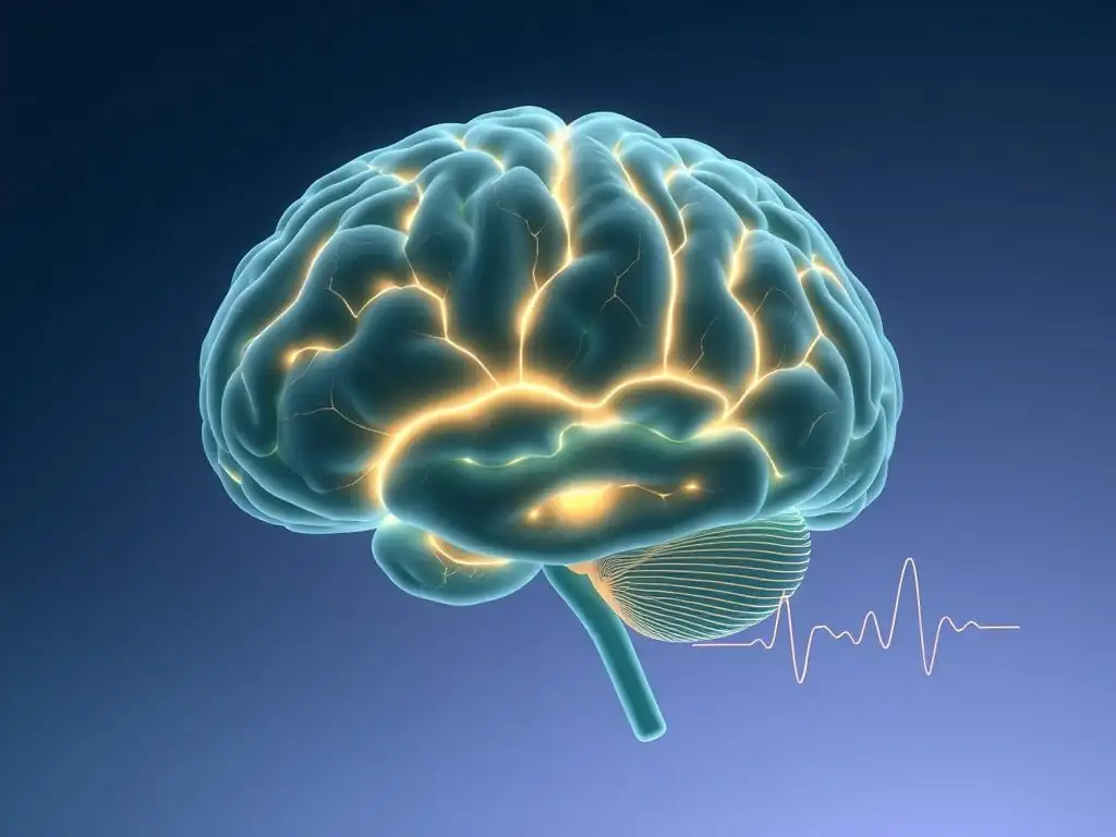
Interpreting brain scan data is a complex process that requires specialized knowledge and sophisticated computational tools. Raw imaging data, whether it's fMRI time series, PET tracer uptake values, or EEG waveforms, needs to be processed, analyzed, and statistically compared to identify meaningful patterns associated with anxiety. The specific anxiety brain scan interpretation techniques used vary depending on the imaging modality and the research question, but they all aim to translate signals from the scanner into insights about brain structure and function in anxious individuals.
These techniques involve multiple steps, including data acquisition, preprocessing (e.g., noise reduction, alignment), analysis (e.g., statistical modeling, connectivity analysis), and visualization. The choice of analysis method can significantly influence the findings, highlighting the importance of rigorous methodology and careful interpretation of results.
Decoding fMRI Data: Identifying Neural Correlates of Anxiety
Decoding fMRI data to identify neural correlates of anxiety typically involves statistical parametric mapping or network analysis. In statistical parametric mapping, researchers compare brain activity voxel-by-voxel (a voxel is a 3D pixel) or within predefined regions of interest (ROIs) between groups (e.g., anxious vs. control) or conditions (e.g., fear vs. neutral stimulus). For instance, a t-test might be used to identify voxels in the amygdala that show significantly higher activity in anxious participants viewing fearful faces compared to controls. This allows researchers to pinpoint specific brain areas involved.
Beyond identifying activated regions, network analysis techniques are increasingly used as part of anxiety brain scan interpretation techniques. These methods examine how different brain regions communicate or are functionally connected. Resting-state fMRI studies, for example, often use techniques like independent component analysis (ICA) or seed-based correlation analysis to identify functional networks (like the default mode network or salience network) and assess how connectivity within or between these networks is altered in anxiety disorders. Studies have shown altered connectivity between the amygdala and prefrontal cortex in various anxiety conditions, suggesting a breakdown in the brain's ability to regulate emotional responses.
Analyzing PET Scan Results: Neurochemical Imbalances in Anxiety Disorders
Analyzing PET scan results involves quantifying the distribution and binding of the radioactive tracer within different brain regions. This often requires techniques to correct for factors like tracer metabolism, blood flow, and individual head shape. For studies examining receptor density or transporter availability, kinetic modeling is often used. This involves measuring how quickly the tracer binds and unbinds from its target over time to estimate parameters like binding potential, which reflects the density of available receptors or transporters.
For example, a PET study investigating serotonin transporters in social anxiety might use a tracer like [11C]DASB. Analysis would involve calculating the binding potential of [11C]DASB in regions like the amygdala, thalamus, or striatum and comparing these values between individuals with social anxiety and controls. Findings from such studies have sometimes shown altered serotonin transporter availability in these regions, although results can be inconsistent across studies.
Metabolic PET scans using [18F]FDG measure glucose uptake as an indicator of neural activity and are analyzed similarly to fMRI data, comparing metabolic rates in different regions. These specific anxiety brain scan interpretation techniques provide crucial insights into the neurochemical landscape of anxiety.
EEG Analysis: Distinguishing Brainwave Patterns Associated with Different Anxiety Types
EEG analysis techniques focus on the temporal dynamics of brain electrical activity. Common methods include spectral analysis, which breaks down the EEG signal into different frequency bands (delta, theta, alpha, beta, gamma) and measures their power or amplitude in different scalp regions. Comparing the power in these bands between anxious individuals and controls, or between different anxiety states, can reveal characteristic patterns. For instance, increased power in the theta band in frontal regions is sometimes associated with excessive worry in GAD.
Another key technique is the analysis of event-related potentials (ERPs). ERPs are small voltage fluctuations in the EEG signal that are consistently associated with specific sensory, cognitive, or motor events. Researchers might present anxious participants with anxiety-provoking stimuli and analyze the amplitude and latency of specific ERP components (like the P300 or N400) that reflect attention, processing, or emotional evaluation.
Differences in these ERP components compared to controls can indicate altered neural processing of information in anxiety. More advanced anxiety brain scan interpretation techniques for EEG include connectivity analysis (measuring synchronization between different brain regions) and source localization (estimating where in the brain the electrical activity is originating), providing a more detailed picture of brain networks involved in anxiety.
Specific Anxiety Disorders and Brain Scan Findings
While general patterns emerge across anxiety disorders, brain imaging studies have also identified specific neural signatures associated with different diagnostic categories. These findings help refine our understanding of the unique biological underpinnings of conditions like GAD, panic disorder, social anxiety, PTSD, and OCD, even though there is considerable overlap and heterogeneity within each disorder. Examining these specific findings illustrates the power of anxiety brain scan interpretation techniques in differentiating conditions.
It is important to note that brain imaging findings are typically group-level differences and cannot currently diagnose an individual with an anxiety disorder. However, they provide valuable insights into the mechanisms driving these conditions.
Generalized Anxiety Disorder (GAD): Brain Scan Insights
Generalized Anxiety Disorder (GAD) is characterized by persistent and excessive worry about various topics. Brain imaging studies in GAD have often focused on the neural circuitry involved in worry and emotional regulation. Findings frequently point to altered activity and connectivity in the prefrontal cortex, amygdala, and insula. For instance, fMRI studies have sometimes shown reduced activation in the ventromedial prefrontal cortex during worry tasks, potentially indicating impaired top-down regulation of emotional responses originating in the amygdala.
Furthermore, resting-state fMRI studies in GAD have reported altered functional connectivity within brain networks, such as increased connectivity within the salience network (involving the insula and anterior cingulate cortex) and decreased connectivity between the salience network and the executive control network (involving parts of the prefrontal cortex). This suggests that the brain's system for detecting potential threats (salience network) might be hyperactive or poorly regulated by the areas responsible for higher-level control (executive control network) in GAD. These specific patterns are key targets for anxiety brain scan interpretation techniques in GAD research.
Panic Disorder: What Brain Scans Reveal About Panic Attacks
Panic disorder is marked by recurrent, unexpected panic attacks – sudden episodes of intense fear accompanied by severe physical symptoms. Brain imaging research on panic disorder has often focused on the fear circuit, particularly the amygdala, insula, and brainstem. Studies have reported increased activity in the amygdala and insula during induced panic attacks or in anticipation of them in individuals with panic disorder. The insula's role in processing interoceptive signals (internal bodily sensations) is particularly relevant here, as panic attacks involve intense physical symptoms.
Some PET studies have investigated neurochemical aspects, finding potential alterations in benzodiazepine receptor binding in brain regions like the orbitofrontal cortex and insula in individuals with panic disorder. These receptors are part of the GABA system, which helps calm neural activity. Findings suggest a possible dysfunction in this inhibitory system contributing to the sudden surge of fear and physical symptoms during a panic attack. Understanding these specific regional activations and neurochemical differences is vital for applying appropriate anxiety brain scan interpretation techniques to panic disorder research.
Social Anxiety Disorder (SAD) involves intense fear of social situations due to worries about being judged or scrutinized. Brain imaging studies in SAD consistently highlight heightened amygdala activity in response to social threat cues, such as critical faces or public speaking tasks. This exaggerated fear response is considered a core neural feature of SAD.
Beyond the amygdala, studies have also shown altered activity in brain regions involved in self-processing and social cognition, such as the medial prefrontal cortex and the temporoparietal junction. For instance, fMRI studies have found increased activity in areas associated with self-referential processing when individuals with SAD anticipate or engage in social interactions. Connectivity analyses have also revealed altered interactions between the amygdala and prefrontal regions, potentially reflecting a failure of cognitive control mechanisms to regulate excessive fear in social contexts. These specific responses to social stimuli are key considerations when applying anxiety brain scan interpretation techniques to SAD.
Post-Traumatic Stress Disorder (PTSD): Brain Scan Findings on Trauma and Anxiety
Post-Traumatic Stress Disorder (PTSD), while classified separately in some diagnostic manuals, shares significant overlap with anxiety disorders and involves dysregulation of fear responses following trauma exposure. Brain imaging studies in PTSD have consistently reported a pattern of altered activity in the amygdala, hippocampus, and medial prefrontal cortex (mPFC). Specifically, findings often show heightened amygdala reactivity to trauma-related cues, reduced volume and/or activity in the hippocampus (involved in contextualizing fear memories), and reduced activity in the mPFC (involved in fear extinction and regulation).
These findings align with the core symptoms of PTSD: exaggerated startle responses (amygdala), difficulty distinguishing safe from unsafe contexts (hippocampus), and impaired ability to inhibit fear responses (mPFC). For example, an fMRI study might show significantly lower mPFC activity when individuals with PTSD are asked to inhibit a fear response learned in a safe context compared to healthy controls. Understanding these consistent patterns is crucial for applying anxiety brain scan interpretation techniques in the context of trauma-related conditions.
Obsessive-Compulsive Disorder (OCD): Imaging the Brain's Compulsive Circuits
Obsessive-Compulsive Disorder (OCD), also sometimes grouped with anxiety disorders due to the distress caused by obsessions, is characterized by intrusive thoughts (obsessions) and repetitive behaviors or mental acts (compulsions). Brain imaging research in OCD has primarily focused on the cortico-striatal-thalamo-cortical (CSTC) circuits, particularly involving the orbitofrontal cortex (OFC), anterior cingulate cortex (ACC), striatum (caudate and putamen), and thalamus.
Studies have consistently reported increased activity or altered connectivity within these circuits in individuals with OCD, especially during symptom provocation tasks (e.g., showing images related to contamination for someone with washing compulsions). For example, fMRI studies often show heightened activity in the OFC and ACC when individuals with OCD are exposed to stimuli triggering their obsessions. These regions are involved in error detection, conflict monitoring, and reward processing.
Altered function in the striatum, involved in habit formation and behavioral control, is thought to contribute to the compulsive behaviors. These findings suggest a hyperactive or dysregulated circuit involved in sensing errors or potential threats and initiating actions, which is a key focus for anxiety brain scan interpretation techniques in OCD.
The Future of Brain Imaging in Anxiety Treatment
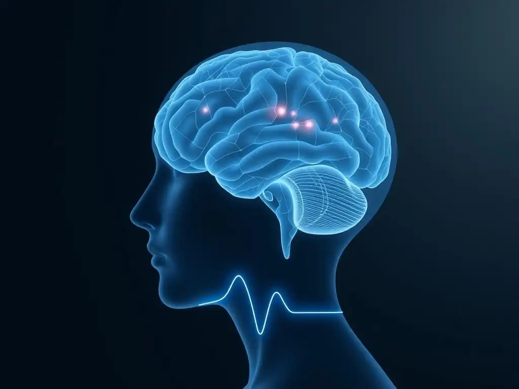
The insights gained from anxiety brain scan interpretation techniques are not just academic; they hold significant promise for improving the treatment of anxiety disorders. Future applications could range from using imaging data to personalize treatment selection to developing novel, image-guided therapies. While still largely in the research phase, the potential to leverage brain imaging to enhance clinical outcomes is substantial.
As imaging technology advances and our understanding of the brain circuits involved in anxiety deepens, brain scans may become increasingly integrated into clinical practice. The goal is to move towards a more precise, biologically informed approach to managing anxiety.
Using Brain Scans to Personalize Anxiety Treatment Approaches
One of the most exciting potential future applications of brain imaging is in personalizing anxiety treatment. Currently, selecting the most effective treatment (e.g., specific type of psychotherapy, medication, or combination) for an individual with anxiety is often a trial-and-error process. Brain imaging could potentially provide biomarkers that predict how well a person might respond to different interventions.
For example, research is exploring whether specific patterns of pre-treatment brain activity or connectivity, as revealed by fMRI or EEG, can predict response to cognitive behavioral therapy (CBT) versus a selective serotonin reuptake inhibitor (SSRI). Preliminary studies have suggested that certain patterns of connectivity in the amygdala-prefrontal circuit might predict CBT outcomes. If validated, such findings could guide clinicians in selecting the most appropriate first-line treatment for an individual, potentially saving time and reducing patient distress associated with ineffective treatments. Advancing anxiety brain scan interpretation techniques is crucial for making such personalized predictions reliable.
Neurofeedback and Brain Stimulation: Image-Guided Therapies for Anxiety
Brain imaging is also playing a role in the development and application of novel, image-guided therapies for anxiety, such as neurofeedback and brain stimulation techniques. Real-time fMRI neurofeedback, for instance, allows individuals to see their own brain activity (e.g., in the amygdala) in real-time while they are in the scanner and learn to voluntarily regulate it. Early studies are exploring whether training individuals with anxiety to reduce amygdala activity using neurofeedback can help alleviate symptoms.
Brain stimulation techniques, such as transcranial magnetic stimulation (TMS) or transcranial direct current stimulation (tDCS), aim to modulate the activity of specific brain regions or networks implicated in anxiety. Brain imaging can help guide the placement of stimulation coils or electrodes to target the most relevant areas identified by research.
For example, applying TMS to the dorsomedial prefrontal cortex, a region involved in emotional regulation, is being investigated as a potential treatment for GAD. Brain imaging findings inform which areas to target and how to optimize stimulation parameters, demonstrating a direct application of anxiety brain scan interpretation techniques to therapy development.
The Promise of Early Detection and Intervention Through Brain Imaging
While currently not used for routine diagnosis, brain imaging holds promise for potentially aiding in the early detection of individuals at risk for developing anxiety disorders or identifying the earliest signs of dysfunction. Research is exploring whether subtle differences in brain structure, function, or connectivity can be detected in individuals with subclinical anxiety or those with a genetic predisposition before a full-blown disorder develops.
Identifying these early neural markers could open the door for preventative interventions. For example, if imaging reveals altered connectivity patterns in adolescents with mild anxiety symptoms but a family history of severe anxiety, targeted early interventions like specific forms of therapy or lifestyle changes could potentially prevent the escalation to a full disorder. This area of research is still nascent, but the potential for brain imaging to contribute to proactive mental healthcare in anxiety is significant, relying heavily on the development of sophisticated anxiety brain scan interpretation techniques for longitudinal data.
Limitations of Brain Scan Interpretation in Anxiety Research
Despite the significant advances and potential of brain imaging, it is crucial to acknowledge the limitations inherent in anxiety brain scan interpretation techniques. The brain is incredibly complex, and anxiety disorders are heterogeneous, meaning they manifest differently in different people. Interpreting imaging data requires navigating technical challenges, ethical considerations, and the need to integrate findings with other levels of analysis.
Brain imaging provides valuable insights, but it is just one piece of the puzzle. It needs to be considered alongside clinical assessments, behavioral observations, genetic information, and environmental factors for a truly comprehensive understanding of anxiety.
Challenges in Interpreting Complex Brain Data
One of the primary challenges in anxiety brain scan interpretation techniques is the sheer complexity of the data. Brain activity involves intricate networks of interconnected regions, and simple interpretations based on single region activity are often insufficient. Analyzing functional connectivity, network dynamics, and the interplay between multiple brain areas requires advanced statistical methods and computational power.
Furthermore, findings across different studies can sometimes be inconsistent due to variations in methodology, sample characteristics (age, gender, severity of symptoms, comorbidity), and the specific tasks or stimuli used during scanning. Replication of findings is essential but not always achieved. Distinguishing between cause and effect is also difficult in cross-sectional imaging studies – does a particular brain pattern cause anxiety, or is it a consequence of having chronic anxiety? Longitudinal studies, which follow individuals over time, are needed to address these questions, but they are expensive and time-consuming.
Ethical Considerations in Using Brain Scans for Anxiety Diagnosis
Currently, brain imaging is not used to diagnose anxiety disorders in clinical practice, and there are significant ethical considerations to address before it could be. Relying solely on a brain scan for diagnosis could lead to misdiagnosis due to the variability in findings and the limitations in interpreting complex brain data. Furthermore, labeling someone based on a scan could potentially lead to stigma or discrimination.
Issues surrounding data privacy and security are also paramount when collecting and storing sensitive brain imaging data. As research progresses towards potential clinical applications, careful consideration must be given to how imaging data will be used, who will have access to it, and how potential biases in interpretation can be mitigated. The development of robust and ethically sound anxiety brain scan interpretation techniques for clinical use is a critical step that requires careful deliberation.
Future Research: Integrating Genetics and Brain Imaging for a Holistic Understanding of Anxiety
Future research in anxiety and brain imaging is moving towards integrating multiple levels of analysis, including genetics, environmental factors, and behavioral data, with imaging findings. Genetic studies have identified numerous genes that confer risk for anxiety disorders, but these genes typically have small effects and interact with environmental factors.
Combining genetic information with brain imaging (a field known as imaging genetics) can help researchers understand how genetic variations influence brain structure and function, and consequently, anxiety susceptibility. For example, researchers might study how a specific genetic variant associated with serotonin processing affects activity in the amygdala or connectivity in the fear circuit as measured by fMRI. This integrated approach, which requires sophisticated techniques for combining diverse datasets and applying multi-modal anxiety brain scan interpretation techniques, holds promise for a more holistic understanding of the complex interplay of factors that contribute to anxiety disorders.
Conclusion: Brain Scans as a Powerful Tool for Understanding and Treating Anxiety
Brain imaging has emerged as a powerful tool in anxiety research, offering unprecedented insights into the neural underpinnings of worry, fear, and related symptoms. Techniques like fMRI, PET, and EEG, coupled with sophisticated anxiety brain scan interpretation techniques, have allowed scientists to identify key brain regions and networks involved in different anxiety disorders, revealing patterns of altered activity, connectivity, and neurochemistry. These findings provide a biological context for the psychological and behavioral manifestations of anxiety, helping to validate the experiences of those affected.
While challenges remain in interpreting complex brain data and translating research findings into routine clinical practice, the potential for brain imaging to revolutionize our approach to anxiety is significant. From personalizing treatment selection based on individual brain profiles to guiding novel therapies like neurofeedback and brain stimulation, brain imaging is paving the way for more effective and targeted interventions. As research continues to integrate imaging with genetic and environmental data, our understanding of anxiety will become even more comprehensive, ultimately leading to better prevention and treatment strategies.
The Importance of Integrating Brain Imaging with Clinical Assessment
It is crucial to emphasize that findings from brain imaging studies should always be integrated with comprehensive clinical assessment. Brain scans provide valuable biological data, but they do not capture the full picture of an individual's subjective experience, their personal history, their specific symptoms, or their functional impairment – all of which are essential for accurate diagnosis and treatment planning.
Currently, brain imaging is a research tool that informs our general understanding of anxiety disorders at a group level. Clinical assessment, including detailed interviews, behavioral observations, and standardized questionnaires, remains the gold standard for diagnosing anxiety and tailoring treatment to the individual's needs. The future likely lies in combining the objective biological insights from brain imaging with the rich, personal information gathered through clinical assessment to create a truly personalized and effective approach to managing anxiety. Mastering anxiety brain scan interpretation techniques is valuable for researchers, but clinicians must integrate these findings with their patient's unique presentation.
The Anxiety & Depression Association of America (ADAA): Resources for Managing Anxiety and Promoting Mental Well-being
For individuals struggling with anxiety, understanding the condition is a critical step towards managing it effectively. Organizations like the Anxiety & Depression Association of America (ADAA) provide invaluable resources, offering evidence-based information on symptoms, diagnosis, and treatment options. They connect individuals with support groups, therapists, and the latest research findings, including accessible summaries of how science, including brain imaging, is advancing our understanding of anxiety.
The ADAA's commitment to promoting mental well-being aligns perfectly with the goal of using scientific advancements, like those derived from anxiety brain scan interpretation techniques, to improve lives. By providing reliable information and support, the ADAA empowers individuals to seek help and navigate their journey towards recovery and improved mental health. For anyone affected by anxiety, exploring the resources offered by the ADAA is a vital step towards finding effective strategies for coping and healing.
Frequently Asked Questions
How do brain scans help us understand anxiety?
Brain scans, such as fMRI and PET scans, allow researchers and clinicians to visualize brain activity and identify areas associated with anxiety. They reveal differences in brain structure and function in individuals with anxiety disorders compared to those without.
What are some common brain scan interpretation techniques used for anxiety?
Common techniques include analyzing regional brain activity, examining connectivity between brain regions, and identifying biomarkers associated with specific anxiety symptoms. These techniques help pinpoint neural correlates of anxiety.
Can a brain scan definitively diagnose an anxiety disorder?
No, a brain scan alone cannot diagnose an anxiety disorder. They provide valuable information but are used in conjunction with clinical assessments and patient history to reach a diagnosis.
What specific brain regions are often examined in anxiety brain scans?
Regions of interest include the amygdala (fear processing), the prefrontal cortex (emotional regulation), and the hippocampus (memory). Activity and connectivity in these regions are often altered in individuals with anxiety.
What are the limitations of using brain scans to study anxiety?
Limitations include the high cost of scans, the complexity of data interpretation, and the fact that brain scans show correlation, not necessarily causation. Also, results must be interpreted in the context of individual variability.
Final Reflection: Unraveling the Anxious Mind Through the Lens of Neuroscience
The intricate dance between our thoughts, feelings, and brain activity is finally yielding its secrets, offering unprecedented insights into the nature of anxiety. Understanding the neural pathways involved is no longer a distant aspiration; it's a crucial step towards developing more effective and personalized treatments. This knowledge empowers us to move beyond simplistic labels and delve into the complex biological reality of anxiety disorders.
The journey through the world of anxiety brain scan interpretation techniques has been profoundly illuminating, highlighting the power of objective measurement in understanding subjective experience. It's a stark reminder that anxiety isn't simply a weakness of character, but a complex neurological process with tangible, measurable manifestations. The mind is not a vessel to be filled, but a fire to be kindled.
– Plutarch. This quote resonates deeply, as the research presented underscores the need to understand the "fire" of anxiety—its underlying mechanisms—to effectively nurture and manage it. The ongoing development of these techniques holds immense promise for refining diagnostic tools and tailoring therapeutic interventions to individual needs.
Now, consider your own experiences with worry and fear. How might this new understanding of the anxious brain reshape your perspective? Perhaps it encourages greater self-compassion, a deeper appreciation for the biological underpinnings of your struggles, or a renewed sense of hope for effective treatment.
Take a moment to reflect on the power of this knowledge and how it can empower you to navigate your own mental landscape with greater understanding and self-awareness. The future of anxiety treatment lies not just in medication or therapy alone, but in the synergistic integration of these approaches with the precise insights provided by advanced neuroimaging techniques. Let's continue to explore, understand, and ultimately alleviate the burden of anxiety, one brain scan at a time.
Unlock Your Brain's Anxiety Code: Take Control Now!
Thanks for diving deep into the world of anxiety with us! You've now gained valuable insights into how your brain responds to worry and fear. Ready to put this knowledge into action?
- Start a mindful breathing practice: Even 5 minutes a day can make a noticeable difference in managing anxious feelings. This is your primary tool to actively combat anxiety.
- Share this article with someone you care about: Spreading awareness and understanding is key to breaking down the stigma surrounding anxiety.
- Explore guided meditation apps: These can provide structured support for your newfound mindful breathing practice.
What simple step will you take today to start managing your anxiety more effectively?