
Imaging the Depressed Brain: What Scans Reveal About Mood Regulation

Welcome to Mind Care Guide, a safe space for understanding and nurturing your mental well-being.
Are you curious about what a depressed brain scan interpretation can reveal? It's more than just an image; it's a window into the intricate workings of mood regulation and the pathways to healing. Join us as we delve into the fascinating neuroscience behind depression and explore how these scans offer crucial insights. What secrets do they hold, and how can this knowledge empower you or someone you care about? Let's begin this journey of discovery together…
Imaging the Depressed Brain: What Scans Reveal About Mood Regulation
- Introduction: Understanding Depression Through Brain Imaging
- Types of Brain Scans Used in Depression Research
- Key Brain Regions Implicated in Depression: Insights from Imaging Studies
- Depressed Brain Scan Interpretation: What to Look For
- How Brain Imaging Helps in Diagnosing and Treating Depression
- Limitations and Challenges of Brain Imaging in Depression Research
- Conclusion: The Power of Brain Imaging in Understanding and Treating Depression
-
Frequently Asked Questions
- What is a brain scan and how does it relate to understanding depression?
- Can a brain scan definitively diagnose depression?
- What specific brain regions are often examined in depressed brain scan interpretation?
- What are some limitations of using brain scans for understanding depression?
- How might brain scan research eventually improve depression treatment?
- Final Reflection: Unveiling the Depressed Brain: A Journey Towards Understanding
- Unveil Your Brain's Potential: Take Charge of Your Mood
Introduction: Understanding Depression Through Brain Imaging
The Role of Brain Scans in Understanding Depression
Major depressive disorder (MDD), often simply called depression, is a complex mental health condition affecting millions worldwide. While clinical assessment based on symptoms remains the primary method for diagnosis, researchers are increasingly turning to neuroscience tools to deepen our understanding of its underlying biological mechanisms. Brain imaging techniques offer a non-invasive window into the living brain, allowing scientists to observe its structure, activity, and connectivity in individuals experiencing depression compared to those who are not. This exploration helps bridge the gap between the subjective experience of depression and its objective neural correlates.
The application of advanced neuroimaging methods, such as MRI and PET scans, has provided compelling evidence that depression is associated with measurable alterations in brain function and structure. These studies contribute significantly to the field of computational psychiatry, aiming to use quantitative data, including brain imaging, to improve diagnosis, predict treatment response, and develop more targeted interventions. Understanding the findings from these scans is a crucial step in unraveling the intricate neural networks involved in mood regulation and dysregulation in conditions like depression.
Why Study the Depressed Brain? Unveiling the Neural Basis of Mood Disorders
Studying the depressed brain is vital for several reasons, fundamentally aimed at improving the lives of those affected by this debilitating disorder. Firstly, it helps to move beyond symptom-based descriptions towards a more biologically informed understanding. By identifying specific brain regions, circuits, and neurotransmitter systems that are altered in depression, researchers can develop more accurate models of the illness. For instance, studies have consistently pointed to abnormalities in areas involved in emotional processing, reward, and executive function, providing concrete targets for investigation and intervention.
Secondly, brain imaging research holds the potential to identify objective biomarkers for depression. Currently, diagnosis relies heavily on self-report and clinical observation, which can be subjective. Identifying neural markers through techniques like depressed brain scan interpretation could eventually lead to more precise diagnostic tools or help differentiate between different subtypes of depression. Furthermore, understanding the neural basis can reduce stigma by demonstrating that depression is a medical illness with tangible biological underpinnings, not merely a personal failing. This research is foundational for developing novel pharmacological and non-pharmacological treatments that target specific brain circuits implicated in the disorder.
Types of Brain Scans Used in Depression Research
Structural Brain Imaging Techniques for Depression
Structural imaging techniques provide detailed anatomical pictures of the brain. They are used in depression research to identify differences in the size, shape, or integrity of specific brain structures between individuals with and without the condition. These methods help determine if there are any observable physical changes in the brain associated with depression.
Understanding the structural landscape of the depressed brain is a key part of depressed brain scan interpretation. While structural scans don't show activity, they can reveal potential vulnerabilities or long-term impacts of the illness on brain tissue. Findings from these studies often highlight specific regions known to be involved in mood and cognition, suggesting that chronic stress or neurobiological processes in depression might lead to observable physical changes over time.
MRI (Magnetic Resonance Imaging) and Depression: Identifying Structural Abnormalities
Magnetic Resonance Imaging (MRI) uses strong magnetic fields and radio waves to create detailed images of the brain's structure. In depression research, high-resolution MRI is used to measure the volume and shape of various brain regions. Studies have frequently reported reduced gray matter volume in specific areas in individuals with depression compared to healthy controls. For example, meta-analyses of MRI studies have suggested a consistent finding of reduced hippocampal volume, often cited as being around 10-15% smaller on average in individuals with recurrent or chronic depression.
Beyond the hippocampus, MRI studies have also shown volume reductions or differences in cortical thickness in areas like the prefrontal cortex (particularly its subregions like the ventromedial prefrontal cortex) and the anterior cingulate cortex. While these findings are often modest at the individual level and can be influenced by factors like age, illness duration, and medication use, they collectively suggest that depression is associated with observable changes in brain structure. Depressed brain scan interpretation using MRI focuses on identifying these potential volumetric or morphological differences.
DTI (Diffusion Tensor Imaging) and Depression: Examining White Matter Integrity
Diffusion Tensor Imaging (DTI) is a specialized MRI technique that measures the diffusion of water molecules in brain tissue. Because water diffuses differently along myelinated axons (white matter tracts) than in other tissues, DTI can provide information about the integrity and organization of these crucial connections between different brain regions. In depression research, DTI is used to investigate abnormalities in the brain's wiring – the white matter pathways that facilitate communication between areas involved in mood regulation.
Studies using DTI have reported altered white matter integrity in several tracts in individuals with depression. For instance, reduced fractional anisotropy (a common DTI measure reflecting the directionality and integrity of white matter) has been observed in pathways connecting the prefrontal cortex, amygdala, and other limbic regions. These findings suggest that communication efficiency between brain areas involved in emotional processing, cognitive control, and self-referential thought might be impaired in depression. Depressed brain scan interpretation with DTI helps researchers understand potential disruptions in the brain's communication network.
Functional imaging techniques measure brain activity by detecting changes in blood flow or metabolic activity. These methods allow researchers to see which parts of the brain are more or less active when individuals are at rest or performing specific tasks related to emotion, cognition, or reward processing.
Investigating brain function is a core component of understanding the neural basis of depression. Functional scans can reveal patterns of activity that correlate with depressive symptoms, such as reduced response to positive stimuli or increased reactivity to negative ones. Depressed brain scan interpretation using functional methods helps identify dysfunctional circuits and potentially pinpoint areas that could be targeted by therapies.
fMRI (Functional Magnetic Resonance Imaging) and Depression: Mapping Brain Activity
Functional Magnetic Resonance Imaging (fMRI) measures brain activity by detecting changes in blood oxygenation level-dependent (BOLD) signals. When a brain region is active, it consumes more oxygen, leading to localized changes in blood flow and oxygenation. fMRI allows researchers to observe which brain areas are more or less active during specific tasks (e.g., viewing emotional faces) or during a resting state. In depression research, fMRI has revealed consistent patterns of altered activity in key brain circuits.
For example, fMRI studies often show hypoactivity (reduced activity) in parts of the prefrontal cortex, particularly areas involved in cognitive control and reward processing, and hyperactivity (increased activity) in limbic regions like the amygdala, especially when processing negative emotional stimuli. Resting-state fMRI studies have also identified altered connectivity within and between large-scale brain networks, such as increased connectivity within the default mode network (associated with self-referential thought, often hyperactive in depression) and decreased connectivity between this network and control networks. Depressed brain scan interpretation using fMRI provides insights into the dynamic functional changes associated with the disorder.
PET Scans (Positron Emission Tomography) and Depression: Assessing Neurotransmitter Function
Positron Emission Tomography (PET) is a nuclear imaging technique that uses small amounts of radioactive tracers injected into the bloodstream. These tracers bind to specific molecules in the brain, such as neurotransmitter receptors or transporters, or reflect metabolic activity (like glucose utilization). PET scans can provide quantitative information about the distribution and density of these molecules or the metabolic rate in different brain regions. In depression research, PET has been particularly valuable for studying neurotransmitter systems implicated in mood, such as serotonin, dopamine, and norepinephrine.
PET studies have provided evidence for abnormalities in serotonin transporter availability and receptor binding in various brain regions of individuals with depression, including the prefrontal cortex, cingulate cortex, and thalamus. For example, some studies have reported reduced serotonin transporter binding in the brainstem and other areas. PET imaging with tracers that bind to dopamine receptors has also suggested alterations in reward pathways. These findings support the neurochemical theories of depression and help refine our understanding of how neurotransmitter system dysfunction contributes to symptoms. Depressed brain scan interpretation using PET focuses on quantifying these molecular targets and metabolic rates.
EEG (Electroencephalography) and Depression: Measuring Brainwave Activity
Electroencephalography (EEG) measures electrical activity in the brain using electrodes placed on the scalp. It provides a direct measure of neuronal activity with very high temporal resolution (milliseconds), allowing researchers to observe rapid changes in brain states. While EEG has lower spatial resolution compared to fMRI or PET, it is useful for studying brainwave patterns and connectivity in different frequency bands (e.g., alpha, theta, gamma waves).
In depression research, EEG studies have identified altered brainwave patterns, particularly asymmetries in frontal lobe activity (e.g., reduced left frontal alpha activity, sometimes interpreted as reflecting reduced approach motivation or increased withdrawal). Abnormalities in event-related potentials (ERPs), which are brain responses time-locked to specific stimuli, have also been observed, such as altered processing of emotional information. EEG can also be used to study functional connectivity by examining the synchronization of electrical activity between different brain regions. Depressed brain scan interpretation using EEG focuses on identifying these temporal and spectral characteristics of brain activity.
Key Brain Regions Implicated in Depression: Insights from Imaging Studies
The Prefrontal Cortex (PFC) and Depression: Executive Function and Emotional Regulation
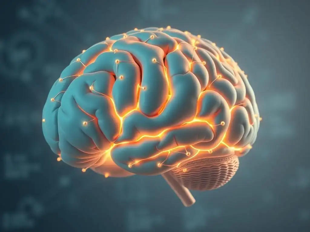
The prefrontal cortex (PFC), located at the front of the brain, is a large and complex area involved in higher-level cognitive functions, including executive control, decision-making, working memory, and regulating emotions. Imaging studies consistently show alterations in the structure and function of the PFC in individuals with depression. Specifically, the ventromedial PFC (vmPFC) and orbitofrontal cortex (OFC), involved in processing rewards and evaluating emotional stimuli, and the dorsolateral PFC (dlPFC), important for cognitive control and planning, are frequently implicated.
Functional MRI studies, for instance, often demonstrate reduced activity in the dlPFC during tasks requiring cognitive effort or emotional regulation in depressed individuals. Conversely, areas like the vmPFC can show increased activity, potentially contributing to excessive self-focus or rumination. Structural MRI studies, as mentioned earlier, have sometimes reported reduced gray matter volume or cortical thickness in parts of the PFC. These findings suggest that impaired function and potential structural changes in the PFC contribute to core depressive symptoms like difficulty concentrating, impaired decision-making, and problems regulating negative emotions. Understanding these regional differences is key to depressed brain scan interpretation.
The Amygdala and Depression: Processing Emotions and Fear Responses
The amygdala is a small, almond-shaped structure located deep within the temporal lobe, playing a central role in processing emotions, particularly fear and threat. Imaging studies in depression have consistently shown altered activity and sometimes altered volume in the amygdala. Functional MRI studies often report increased amygdala reactivity to negative emotional stimuli, such as sad or fearful faces, in individuals with depression compared to healthy controls. This heightened response may contribute to the increased experience of negative affect and anxiety often seen in depression.
While findings on amygdala volume in depression are less consistent than those for the hippocampus, some studies have reported increased amygdala volume, particularly in individuals with early-onset or severe depression. The amygdala is also part of complex emotional circuits, and its connectivity with other regions, such as the PFC, is often found to be altered in depression. For example, reduced connectivity between the PFC and amygdala might impair the ability of cognitive control regions to downregulate excessive amygdala activity. Depressed brain scan interpretation often highlights the amygdala's role in the hyper-responsiveness to negative emotional cues.
The Hippocampus and Depression: Memory and Stress Response
The hippocampus, part of the limbic system, is crucial for learning, memory formation, and regulating the body's stress response (the hypothalamic-pituitary-adrenal, or HPA, axis). It is one of the most frequently implicated brain regions in structural imaging studies of depression. As noted earlier, reductions in hippocampal volume, often ranging from 5% to 15% compared to healthy controls, are a common finding, particularly in individuals with chronic or recurrent depression.
This structural change is thought to be related to the neurotoxic effects of prolonged stress, mediated by elevated levels of stress hormones like cortisol, which the hippocampus is sensitive to. Impaired hippocampal function may contribute to cognitive symptoms of depression, such as memory problems and difficulty concentrating, as well as disruptions in the stress response system. Functional imaging studies have also shown altered hippocampal activity during memory or emotional tasks. Depressed brain scan interpretation often pays close attention to hippocampal volume and activity as potential indicators of stress-related changes and cognitive impairment.
The Anterior Cingulate Cortex (ACC) and Depression: Error Detection and Motivation
The anterior cingulate cortex (ACC) is a region located in the front of the brain, surrounding the corpus callosum. It is involved in a wide range of functions, including conflict monitoring, error detection, decision-making, motivation, and emotional regulation. The ACC can be broadly divided into a dorsal cognitive subdivision and a ventral affective subdivision, both of which are implicated in depression.
Imaging studies have shown altered activity in the ACC in depressed individuals. The ventral ACC, involved in processing emotional information and regulating responses, often shows increased activity at rest or when processing emotional stimuli. Conversely, the dorsal ACC, involved in cognitive control and task performance, may show reduced activity, potentially contributing to symptoms like apathy, difficulty concentrating, and problems with goal-directed behavior.
Structural studies have also sometimes reported altered volume or thickness in parts of the ACC. These findings highlight the ACC's role in the complex interplay between cognitive and emotional dysfunction in depression, making it a significant area for depressed brain scan interpretation.
Depressed Brain Scan Interpretation: What to Look For
Interpreting Structural Changes in the Depressed Brain: Volume Loss and Connectivity
Interpreting structural changes in depressed brain scans involves looking for consistent patterns of altered brain anatomy. The most common findings include reduced gray matter volume in specific regions. As discussed, areas like the hippocampus, prefrontal cortex (particularly vmPFC and dlPFC), and anterior cingulate cortex are frequently reported to have smaller volumes in groups of depressed individuals compared to healthy controls. These volume differences are often modest and can vary depending on factors like age, severity, and duration of depression, as well as medication use.
Beyond volume, structural interpretation also involves assessing white matter integrity using techniques like DTI. Reduced fractional anisotropy (FA) or increased radial diffusivity (RD) in specific white matter tracts, such as those connecting frontal-limbic regions, can indicate damage or disorganization of nerve fibers. This suggests impaired communication efficiency between critical brain areas. Interpreting these structural findings requires comparing individual scans to normative databases or control groups and considering the clinical context, as structural changes are not universal across all depressed individuals and can overlap with other conditions.
Interpreting Functional Changes in the Depressed Brain: Hypoactivity and Hyperactivity
Interpreting functional changes in depressed brain scans, particularly using fMRI or PET, involves identifying patterns of altered neural activity or metabolism. A common pattern observed in depression is a combination of hypoactivity (reduced activity) in certain brain regions and hyperactivity (increased activity) in others. For example, depressed brain scan interpretation often reveals hypoactivity in areas associated with cognitive control and reward, such as the dorsolateral prefrontal cortex and ventral striatum, which may correlate with symptoms like anhedonia (loss of pleasure) and impaired executive function.
Conversely, hyperactivity is frequently observed in regions involved in emotional processing and self-referential thought, such as the amygdala (especially during emotional tasks) and the default mode network (DMN) at rest. Increased connectivity within the DMN and altered connectivity between the DMN and other networks (like the central executive network) are also common functional findings. These patterns suggest an imbalance in brain circuitry, where emotional and self-referential processing is heightened, while cognitive control and reward processing are diminished. Functional interpretation is complex as activity patterns can change depending on the task or resting state conditions.
Identifying Biomarkers of Depression Using Brain Imaging
A major goal of depressed brain scan interpretation is the identification of reliable biomarkers that could aid in the diagnosis, prognosis, or treatment selection for depression. While no single brain imaging marker is currently used for routine clinical diagnosis of depression, research is actively exploring potential candidates. Structural measures, such as hippocampal volume, or functional measures, like resting-state connectivity patterns within specific networks, are being investigated as potential indicators.
For instance, some studies have explored whether specific patterns of prefrontal or ACC activity predict response to antidepressant medication or psychotherapy. Others are looking at whether PET measures of neurotransmitter receptor availability correlate with symptom severity or treatment outcome. The challenge lies in developing imaging markers that are sensitive and specific enough to be clinically useful, accounting for the significant heterogeneity within the depressed population. Identifying robust imaging biomarkers remains a key area of research, aiming to move towards more objective and personalized approaches to depression care.
How Brain Imaging Helps in Diagnosing and Treating Depression
Using Brain Scans to Differentiate Depression Subtypes
Depression is a heterogeneous disorder, meaning it presents with a wide range of symptoms and potential underlying causes. Brain imaging holds promise as a tool to help differentiate between these subtypes, which could have implications for treatment. For example, research is exploring whether distinct patterns of brain activity or connectivity, observable through techniques like fMRI, characterize different presentations of depression, such as melancholic depression versus atypical depression, or depression with high anxiety versus low anxiety.
Studies are also investigating whether imaging can help distinguish depression from other conditions with overlapping symptoms, such as bipolar disorder or certain neurological conditions. For instance, differences in specific white matter tracts or patterns of regional gray matter loss might help differentiate MDD from bipolar depression in some cases. While this is still largely an area of active research and not yet a standard clinical practice, the potential for depressed brain scan interpretation to provide objective data supporting differential diagnosis is significant.
Brain Imaging as a Tool for Predicting Treatment Response in Depression
Predicting which treatment will work best for a particular individual with depression is a major clinical challenge. Brain imaging is being explored as a potential tool to help personalize treatment selection by identifying neural patterns that predict response to specific interventions. For example, studies have investigated whether baseline activity levels or connectivity patterns in certain brain regions predict response to antidepressant medications (like SSRIs), psychotherapy (like CBT), or neuromodulation techniques (like transcranial magnetic stimulation, TMS).
Some research suggests that individuals with lower activity in certain prefrontal areas might be less likely to respond to psychotherapy but potentially more likely to respond to antidepressant medication. Conversely, specific patterns of ACC activity have been explored as predictors of response to different types of treatment. While these findings are not yet robust enough for routine clinical use, the ability to use depressed brain scan interpretation to guide treatment decisions could revolutionize personalized medicine for depression, saving individuals from lengthy trials and errors with different therapies.
Personalized Medicine and Depression: Tailoring Treatment Based on Brain Scan Results
The ultimate goal of integrating brain imaging into clinical practice for depression is to move towards personalized medicine. This involves using data from an individual's brain scan, potentially combined with other biological and clinical information, to select the most effective treatment strategy for them. Instead of a one-size-fits-all approach, treatment could be tailored based on the specific neural profile identified through techniques like depressed brain scan interpretation.
For example, if a scan reveals particularly pronounced hypoactivity in reward circuits, treatments targeting the dopaminergic system might be prioritized. If altered connectivity patterns suggest a problem with cognitive control over emotional processing, therapies focusing on strengthening these pathways, such as certain types of psychotherapy or neuromodulation, might be considered. While significant research is still needed to establish the clinical utility and cost-effectiveness of this approach, the vision is that brain imaging could become a valuable tool in the clinician's arsenal for optimizing treatment outcomes for individuals suffering from depression.
Limitations and Challenges of Brain Imaging in Depression Research
Ethical Considerations in Brain Imaging Research
Brain imaging research in depression, like all human research, involves significant ethical considerations. Ensuring informed consent is paramount, particularly when studying individuals who may have impaired cognitive function or judgment due to their illness. Researchers must clearly explain the procedures, risks (though minimal with non-invasive techniques like MRI/fMRI/EEG), benefits, and the voluntary nature of participation. Privacy and data security are also critical, as brain scans generate sensitive personal information.
Furthermore, there is an ethical responsibility in how findings from brain imaging studies are communicated to participants and the public. Oversimplification or deterministic interpretations of complex findings could lead to misconceptions about depression being solely a "brain disease" that removes personal agency, potentially impacting stigma or self-perception. Researchers must be cautious about attributing causality based solely on correlational imaging findings and avoid presenting preliminary results as definitive diagnostic tools. Ensuring equitable access to potential future benefits derived from this research is also an important consideration.
The Future of Brain Imaging in Depression: Advances and Opportunities
The field of brain imaging in depression is rapidly evolving, driven by technological advancements and sophisticated analytical methods. Future opportunities include the development of higher-resolution imaging techniques, faster scanning sequences, and novel tracers for PET that can target a wider range of neurotransmitter systems and molecular processes. The integration of different imaging modalities (multimodal imaging), such as combining structural MRI, fMRI, and DTI data, offers a more comprehensive picture of brain alterations.
Advanced computational approaches, including machine learning and artificial intelligence, are increasingly being used to analyze large datasets from imaging studies. These methods have the potential to identify subtle patterns in depressed brain scan interpretation that are not apparent through traditional statistical analyses, potentially leading to more robust biomarkers for diagnosis or treatment prediction. The future holds promise for brain imaging to contribute significantly to a deeper understanding of depression, leading to more effective and personalized interventions.
Conclusion: The Power of Brain Imaging in Understanding and Treating Depression
The Importance of Integrating Brain Imaging with Clinical Assessment
Brain imaging provides invaluable insights into the neural underpinnings of depression, revealing structural, functional, and neurochemical alterations associated with the disorder. Techniques like MRI, fMRI, PET, and EEG have significantly advanced our understanding of the key brain regions and circuits involved in mood dysregulation. However, it is crucial to emphasize that brain imaging is currently a research tool and not a standalone diagnostic test for depression in clinical practice.
The power of brain imaging lies in its integration with comprehensive clinical assessment. Symptom profiles, personal history, genetic factors, and environmental influences all contribute to the complexity of depression. Brain imaging data can provide a biological context for these clinical observations, helping researchers develop more accurate models of the illness and identify potential targets for intervention. For clinical diagnosis and treatment planning today, the detailed information gathered through patient interviews and standardized assessments remains primary.
Future Directions: Advancing Computational Psychiatry Through Neuroimaging
The future of understanding and treating depression will likely involve a more integrated approach, where data from various sources, including neuroimaging, genetics, wearable sensors, and clinical assessments, are combined using advanced computational methods. This field, known as computational psychiatry, aims to use quantitative data to build predictive models for individual patients. Depressed brain scan interpretation will play a crucial role within this framework.
By leveraging large datasets and machine learning, researchers hope to identify complex patterns in brain imaging data that can predict an individual's risk of developing depression, the likely course of their illness, or their response to specific treatments. This could pave the way for truly personalized interventions, moving beyond trial-and-error approaches. While significant challenges remain in standardizing imaging protocols, analyzing large datasets, and ensuring the clinical utility of findings, the potential for neuroimaging to transform the understanding and treatment of depression is substantial, offering hope for more effective care in the future.
Frequently Asked Questions
What is a brain scan and how does it relate to understanding depression?
Brain scans, such as fMRI and PET scans, provide visual representations of brain activity and structure. In the context of depression, these scans can help researchers and clinicians identify neurological differences compared to individuals without depression.
Can a brain scan definitively diagnose depression?
No, a brain scan alone cannot definitively diagnose depression. Diagnosis relies on a clinical evaluation, including interviews and symptom assessment. Brain scans are primarily used in research settings to understand the neurological basis of depression.
What specific brain regions are often examined in depressed brain scan interpretation?
Scans often focus on regions like the prefrontal cortex, amygdala, and hippocampus, as these areas are known to play crucial roles in mood regulation, emotional processing, and memory. Dysfunction in these regions is often observed in individuals with depression.
What are some limitations of using brain scans for understanding depression?
Limitations include the high cost of scans, the complexity of interpreting results, and the fact that scans show correlations, not necessarily causal relationships. There's also significant individual variability in brain structure and function.
How might brain scan research eventually improve depression treatment?
By identifying specific neural circuits involved in depression, researchers can develop more targeted treatments. This could lead to personalized interventions, such as medications or therapies, designed to address individual brain function profiles.
Final Reflection: Unveiling the Depressed Brain: A Journey Towards Understanding
The intricate relationship between brain structure, function, and mood disorders remains a critical area of ongoing research, with implications far beyond the clinical setting. Understanding how neuroimaging techniques illuminate the depressed brain is not merely an academic pursuit; it's a vital step towards developing more effective treatments and fostering a more compassionate understanding of mental illness.
The journey to interpret brain scans in depression has been a powerful one, revealing the complex interplay of neurochemical imbalances, structural changes, and functional connectivity disruptions. This exploration has profoundly shifted our understanding of depression, moving beyond simplistic explanations towards a more nuanced appreciation of its biological underpinnings. The brain is not a machine; it is an organ of immense complexity, capable of both profound suffering and remarkable resilience.
This quote underscores the importance of continued research and the need to approach the interpretation of brain scans with sensitivity and a holistic perspective.
The insights gained from imaging the depressed brain are not just for researchers and clinicians. They offer hope and a path towards self-understanding for individuals struggling with depression. Consider the implications: a clearer picture of the biological processes involved can reduce stigma, personalize treatment approaches, and empower individuals to advocate for their own mental healthcare. You, the reader, are now equipped with a deeper understanding of this complex landscape. Use this knowledge to foster empathy, challenge misconceptions, and advocate for improved mental health services.
Unveil Your Brain's Potential: Take Charge of Your Mood
Thanks for exploring the fascinating world of brain imaging and mood regulation with us! You've gained valuable insights into how your brain works, and now it's time to put that knowledge into action.
Here's how you can continue your journey towards better mental well-being:
- Prioritize self-care strategies: Identify one self-care practice (exercise, mindfulness, healthy eating) that resonates with you most and commit to incorporating it into your daily routine this week.
- Share this knowledge: Discuss your key takeaways with a friend or family member who might also benefit from understanding mood regulation better. Perhaps a conversation could spark a positive change for both of you.
- Explore resources: Dive deeper into the topic by researching additional reputable sources on brain health and mood management. Consider searching for local support groups or online communities.
What small step will you take today to nurture your brain's well-being, inspired by what you've learned?