
PET Scans: Illuminating Brain Chemistry in Mental Health Research

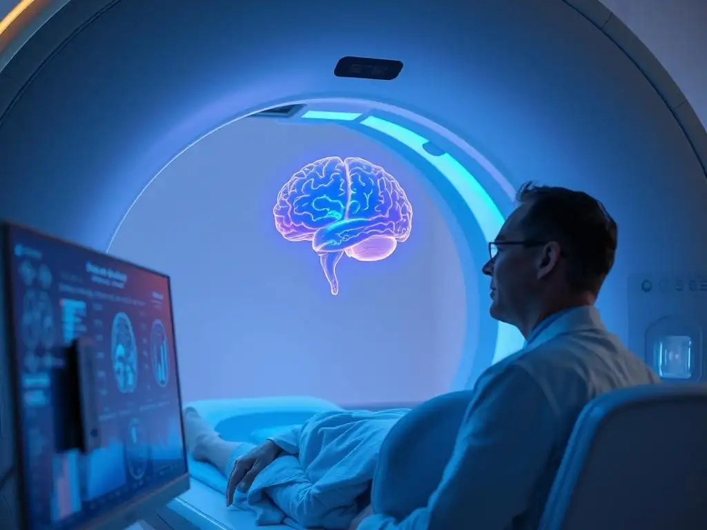
Welcome to Mind Care Guide, a safe space dedicated to understanding and nurturing your mental well-being. Ever wondered how we really see inside the brain to unravel the mysteries of mental health? Our groundbreaking article delves into the fascinating world of PET scans mental health research, offering a glimpse into the cutting-edge technology helping us understand brain chemistry like never before. Ready to explore how these scans are illuminating the path to better treatments and deeper insights? Let's begin...
PET Scans: Illuminating Brain Chemistry in Mental Health Research
- Introduction to PET Scans in Mental Health Research
- How PET Scans Work: A Detailed Explanation
-
Applications of PET Scans in Mental Health Research
- PET Scans and Depression Research: Unveiling Neurochemical Imbalances
- Investigating Schizophrenia with PET Scans: Dopamine and Beyond
- PET Scans in Alzheimer's Disease Research: Identifying Early Amyloid Plaques
- Exploring Anxiety Disorders with PET Imaging: Serotonin Transporter Availability
- Studying Addiction with PET Scans: The Reward System and Brain Changes
- Advantages of Using PET Scans in Mental Health
- Limitations and Challenges of PET Scans
- The Future of PET Scans in Mental Health Research
- Real-World Examples of PET Scan Studies in Mental Health
- Understanding Neurotransmitters and PET Scan Imaging
- How PET Scans Contribute to the Development of New Treatments
- Conclusion: The Power of PET Scans in Understanding Mental Health
-
Frequently Asked Questions
- What are PET scans and how are they used in mental health research?
- How do PET scans contribute to our understanding of mental illnesses?
- What kind of brain activity can PET scans reveal in the context of mental health?
- Are there any risks associated with undergoing a PET scan for mental health research?
- How is PET scan data used to develop new treatments for mental health conditions?
- Final Reflection: The Unfolding Promise of PET Scans in Mental Health
- Unlock Your Understanding: Deep Dive into Brain Chemistry
Introduction to PET Scans in Mental Health Research
Understanding the Role of Brain Imaging in Mental Health
Brain imaging techniques play a pivotal role in advancing our understanding of mental health disorders. They allow researchers and clinicians to visualize the structure, function, and chemical activity within the living brain, providing crucial insights that were previously unattainable. By observing how different brain regions interact or how specific neurochemicals are distributed, scientists can begin to unravel the complex biological underpinnings of conditions like depression, schizophrenia, anxiety disorders, and Alzheimer's disease. These non-invasive (or minimally invasive) methods offer a window into the brain's intricate network, helping to identify biomarkers, track disease progression, and evaluate the effectiveness of treatments.
Historically, studying the brain's involvement in mental illness relied heavily on post-mortem analysis or behavioral observations. The advent of advanced imaging technologies, such as Magnetic Resonance Imaging (MRI), Electroencephalography (EEG), and crucially, Positron Emission Tomography (PET), revolutionized the field. These tools provide objective data on brain activity and structure in real-time or near real-time, allowing for correlations to be drawn between specific brain characteristics and psychological symptoms. This shift towards a more biologically informed perspective is essential for developing more accurate diagnostic tools and targeted therapeutic interventions in mental healthcare.
PET Scans: A Powerful Tool for Investigating Brain Function
Positron Emission Tomography (PET) stands out among brain imaging techniques for its unique ability to map biochemical processes within the brain. Unlike MRI, which primarily visualizes structure, or fMRI, which measures blood flow (an indirect measure of neural activity), PET scans directly trace the distribution and concentration of specific molecules.
This is achieved by using radioactive tracers designed to bind to particular receptors, transporters, or enzymes, or to track metabolic activity like glucose consumption. This direct measurement of neurochemical dynamics makes PET scans invaluable for exploring the complex interplay of neurotransmitters and other biological pathways implicated in mental health conditions.
The power of PET scans lies in their specificity. Researchers can design tracers that target, for instance, dopamine receptors, serotonin transporters, or amyloid plaques, providing a quantitative measure of their presence and activity in different brain regions. This capability is critical for testing hypotheses about neurochemical imbalances or pathological protein accumulations associated with various disorders.
For example, PET scans have been used to demonstrate altered dopamine signaling in schizophrenia or reduced serotonin transporter availability in depression. This level of detail about brain chemistry is unparalleled by other commonly used imaging modalities, making PET scans a cornerstone of modern mental health research.
Why PET Scans Are Crucial for Mental Health Research
PET scans are crucial for mental health research because they offer a direct window into the neurochemical landscape of the brain, which is often hypothesized to be dysregulated in psychiatric disorders. While structural imaging like MRI can show if there are physical changes or lesions, and functional MRI can show areas of activity, only PET can typically quantify the density or activity of specific molecular targets.
This allows researchers to test specific hypotheses about the roles of neurotransmitters, enzymes, and receptors in the etiology and progression of mental illnesses. For instance, understanding the distribution and availability of dopamine receptors is vital for studying conditions like addiction and psychosis, and PET is the primary tool for this.
Furthermore, PET scans enable researchers to track changes in brain chemistry over time or in response to treatment. This longitudinal capability is essential for understanding disease trajectories and evaluating the efficacy of new drugs or therapeutic interventions. By performing PET scans before and after a course of treatment, researchers can observe whether the intervention has altered the targeted neurochemical system.
For example, a study might use PET to see if a new antidepressant increases serotonin transporter availability or if a treatment for addiction alters dopamine receptor binding. This provides objective evidence of biological changes associated with clinical improvement, significantly advancing the field of PET scans mental health research.
How PET Scans Work: A Detailed Explanation
The Science Behind Positron Emission Tomography (PET)
Positron Emission Tomography (PET) is a nuclear medicine imaging technique that utilizes radioactive substances, known as radiotracers, to visualize and measure metabolic processes or molecular activity within the body. The fundamental principle involves introducing a radiotracer into the bloodstream, which then accumulates in specific tissues or organs depending on the molecule it is bound to. The radioisotope attached to the tracer is a positron emitter.
When a positron is emitted, it travels a short distance and then annihilates with an electron it encounters in the surrounding tissue. This annihilation event produces two gamma rays that travel in opposite directions (approximately 180 degrees apart).
These pairs of gamma rays are detected by a ring of detectors surrounding the patient. By simultaneously detecting these paired gamma rays and tracing their paths back to their origin, a computer can reconstruct a 3D image showing the spatial distribution of the radiotracer within the body.
The intensity of the signal in different areas of the reconstructed image corresponds to the concentration of the radiotracer, which in turn reflects the level of the metabolic process or molecular target being investigated. This sophisticated process allows PET scans to provide functional information about organs and tissues, making them invaluable in fields like oncology, cardiology, and, critically, PET scans mental health research.
Radioactive Tracers: The Key to Visualizing Brain Activity
Radioactive tracers, also known as radiopharmaceuticals or radioligands, are the cornerstone of PET imaging. These are molecules that are biologically active and designed to target specific processes or structures within the body, to which a short-lived radioisotope that emits positrons is attached. Common radioisotopes used in PET include Fluorine-18 (¹⁸F), Carbon-11 (¹¹C), Nitrogen-13 (¹³N), and Oxygen-15 (¹⁵O).
The choice of radioisotope depends on the half-life required and the specific tracer molecule being synthesized. For instance, ¹⁸F has a half-life of approximately 110 minutes, making it suitable for synthesizing various tracers like FDG (fluorodeoxyglucose) for glucose metabolism studies or tracers targeting amyloid plaques. ¹¹C has a shorter half-life (about 20 minutes), which is useful for tracers that bind rapidly to targets like neurotransmitter receptors.
The tracer molecule itself is designed to mimic naturally occurring substances or to bind specifically to targeted receptors, transporters, or enzymes. For example, [¹⁸F]FDG is a glucose analog used to measure glucose metabolism, which is an indicator of neuronal activity. Tracers like [¹¹C]raclopride are used to study dopamine D2/D3 receptors, while [¹¹C]WAY-100635 targets serotonin 5-HT1A receptors.
The tracer is administered intravenously, travels through the bloodstream, and accumulates in the brain based on its affinity for the target molecule. The concentration of the tracer in different brain regions, as measured by the PET scanner, provides quantitative information about the density, availability, or activity of the target, offering unique insights into brain chemistry relevant to PET scans mental health research.
The PET Scan Procedure: What to Expect
Undergoing a PET scan typically involves several steps. First, the patient meets with a technologist who explains the procedure and answers any questions. Depending on the specific tracer being used, there might be preparation instructions, such as fasting for a certain period (especially for FDG scans which measure glucose metabolism).
The radiotracer is then administered, usually through an intravenous (IV) line inserted into a vein in the arm. There is usually a waiting period after injection, ranging from 30 minutes to over an hour, to allow the tracer to distribute throughout the body and accumulate in the target tissue or organ.
During the scan itself, the patient lies on a comfortable table that slides into the bore of the PET scanner, which is a large, donut-shaped machine. It is important for the patient to remain as still as possible during the scanning process to ensure clear images. The scan itself typically takes between 20 minutes and an hour, depending on the specific protocol.
The process is generally painless, although some people may experience mild discomfort from lying still for an extended period. After the scan is complete, the IV line is removed, and the patient is usually free to leave, with instructions to drink plenty of fluids to help flush the small amount of radioactivity from their system. The level of radiation exposure is generally considered safe for diagnostic purposes.
Interpreting PET Scan Results: A Complex Process
Interpreting PET scan results requires specialized expertise, typically by a nuclear medicine physician or radiologist with experience in neuroimaging. The raw data from the PET scanner is processed by computer software to reconstruct cross-sectional or 3D images of the brain. These images show the distribution and intensity of the radiotracer throughout different brain regions, often displayed in color maps where different colors represent varying levels of tracer uptake. High tracer uptake in a specific region might indicate high metabolic activity (with FDG) or a high density of the targeted receptor or transporter (with specific radioligands).
However, interpreting these images in the context of mental health conditions is complex. It involves comparing the patient's scan results to normative databases, considering the patient's clinical history, symptoms, and other diagnostic information. For example, in depression research using PET scans, reduced binding of a serotonin transporter tracer in certain brain regions might be observed compared to healthy controls.
In Alzheimer's research, increased uptake of an amyloid tracer in specific cortical areas is indicative of amyloid plaque deposition. The interpretation must account for factors like age, sex, medication use, and potential motion artifacts. Researchers and clinicians use sophisticated quantitative analysis techniques to measure tracer binding or uptake in specific volumes of interest (VOIs) or across the entire brain, providing objective data points for diagnosis, prognosis, and monitoring treatment response in PET scans mental health research.
Applications of PET Scans in Mental Health Research
PET Scans and Depression Research: Unveiling Neurochemical Imbalances
PET scans have been instrumental in investigating the neurochemical underpinnings of major depressive disorder (MDD). Research using PET imaging has focused on understanding the roles of various neurotransmitter systems, particularly serotonin, dopamine, and norepinephrine, which are targeted by many antidepressant medications. For example, studies using tracers for the serotonin transporter (SERT), such as [¹¹C]DASB or [¹¹C]MADAM, have consistently shown reduced SERT availability in several brain regions, including the brainstem, thalamus, and limbic areas, in individuals with MDD compared to healthy controls. This finding supports the hypothesis that alterations in serotonin reuptake mechanisms are involved in the pathophysiology of depression.
Beyond serotonin, PET has explored other pathways. Studies using tracers for dopamine receptors (e.g., [¹¹C]raclopride for D2/D3 receptors) and dopamine transporters (DAT) have provided mixed results but suggest potential alterations in dopamine signaling, particularly in reward and motivation circuits, which are often impaired in depression. Furthermore, PET studies investigating glutamate and GABA systems, which are involved in excitatory and inhibitory neurotransmission respectively, are ongoing.
For instance, research with [¹¹C]FMZ, a tracer for GABA-A receptors, has explored potential changes in inhibitory tone in depression. These investigations using PET scans mental health research are vital for developing a more comprehensive understanding of the multifaceted neurochemical dysregulations in MDD.
Investigating Schizophrenia with PET Scans: Dopamine and Beyond
Schizophrenia is a complex psychiatric disorder, and PET scans have played a crucial role in shaping our understanding of its neurobiological basis, particularly the dopamine hypothesis. Early PET studies using tracers like [¹¹C]raclopride provided strong evidence for altered dopamine function in the striatum of individuals with schizophrenia, showing increased dopamine synthesis capacity and release, especially in those experiencing positive symptoms like hallucinations and delusions.
This excess dopamine activity in the striatum is thought to contribute significantly to psychotic symptoms. PET has also been used to study dopamine receptor density and occupancy by antipsychotic medications, helping to understand why these drugs are effective and why side effects occur.
While dopamine remains a key focus, PET research in schizophrenia has expanded to investigate other neurotransmitter systems and pathological processes. Studies have explored glutamate receptors (e.g., NMDA receptors) using tracers like [¹¹C]CNS-5161, given the hypothesis of glutamatergic dysfunction in schizophrenia. Investigations into serotonin receptors, particularly 5-HT2A receptors (targeted by atypical antipsychotics), using tracers like [¹⁸F]altanserin, have also provided insights into the mechanisms of action of these medications.
Furthermore, emerging PET research is exploring neuroinflammation in schizophrenia using tracers that target activated microglia, potentially identifying a new avenue for therapeutic intervention. These diverse applications highlight the power of PET scans in schizophrenia research, moving beyond a singular focus on dopamine to encompass a broader range of neurobiological factors.
PET Scans in Alzheimer's Disease Research: Identifying Early Amyloid Plaques
Alzheimer's disease (AD) is characterized by the accumulation of amyloid-beta (Aβ) plaques and tau tangles in the brain, leading to progressive cognitive decline. For many years, definitive diagnosis of AD required post-mortem examination. However, the development of specific PET radiotracers capable of binding to Aβ plaques revolutionized AD research and diagnosis.
Tracers like [¹¹C]PIB, [¹⁸F]florbetapir, [¹⁸F]flutemetamol, and [¹⁸F]florbetaben can now visualize and quantify Aβ burden in the living brain, often years before the onset of significant clinical symptoms. This allows for earlier and more accurate diagnosis, differentiation from other forms of dementia, and identification of individuals at risk for clinical trials.
The ability to detect Aβ plaques in vivo using PET scans has been critical for testing the amyloid cascade hypothesis and evaluating anti-amyloid therapies. Clinical trials for AD drugs often use amyloid PET imaging as a key endpoint to determine if a treatment can reduce plaque burden.
More recently, PET tracers targeting tau pathology (e.g., [¹⁸F]flortaucipir) have been developed, allowing researchers to visualize the distribution and spread of tau tangles, which correlates more closely with cognitive decline than amyloid burden. These advancements in PET imaging for AD, including both amyloid and tau tracers, are transforming our understanding of disease progression and accelerating the search for effective treatments, making PET scans mental health research in the context of neurodegenerative disorders particularly impactful.
Exploring Anxiety Disorders with PET Imaging: Serotonin Transporter Availability
Anxiety disorders, such as generalized anxiety disorder, panic disorder, and social anxiety disorder, are often linked to dysregulation of neurotransmitter systems, particularly serotonin. PET scans using radiotracers that bind to the serotonin transporter (SERT) have been employed to investigate its role in these conditions. The SERT is responsible for reuptake of serotonin from the synaptic cleft, and its availability is a key indicator of serotonergic system function. Studies have explored whether alterations in SERT density or binding are associated with different anxiety subtypes.
While findings have been somewhat variable depending on the specific anxiety disorder and study population, some research using tracers like [¹¹C]DASB has suggested altered SERT binding in regions like the amygdala, hippocampus, and prefrontal cortex in individuals with anxiety disorders compared to healthy controls. For example, some studies have reported reduced SERT availability in certain brain regions, while others have found no significant differences or even increases in specific areas.
This variability may reflect the heterogeneity of anxiety disorders or the influence of factors like medication history. Nevertheless, PET imaging of the SERT and other neurotransmitter systems (such as GABA and benzodiazepine receptors) continues to be a valuable tool for researchers seeking to understand the neurobiological basis of anxiety and identify potential targets for therapeutic intervention through PET scans mental health research.
Studying Addiction with PET Scans: The Reward System and Brain Changes
PET scans have provided profound insights into the neurobiological mechanisms underlying substance use disorders and addiction, particularly focusing on the brain's reward system. This system heavily involves dopamine pathways, originating in the ventral tegmental area (VTA) and projecting to areas like the nucleus accumbens and prefrontal cortex.
Drugs of abuse, such as cocaine, amphetamines, and alcohol, significantly impact dopamine signaling, leading to the euphoric effects and reinforcing the addictive behavior. PET studies using dopamine receptor tracers (e.g., [¹¹C]raclopride for D2/D3 receptors) and dopamine transporter tracers (e.g., [¹¹C]CFT or [¹¹C]PE2I) have consistently shown alterations in the dopamine system in individuals with addiction.
A common finding across various types of addiction is a reduction in the availability of dopamine D2/D3 receptors in the striatum. This reduction is thought to contribute to the diminished ability to experience pleasure from natural rewards and may drive compulsive drug-seeking behavior. PET has also been used to study the release of dopamine in response to drug cues or drug administration, showing exaggerated release in addicted individuals.
Furthermore, PET imaging has explored changes in brain metabolism ([¹⁸F]FDG) and other neurotransmitter systems (e.g., opioid receptors, cannabinoid receptors) in addiction. This body of PET scans mental health research has significantly advanced our understanding of the persistent brain changes associated with addiction, informing the development of pharmacological and behavioral therapies.
Advantages of Using PET Scans in Mental Health
High Sensitivity for Detecting Subtle Brain Changes
One of the primary advantages of PET scans in mental health research is their high sensitivity, particularly at the molecular level. While other imaging techniques like MRI can detect structural abnormalities or large-scale functional changes, PET can detect and quantify the presence and activity of specific molecules, such as neurotransmitter receptors or metabolic enzymes, even when they are present in relatively low concentrations.
This sensitivity allows researchers to identify subtle neurochemical alterations that may underlie the early stages of a mental health disorder or represent specific biological subtypes within a diagnostic category. For instance, PET can detect small changes in receptor density that might not be evident through indirect measures of brain activity.
This molecular sensitivity is crucial for understanding the biological mechanisms of mental illness, which often involve complex and subtle dysregulations of neurochemical pathways. By providing quantitative data on the distribution and availability of specific molecular targets, PET scans allow researchers to investigate hypotheses about the role of particular genes, proteins, or metabolic processes in the development and maintenance of psychiatric conditions.
This level of detail goes beyond simply observing which brain regions are active during a task; it delves into the underlying biochemical machinery, making PET an indispensable tool for uncovering the intricate biological basis that informs PET scans mental health research.
Ability to Measure Neurotransmitter Activity Directly
A key strength of PET imaging is its unique ability to directly measure aspects of neurotransmitter system activity in vivo. While fMRI can correlate blood flow changes with neural activity driven by neurotransmitters, PET can use specific radioligands to bind to receptors, transporters, or enzymes involved in neurotransmitter synthesis or breakdown. This allows for the quantification of their density, affinity, or even dynamic release (in some cases). For example, using a suitable radiotracer, researchers can measure the density of dopamine D2 receptors in the striatum or the availability of serotonin transporters in the brainstem.
This direct measurement capability is invaluable for testing hypotheses related to neurochemical imbalances in mental health disorders. For example, if a hypothesis suggests that reduced dopamine receptor binding contributes to anhedonia in depression, a PET scan using a D2/D3 receptor tracer can directly assess this.
Similarly, if a new drug is designed to block a specific serotonin receptor, PET can be used to confirm that the drug is binding to the target receptor in the human brain and to what extent. This direct biochemical information provided by PET scans is unparalleled by other common neuroimaging techniques, making it essential for advancing our understanding of the neurochemical basis of mental illness and for developing targeted therapies in PET scans mental health research.
Providing Quantitative Data for Research Analysis
PET scans provide quantitative data, meaning they yield numerical values representing the concentration or binding potential of the radiotracer in specific brain regions. This quantitative nature is a significant advantage for research analysis. Instead of relying solely on visual inspection of images, researchers can extract precise measurements from defined volumes of interest (VOIs) or use voxel-wise analysis across the entire brain. These numerical values can then be subjected to rigorous statistical analysis to compare groups (e.g., patients vs. controls), correlate findings with clinical symptoms or cognitive performance, and track changes over time or in response to treatment.
For instance, a study investigating schizophrenia might report that the average binding potential of a dopamine D2/D3 receptor tracer is 20% higher in the striatum of patients compared to controls, with a specific statistical p-value. This provides an objective, quantifiable piece of evidence supporting the hypothesis of altered dopamine function. This quantitative capability allows for greater precision and objectivity in research findings compared to purely qualitative or semi-quantitative methods. It facilitates meta-analyses across different studies and enables researchers to build predictive models based on neurochemical profiles, thereby strengthening the scientific rigor of PET scans mental health research.
Potential for Early Diagnosis and Personalized Treatment
The ability of PET scans to detect subtle molecular changes associated with disease processes holds significant potential for early diagnosis and the development of personalized treatment strategies in mental health. In some cases, neurochemical alterations may precede the onset of overt clinical symptoms, particularly in neurodegenerative conditions like Alzheimer's disease where amyloid and tau pathology can be detected years before significant cognitive decline. Identifying these biomarkers early could allow for interventions at a stage where they might be most effective in slowing or preventing disease progression.
Furthermore, PET scans can provide insights into the specific neurobiological profile of an individual's disorder, which could inform personalized treatment decisions. For example, if PET imaging reveals that a patient with depression has particularly low serotonin transporter availability, this might suggest that an SSRI (Selective Serotonin Reuptake Inhibitor) medication, which targets the SERT, could be a suitable treatment.
Conversely, if another patient shows more prominent alterations in dopamine pathways, a different class of medication might be considered. By providing objective, patient-specific neurochemical data, PET scans have the potential to move mental healthcare towards a more precision medicine approach, optimizing treatment selection based on individual biological profiles, a key area of focus in PET scans mental health research.
Limitations and Challenges of PET Scans
Radiation Exposure: Weighing the Risks and Benefits
One significant limitation of PET scans is that they involve the administration of a small dose of radioactive material. While the radiotracers used have short half-lives and the radiation dose is generally considered low and safe for diagnostic purposes, it is still a factor that must be carefully considered, especially for studies involving children, pregnant women, or repeated scans on the same individual.
The potential long-term risks associated with low-dose radiation exposure, although small, necessitate a careful weighing of the benefits of obtaining the imaging information against the potential risks, particularly in research settings where scans may be performed solely for scientific inquiry rather than immediate clinical necessity.
Researchers and clinicians adhere to strict safety protocols to minimize radiation exposure, following the ALARA principle (As Low As Reasonably Achievable). This includes using the lowest effective dose of radiotracer, limiting the duration of exposure, and ensuring proper shielding.
However, the need to manage radiation exposure restricts the frequency with which PET scans can be performed on an individual and can be a barrier to conducting longitudinal studies requiring many scans over time. This inherent aspect of PET imaging is a crucial consideration when designing studies and evaluating its suitability compared to non-radioactive imaging modalities in PET scans mental health research.
Limited Spatial Resolution Compared to Other Imaging Techniques
While PET scans excel at providing molecular and functional information, their spatial resolution is typically lower than that of structural imaging techniques like MRI. Spatial resolution refers to the ability to distinguish between two separate points or structures in the image. Standard clinical PET scanners typically have a spatial resolution in the range of 4-6 millimeters, although high-resolution research scanners can achieve better resolution (around 2-3 mm). In contrast, modern MRI scanners can achieve resolutions of 1 millimeter or less.
This limitation in spatial resolution means that PET images may not clearly delineate very small brain structures or precisely localize activity to fine anatomical details. For instance, distinguishing between activity in adjacent small nuclei can be challenging with PET alone.
This is why PET scans are often combined with MRI scans (either performed separately and fused, or acquired simultaneously using a hybrid PET/MRI scanner). The MRI provides high-resolution anatomical context onto which the PET functional or molecular data can be overlaid, compensating for the spatial resolution limitation of PET and providing a more complete picture of the brain in PET scans mental health research.
Cost and Accessibility of PET Scan Technology
PET scan technology is expensive, both in terms of the initial cost of the scanner equipment and the ongoing operational costs. PET scanners are complex machines requiring specialized facilities, including a cyclotron (to produce certain short-lived radioisotopes like ¹¹C and ¹⁵O) or access to nearby radiopharmacies, and highly trained personnel (nuclear medicine physicians, technologists, radiochemists, medical physicists). The production of radiotracers is also a costly and logistically challenging process, as many have very short half-lives and must be synthesized and transported rapidly.
These high costs and the need for specialized infrastructure limit the accessibility of PET scanning. Many hospitals and research centers, particularly outside of major metropolitan areas or in lower-resource settings, may not have access to PET technology. This can create disparities in access to advanced diagnostic imaging and can make it challenging for researchers at institutions without PET facilities to conduct studies utilizing this technique. The financial investment required for PET significantly impacts its widespread availability and integration into routine clinical practice for mental health conditions, posing a challenge for expanding the reach of PET scans mental health research.
Ethical Considerations in Using PET Scans for Research
Using PET scans in mental health research raises several ethical considerations. As the technique involves administering radioactive substances, researchers must ensure that the potential benefits of the study outweigh the risks of radiation exposure to participants. This is particularly important when recruiting vulnerable populations, such as individuals with severe mental illness, children, or those with impaired decision-making capacity. Informed consent procedures must be rigorous, ensuring participants fully understand the nature of the procedure, the risks involved, and their right to withdraw at any time.
Furthermore, the interpretation and potential implications of PET findings in mental health research require careful ethical consideration. Findings related to specific biological markers or predicted treatment responses must be communicated responsibly to participants, avoiding deterministic language or overstating the clinical utility of research findings that are not yet validated for individual patient care.
There are also ethical discussions around the use of PET in diagnosing conditions like Alzheimer's disease in asymptomatic individuals and the potential psychological impact of such findings. Researchers must navigate these complex ethical landscapes to ensure that studies are conducted responsibly and that participant well-being is prioritized in PET scans mental health research.
The Future of PET Scans in Mental Health Research
Advancements in Tracer Development for Specific Mental Disorders
The future of PET scans in mental health research is closely tied to the ongoing development of novel and more specific radiotracers. While tracers for dopamine, serotonin, and amyloid are well-established, there is a significant need for tracers that target other neurotransmitter systems, receptors, enzymes, or pathological processes implicated in mental disorders. For example, researchers are actively developing tracers for specific glutamate receptor subtypes (like mGluR5), neuroinflammatory markers (like TSPO for microglia), and other protein aggregates beyond amyloid and tau that might play a role in psychiatric conditions.
The development of tracers with higher specificity, affinity, and lower non-specific binding is crucial for obtaining clearer and more reliable signals. Furthermore, tracers that can measure dynamic processes, such as neurotransmitter release or receptor occupancy changes in response to stimuli or medication, are highly desirable. Advances in radiochemistry and molecular biology are facilitating the design and synthesis of these new ligands. A robust pipeline of innovative tracers will unlock new possibilities for investigating the intricate neurobiological mechanisms of mental illness and identifying novel therapeutic targets, propelling the field of PET scans mental health research forward.
Combining PET Scans with Other Imaging Modalities (e.g., MRI)
The integration of PET scans with other imaging modalities, particularly Magnetic Resonance Imaging (MRI), represents a major trend and future direction in neuroimaging. Hybrid PET/MRI scanners, which acquire PET and MRI data simultaneously, offer several advantages. They allow for perfect spatial and temporal co-registration of structural (MRI) and functional/molecular (PET) data, eliminating potential errors from separate scanning and image fusion. This provides a more comprehensive picture of brain structure, function, and chemistry within a single imaging session.
Combining PET with MRI leverages the strengths of both techniques: MRI provides high-resolution anatomical images and can assess structural integrity, white matter tracts (using diffusion tensor imaging - DTI), or blood flow (using arterial spin labeling - ASL), while PET provides quantitative molecular information. This multimodal approach is particularly powerful for studying complex brain disorders where multiple aspects of brain health are affected.
For example, combining amyloid PET with structural MRI can assess both plaque burden and associated brain atrophy in Alzheimer's disease. The continued development and wider availability of hybrid PET/MRI systems will significantly enhance our ability to conduct comprehensive PET scans mental health research.
Developing New PET Imaging Protocols for Improved Diagnostic Accuracy
Beyond technological advancements in hardware and tracers, future progress in PET scans mental health research will involve the development of new and refined imaging protocols to improve diagnostic accuracy and sensitivity. This includes optimizing tracer administration methods, scanning durations, and data acquisition parameters to maximize signal-to-noise ratio and minimize motion artifacts. Furthermore, the development of dynamic PET protocols, which acquire data continuously over time after tracer injection, allows for the measurement of kinetic parameters related to tracer uptake, binding, and clearance, providing more detailed information about the underlying biological processes than static scans.
Advanced computational methods and machine learning algorithms are also being developed to analyze PET data more effectively. These tools can identify complex patterns in tracer distribution that may not be obvious from visual inspection or simple regional analysis. By integrating PET data with other types of data (e.g., genetic information, clinical assessments, cognitive test results), researchers are working towards developing predictive models that could aid in diagnosis, prognosis, and treatment selection. These efforts to refine protocols and analysis techniques are crucial for translating research findings from PET scans mental health research into clinically useful tools.
The Potential of PET Scans for Guiding Treatment Strategies
One of the most exciting future potentials of PET scans in mental health is their use in guiding individualized treatment strategies. As our understanding of the specific neurochemical profiles associated with different disorders and individual variations within those disorders grows, PET imaging could potentially help clinicians select the most appropriate treatment for a patient. For example, PET could be used to confirm target engagement of a prescribed medication (i.e., whether the drug is binding to the intended receptor in the brain) or to assess changes in neurochemical markers in response to therapy.
If PET scans can reliably identify biological subtypes of depression or schizophrenia, for instance, patients could be directed towards treatments that are most likely to be effective for their specific subtype. PET could also help predict response to certain therapies or identify individuals who are unlikely to respond, potentially saving time and reducing the burden of ineffective treatments.
While still largely in the research phase, the vision is that PET imaging could become a tool in the clinician's arsenal, providing objective biological information to complement clinical assessment and move towards truly personalized medicine in mental healthcare, driven by insights from PET scans mental health research.
Real-World Examples of PET Scan Studies in Mental Health
Case Study 1: Reduced Serotonin 1A Receptor Binding in Depression
A landmark area of PET scans mental health research has focused on serotonin 1A (5-HT1A) receptors, which are inhibitory autoreceptors in the raphe nuclei (where serotonin neurons originate) and postsynaptic receptors in regions like the hippocampus and prefrontal cortex. Studies using tracers like [¹¹C]WAY-100635 have consistently reported reduced binding potential of 5-HT1A receptors in several brain regions of individuals with major depressive disorder compared to healthy controls. For example, a meta-analysis combining data from multiple studies found significantly lower 5-HT1A receptor binding in the raphe nuclei, prefrontal cortex, and hippocampus in depressed patients.
This finding is significant because the 5-HT1A receptor is involved in regulating serotonin release and neuronal excitability. Reduced binding in the raphe nuclei might lead to increased serotonin release, initially. However, chronic stress or other factors could potentially lead to desensitization or downregulation of these receptors, impacting overall serotonergic tone.
Reduced postsynaptic 5-HT1A receptor binding in areas like the prefrontal cortex and hippocampus, regions critical for mood regulation and cognitive function, could contribute to the symptoms of depression. These PET findings have helped to refine hypotheses about serotonin system dysfunction in MDD and have informed the development of antidepressant drugs that target these receptors, showcasing the direct impact of PET scans mental health research.
Case Study 2: Elevated Striatal Dopamine Release in Schizophrenia
PET imaging has provided compelling evidence supporting the dopamine hypothesis of schizophrenia, particularly regarding excessive dopamine activity in the striatum. Studies using tracers like [¹¹C]raclopride, which competes with endogenous dopamine for binding to D2/D3 receptors, have shown that individuals with schizophrenia, especially those with prominent positive symptoms, exhibit elevated dopamine release in the striatum. This is often demonstrated using amphetamine challenge studies, where participants receive a dose of amphetamine (which stimulates dopamine release) during the PET scan. The amphetamine-induced change in raclopride binding is used as an index of dopamine release.
For instance, a seminal study by Laruelle et al. in 1996 used this method and found that amphetamine administration induced a significantly greater decrease in striatal [¹¹C]raclopride binding in patients with schizophrenia compared to controls, indicating greater dopamine release.
Subsequent studies have replicated this finding and extended it to first-episode psychosis patients, suggesting that this hyperdopaminergic state in the striatum is present early in the illness. This evidence from PET scans has strongly supported the use of dopamine D2 receptor blocking antipsychotic medications as the primary treatment for positive symptoms in schizophrenia and continues to inform research into the pathophysiology of the disorder, demonstrating the power of PET scans mental health research in understanding psychosis.
Case Study 3: Amyloid-Beta Deposition in Alzheimer's Disease
The development of amyloid-beta (Aβ) specific PET tracers has revolutionized the study of Alzheimer's disease (AD). Tracers like [¹¹C]PIB and later [¹⁸F]-labeled tracers such as florbetapir, flutemetamol, and florbetaben, bind specifically to Aβ plaques in the brain. PET studies using these tracers have demonstrated that Aβ pathology begins accumulating in the brain years, sometimes even decades, before the onset of clinical symptoms of dementia. For example, studies of cognitively normal older adults at high genetic risk for AD have shown detectable amyloid deposition via PET.
These studies have shown a characteristic pattern of Aβ deposition, starting in neocortical areas and progressing to other regions. Quantitative analysis of amyloid PET scans provides a measure of the overall amyloid burden in the brain. This has been crucial for diagnosing AD in vivo, differentiating it from other dementias, and selecting participants for clinical trials of anti-amyloid therapies.
For example, trials for drugs like aducanumab and lecanemab used amyloid PET as a key biomarker to demonstrate drug efficacy in reducing amyloid plaques. The ability to visualize and quantify this key pathological hallmark using PET scans mental health research has fundamentally changed the landscape of AD research and clinical management.
Understanding Neurotransmitters and PET Scan Imaging
Dopamine: Imaging the Reward System
Dopamine is a key neurotransmitter involved in reward, motivation, pleasure, motor control, and executive functions. Dysregulation of dopamine signaling is implicated in several mental health conditions, including schizophrenia (excess striatal dopamine), addiction (alterations in reward pathway dopamine), depression (potential deficits in reward-related dopamine), and Parkinson's disease (neurodegenerative loss of dopaminergic neurons, often associated with mood symptoms). PET scans are a primary tool for studying the dopamine system in vivo.
Various PET radiotracers have been developed to target different components of the dopamine system. For example, [¹¹C]raclopride and [¹⁸F]fallypride are commonly used to measure the availability of dopamine D2/D3 receptors, which are particularly abundant in the striatum.
Tracers like [¹¹C]CFT (also known as WIN 35,428) and [¹¹C]PE2I target the dopamine transporter (DAT), which is responsible for reuptake of dopamine from the synapse and is primarily located on presynaptic terminals, especially in the striatum. By measuring the binding of these tracers in different brain regions, PET allows researchers to quantify receptor density, transporter availability, and even estimate dopamine release (using challenge paradigms), providing critical insights into the role of dopamine in health and disease, a central theme in PET scans mental health research.
Serotonin: Visualizing Mood Regulation
Serotonin, also known as 5-hydroxytryptamine (5-HT), is a neurotransmitter widely involved in regulating mood, sleep, appetite, and cognition. Dysfunction of the serotonin system is strongly implicated in mood disorders like depression and anxiety disorders. PET imaging has been extensively used to investigate various components of the serotonin system in the living brain.
Radiotracers have been developed to target the serotonin transporter (SERT), which is the site of action for SSRI antidepressants, and various serotonin receptor subtypes (e.g., 5-HT1A, 5-HT1B, 5-HT2A, 5-HT4, 5-HT6, 5-HT7). Tracers like [¹¹C]DASB and [¹¹C]MADAM are used to measure SERT availability, often found to be reduced in depression. Tracers like [¹¹C]WAY-100635 target 5-HT1A receptors, and studies have shown altered binding in depression and anxiety.
[¹⁸F]altanserin is used to image 5-HT2A receptors, which are targets of some atypical antipsychotics and hallucinogens. By selectively binding to these targets, PET allows researchers to visualize their distribution and quantify their availability in different brain regions, providing valuable data on the state of the serotonin system in individuals with mental health conditions and assessing the impact of treatments that modulate serotonin signaling, a key area for PET scans mental health research.
Glutamate: Its Role in Brain Activity and Mental Disorders
Glutamate is the primary excitatory neurotransmitter in the mammalian central nervous system, playing a critical role in synaptic plasticity, learning, and memory. Dysregulation of glutamatergic neurotransmission is increasingly implicated in a range of mental health disorders, including schizophrenia, depression, anxiety, and addiction. While imaging the dynamics of glutamate itself with PET is challenging, tracers targeting specific glutamate receptor subtypes or related enzymes have been developed to investigate glutamatergic system function.
Tracers targeting the metabotropic glutamate receptor subtype 5 (mGluR5), such as [¹¹C]ABP688 and [¹⁸F]FPEB, have been used to explore potential alterations in glutamatergic signaling in conditions like depression and schizophrenia. For example, some studies have suggested altered mGluR5 binding in certain brain regions in depressed patients.
PET imaging of the enzyme glutamic acid decarboxylase (GAD), which synthesizes GABA from glutamate, is also an area of research. While imaging the glutamate system with PET is more complex than imaging monoamines like dopamine and serotonin, the development of new, specific tracers is opening up new avenues for understanding the role of this crucial excitatory system in the pathophysiology of mental illness and advancing PET scans mental health research.
GABA: Understanding Inhibitory Processes
Gamma-aminobutyric acid (GABA) is the primary inhibitory neurotransmitter in the central nervous system, counterbalancing the excitatory effects of glutamate. GABA plays a crucial role in regulating neuronal excitability, anxiety levels, and sleep. Dysregulation of the GABAergic system is implicated in anxiety disorders, epilepsy, and potentially other conditions like depression and schizophrenia. PET imaging offers a way to investigate aspects of the GABAergic system in vivo.
PET tracers have been developed to target GABA-A receptors, which are the binding sites for benzodiazepines and other sedative/anxiolytic drugs. Tracers like [¹¹C]flumazenil ([¹¹C]FMZ) bind to the benzodiazepine site on the GABA-A receptor complex.
Studies using [¹¹C]FMZ have explored potential changes in GABA-A receptor binding in anxiety disorders and depression, although findings have been somewhat inconsistent, possibly due to tracer limitations or the complexity of the GABA-A receptor subtypes. Research is ongoing to develop more selective and sensitive tracers for different components of the GABAergic system, including enzymes involved in GABA synthesis or degradation, to gain a clearer picture of how alterations in inhibition contribute to mental health conditions, expanding the scope of PET scans mental health research.
How PET Scans Contribute to the Development of New Treatments
Identifying Potential Drug Targets Using PET Imaging
PET scans play a crucial role in the early stages of drug discovery and development for mental health disorders by helping to identify and validate potential molecular targets. If research using PET imaging reveals that a specific receptor, transporter, or enzyme is altered in a particular mental illness (e.g., reduced serotonin transporter availability in depression, elevated dopamine release in schizophrenia), this molecule becomes a prime candidate for therapeutic intervention. A drug designed to modulate the activity of this target could potentially correct the underlying neurochemical imbalance.
For example, the finding from PET studies that dopamine D2 receptors are upregulated or dopamine release is increased in schizophrenia directly supported the development and use of antipsychotic drugs that block these receptors. Similarly, observing reduced serotonin transporter availability in depression reinforced the rationale for developing and prescribing SSRIs.
PET can also be used in preclinical studies (in animal models) and early-phase clinical trials to assess the distribution and binding of novel drug candidates to their intended targets in the brain, providing evidence of target engagement and helping to determine appropriate dosing, thus accelerating the process of finding effective treatments through PET scans mental health research.
Monitoring Treatment Response with PET Scans
PET scans can be used to objectively monitor the neurobiological effects of treatments for mental health disorders. By performing PET scans before and after a course of medication, psychotherapy, or neuromodulation technique (like transcranial magnetic stimulation - TMS), researchers can assess whether the treatment has altered the targeted neurochemical system or metabolic activity. This provides biological validation for the treatment's mechanism of action and can help correlate biological changes with clinical improvements.
For instance, PET studies using dopamine D2 receptor tracers can confirm that an antipsychotic medication is binding to its target receptors in the brain and to what extent (receptor occupancy). Studies using serotonin transporter tracers can assess whether an SSRI is effectively blocking serotonin reuptake.
In the context of Alzheimer's disease, amyloid PET scans are used in clinical trials to demonstrate whether a new drug can reduce the burden of amyloid plaques. This ability to visualize and quantify treatment-induced changes at the molecular level using PET scans is invaluable for evaluating the efficacy of new therapies and understanding why some patients respond while others do not, significantly contributing to PET scans mental health research aimed at improving treatment outcomes.
Personalized Medicine: Tailoring Treatment Based on PET Scan Results
The quantitative information provided by PET scans holds promise for moving towards personalized medicine in mental healthcare. While this is still largely an area of active research rather than routine clinical practice, the potential is significant. If PET imaging can identify specific neurochemical or metabolic profiles that characterize different subtypes of a disorder or predict response to certain treatments, this information could be used to tailor therapeutic interventions to individual patients.
For example, if research shows that patients with depression who have a specific pattern of glutamate receptor binding are more likely to respond to a particular novel antidepressant, a PET scan could potentially help identify which patients might benefit most from that drug. Similarly, in addiction treatment, PET could potentially help identify individuals with specific dopamine system abnormalities who might respond better to certain pharmacotherapies.
While challenges remain in establishing the clinical utility and cost-effectiveness of using PET scans for routine treatment selection, the ability to obtain objective, patient-specific biological data offers an exciting path towards more precise and effective mental healthcare, driven by insights from PET scans mental health research.
Conclusion: The Power of PET Scans in Understanding Mental Health
PET Scans: A Vital Tool for Advancing Mental Health Research
PET scans have unequivocally established themselves as a vital and indispensable tool in the complex landscape of mental health research. Their unique ability to visualize and quantify specific molecular targets and metabolic processes within the living brain provides insights that are often unattainable with other imaging modalities.
From unraveling the intricate roles of neurotransmitters like dopamine and serotonin in conditions ranging from schizophrenia to depression and addiction, to detecting the pathological protein aggregates characteristic of neurodegenerative disorders like Alzheimer's, PET has consistently pushed the boundaries of our understanding of the biological underpinnings of mental illness. The quantitative nature of PET data allows for rigorous scientific analysis, enabling researchers to identify biomarkers, track disease progression, and investigate the mechanisms of action of therapeutic interventions.
The contributions of PET scans mental health research extend from basic science, helping us to build more accurate models of brain function and dysfunction, to translational research, aiding in the development and evaluation of novel treatments. The ability to directly measure neurochemical parameters in vivo has been critical for testing hypotheses about the roles of specific molecular pathways in psychiatric and neurological disorders. While challenges related to cost, accessibility, and spatial resolution exist, ongoing advancements in radiotracer development, hybrid imaging systems, and data analysis techniques continue to enhance the power and potential of PET.
The Importance of Continued Investment in PET Scan Technology
Given the profound impact that PET scans have had on advancing our understanding of mental health and accelerating the development of new treatments, continued investment in PET scan technology and research is of paramount importance. This includes funding for the development of novel radiotracers that can target a wider range of molecules and processes relevant to mental illness, including those involved in neuroinflammation, synaptic function, and genetic expression. Investment is also needed for the development and dissemination of advanced PET/MRI hybrid systems, which offer superior data quality and a more comprehensive picture of brain health.
Furthermore, supporting the infrastructure and personnel required to operate PET facilities and conduct complex PET studies is crucial. This involves training the next generation of radiochemists, medical physicists, technologists, and researchers who can effectively utilize this technology. Continued investment in large-scale, multi-center studies using PET imaging can help to build robust databases, validate findings across diverse populations, and accelerate the translation of research discoveries into clinical practice. Sustained commitment to funding PET scans mental health research is essential for unlocking further insights into the brain and developing more effective strategies for preventing, diagnosing, and treating mental health disorders.
Improving Mental Health Outcomes Through Brain Imaging
Ultimately, the goal of utilizing powerful tools like PET scans in mental health research is to improve outcomes for individuals affected by these challenging conditions. By providing a deeper understanding of the biological mechanisms underlying mental illness, PET imaging facilitates the development of more accurate diagnostic methods, helps in identifying individuals at risk, and accelerates the discovery of novel therapeutic targets. The potential to use PET scans to guide personalized treatment selection based on individual neurobiological profiles offers a future where mental healthcare is more precise and effective.
Insights gained from PET scans mental health research inform the development of new medications, guide the application of neuromodulation techniques, and help refine psychotherapeutic approaches by clarifying the brain circuits they impact. As the technology continues to evolve and become more accessible, PET imaging is poised to play an even greater role in both research and, eventually, clinical decision-making. The pursuit of knowledge through advanced brain imaging techniques like PET is a critical endeavor in our collective effort to alleviate the burden of mental illness and improve the lives of millions worldwide.
Frequently Asked Questions
What are PET scans and how are they used in mental health research?
PET scans, or Positron Emission Tomography scans, are imaging techniques that use radioactive tracers to visualize and measure brain activity. In mental health research, they help scientists understand how the brain's chemistry and function are altered in different mental disorders.
How do PET scans contribute to our understanding of mental illnesses?
PET scans allow researchers to directly observe neurochemical processes in the brain, like neurotransmitter activity. This helps identify biological markers associated with specific mental illnesses, leading to better diagnostic tools and treatments.
What kind of brain activity can PET scans reveal in the context of mental health?
PET scans can reveal information about glucose metabolism, blood flow, and the distribution and activity of neurotransmitters like dopamine and serotonin. These insights are crucial for understanding the underlying mechanisms of mental disorders.
Are there any risks associated with undergoing a PET scan for mental health research?
The primary risk is exposure to a small amount of radiation from the radioactive tracer. However, the levels are generally considered safe and the potential benefits in advancing mental health research outweigh the risks.
How is PET scan data used to develop new treatments for mental health conditions?
By identifying specific neurochemical imbalances, PET scans can help researchers design drugs that target these imbalances. This allows for the development of more effective and personalized treatments for various mental health conditions.
Final Reflection: The Unfolding Promise of PET Scans in Mental Health
The intricate dance of neurochemicals within the brain remains one of the greatest mysteries in medicine, yet understanding this dance holds the key to unlocking effective treatments for mental illness. PET scans, with their ability to visualize brain activity at a molecular level, offer an unprecedented window into this complex world, paving the way for more precise diagnoses and personalized therapies. Their impact is only beginning to be felt.
The journey of mental health research, from the stigma-laden past to the burgeoning field of personalized medicine, has been profoundly shaped by advancements in neuroimaging, with PET scans playing a pivotal role. These scans aren't just tools; they are bridges connecting the abstract world of neurochemistry to the tangible experiences of individuals struggling with mental illness. The greatest advances in medicine are those that bring hope to the hopeless.
This quote resonates deeply as we consider the potential of PET scans to offer hope where it was once scarce.
The future of mental health care hinges on our ability to translate the insights gleaned from PET scans into tangible improvements in patient lives. Consider the implications: more accurate diagnoses, tailored treatment plans, and a deeper understanding of the underlying mechanisms of mental illness. You, the reader, are now part of this story.
Take a moment to reflect on the power of this technology, and consider how you can contribute to the ongoing conversation surrounding mental health research and the innovative tools that are shaping its future. Let us work together to illuminate the path towards a healthier and more compassionate world.
Unlock Your Understanding: Deep Dive into Brain Chemistry
Thanks for taking the time to explore the fascinating world of PET scans and their role in mental health research! You've gained valuable insights into a cutting-edge field impacting diagnosis and treatment. Now, let's take that understanding further.
- Dive deeper: Research a specific mental health condition and explore how PET scans are contributing to its understanding. Find studies related to the condition that you are interested in and share your thoughts with other readers in the comments below.
- Share the knowledge: Pass this article along to a friend or family member who might find it helpful. Spark a conversation about mental health and the advancements in brain imaging.
- Challenge your assumptions: Consider how advancements in PET scan technology might transform mental health care in the future. What new possibilities can we imagine?
What innovative applications of PET scan technology do you foresee for advancing mental health treatment in the next decade?