
Quantitative EEG (qEEG): Using Electrical Signals to Assess Brain Function in Mental Health

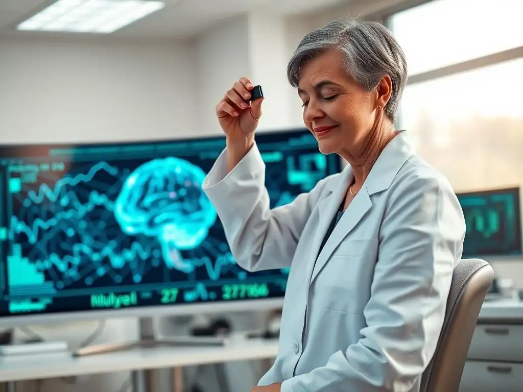
Welcome to Mind Care Guide, a space dedicated to understanding and nurturing your mental well-being. Ever wondered how technology can unlock the secrets of your brain and offer a deeper understanding of your mental state? Dive in to explore the fascinating world of qEEG for mental health assessment, and discover how this innovative technique uses electrical signals to illuminate brain function. Curious to learn more about how qEEG might revolutionize mental health care? Let's embark on this journey together.
Quantitative EEG (qEEG): Assessing Brain Function in Mental Health
Quantitative Electroencephalography, or qEEG, is an advanced method used to analyze electrical activity in the brain. Unlike traditional EEG, which relies heavily on visual inspection of raw brainwave patterns, qEEG employs sophisticated computer analysis to quantify and map these patterns. This technological leap allows for a more objective and detailed assessment of brain function, providing insights that can be invaluable in understanding various mental health conditions. The application of qEEG for mental health assessment is growing as clinicians and researchers seek more precise tools.
The brain's electrical signals, generated by the synchronized activity of millions of neurons, reflect its functional state. Different patterns of electrical activity, known as brainwaves (delta, theta, alpha, beta, gamma), are associated with different states of consciousness and cognitive processes. By measuring and analyzing these patterns using qEEG, practitioners can identify deviations from typical brain activity that may correlate with specific mental health challenges, offering a complementary perspective alongside clinical evaluation.
- Introduction to qEEG in Mental Health
- How qEEG Works
- qEEG vs. Traditional EEG
- Applications of qEEG in Mental Health Assessment
- Benefits of qEEG in Mental Health Practice
- Limitations and Considerations of qEEG
- The Future of qEEG in Mental Health
- qEEG and Brain Imaging
- Conclusion: Leveraging qEEG for Better Mental Health Outcomes
- Frequently Asked Questions
- Final Reflection: Unlocking the Brain's Electrical Language for Mental Health
- Unlock Your Brain's Potential: Take Charge of Your Mental Wellbeing
Introduction to qEEG in Mental Health
Quantitative EEG has emerged as a significant tool within the field of neuroscience and clinical practice for its capacity to provide objective data on brain function. This method moves beyond the qualitative analysis of traditional EEG recordings, transforming complex brainwave data into statistical maps and indices. These quantitative measures allow for comparisons against normative databases, helping to identify patterns that may be linked to neurological or psychological conditions. The integration of qEEG for mental health assessment represents a step towards more data-driven approaches in understanding the biological underpinnings of these conditions.
In the context of mental health, qEEG offers a window into the neural circuits involved in mood regulation, cognitive processing, attention, and emotional responses. By pinpointing areas or networks with atypical electrical activity, qEEG can potentially help in differentiating between conditions that might present with similar symptoms, such as depression and anxiety, or different subtypes of ADHD. This objective information complements subjective patient reports and clinical observations, contributing to a more comprehensive understanding of an individual's mental state and potential treatment pathways.
Understanding qEEG Technology
qEEG technology involves placing electrodes on the scalp according to a standardized system, typically the 10-20 system, to record the electrical potentials generated by the brain. These raw electrical signals, or EEG data, are then digitized and subjected to rigorous mathematical and statistical analysis.
This process transforms the continuous waveform data into quantitative metrics, such as power spectral density (measuring the strength of different brainwave frequencies), coherence (measuring the degree of synchronization between different brain regions), and asymmetry (comparing activity between hemispheres). These metrics are then often visualized as brain maps, or "topographical maps," which provide a color-coded representation of brain activity across different areas.
The core of qEEG lies in comparing an individual's brain activity patterns to a large normative database of healthy individuals, often stratified by age and sex. Statistical analyses, such as z-score comparisons, highlight areas where an individual's brain activity significantly deviates from the norm. For instance, a study might reveal elevated theta activity in frontal regions compared to the normative database, a pattern that has been associated with conditions affecting attention and executive function. This objective comparison helps in identifying potential biomarkers or correlates of mental health conditions, supporting the utility of qEEG for mental health assessment.
The Role of qEEG in Neuroscience
Neuroscience research heavily utilizes qEEG to investigate the neural correlates of various cognitive processes, emotional states, and neurological disorders. By providing a high temporal resolution – meaning it can capture brain activity changes that occur in milliseconds – qEEG is excellent for studying the dynamic nature of brain function. Researchers can analyze how brainwave patterns change during specific tasks, in response to stimuli, or in different clinical populations. This allows for a deeper understanding of how brain networks operate and how their dysfunction might manifest in mental health symptoms.
Furthermore, qEEG contributes to the development of computational models of brain function. By analyzing complex patterns of connectivity and activity across multiple brain regions, neuroscientists can build models that simulate brain processes and predict how alterations might lead to specific clinical presentations. Research using qEEG has, for example, explored differences in alpha power asymmetry related to emotional processing or changes in connectivity patterns in individuals with schizophrenia compared to control groups. This research solidifies the role of qEEG as a valuable tool for advancing our understanding of the brain's role in mental health.
How qEEG Works
The process of conducting a qEEG assessment involves several key steps, starting with the recording of raw EEG data and culminating in sophisticated analysis and interpretation. The fundamental principle is to capture the brain's electrical activity accurately and then apply mathematical methods to extract meaningful, quantifiable information. This quantitative approach is what distinguishes qEEG from traditional visual EEG analysis and makes it particularly useful for objective assessments, including qEEG for mental health assessment.
The technology relies on the fact that neuronal communication involves electrical signals. When large populations of neurons fire in a synchronized manner, these electrical signals are strong enough to be detected by electrodes placed on the scalp. The patterns of these signals vary depending on the state of the brain (e.g., alert, relaxed, asleep) and the specific cognitive or emotional processes occurring. qEEG provides the tools to dissect these complex patterns into their constituent frequencies and spatial distributions, allowing for a detailed "brain map" of electrical activity.
Recording Brain Electrical Activity
The initial step in a qEEG assessment is the recording of the raw electroencephalogram (EEG). This involves preparing the scalp and attaching multiple electrodes, typically ranging from 19 to 128 or more, using a conductive gel or paste to ensure good electrical contact. The electrodes are placed at standardized positions on the scalp according to systems like the international 10-20 system. The patient is usually asked to sit quietly with their eyes closed or open, and sometimes specific tasks are administered to assess brain function during different states.
The recording device, an electroencephalograph, amplifies the tiny electrical signals picked up by the electrodes. These signals are then digitized, converting the continuous analog waveforms into a digital format that can be processed by a computer. The quality of the recording is crucial; artifacts from eye movements, muscle tension, or electrical interference must be minimized or removed during subsequent processing to ensure accurate data. A typical recording session might last between 20 and 40 minutes, collecting millions of data points representing the brain's electrical activity.
Transforming EEG Data into Quantitative Measures
Once the raw EEG data is digitized and cleaned of artifacts, specialized software is used to transform it into quantitative measures. A primary method is the Fast Fourier Transform (FFT), which decomposes the complex EEG waveform into its constituent frequency components (delta, theta, alpha, beta, gamma). For each frequency band, the software calculates metrics like power (the amplitude squared, indicating the strength of the signal at that frequency) at each electrode location.
Beyond power, other quantitative measures are computed. Coherence measures the functional connectivity between different brain regions by assessing the consistency of phase differences between signals from two electrodes at a specific frequency. Asymmetry compares the power or coherence between homologous regions in the left and right hemispheres. Additional metrics might include phase lag, amplitude asymmetry, and various connectivity indices. These numerical values provide an objective description of the brain's electrical activity patterns, forming the basis for the qEEG analysis and potential qEEG for mental health assessment.
qEEG Analysis and Interpretation
The quantitative measures derived from the EEG data are then analyzed, often by comparing them to age- and sex-matched normative databases. This comparison identifies deviations from typical brain activity patterns, often expressed as z-scores, which indicate how many standard deviations an individual's measure is away from the mean of the normative group. These deviations can be visualized as topographical brain maps, where different colors represent the magnitude of the deviation (e.g., red for significantly elevated activity, blue for significantly reduced activity).
Interpretation of the qEEG analysis requires expertise, combining the statistical findings from the brain maps and quantitative indices with the individual's clinical history, symptoms, and other assessment data. Certain patterns of deviation have been associated with specific mental health conditions in research literature.
For example, elevated theta/beta ratios in frontal areas are often reported in individuals with ADHD, while alpha asymmetry might be observed in some forms of depression. However, it's critical to remember that qEEG findings are not diagnostic on their own but serve as valuable pieces of information to inform clinical understanding and treatment planning, supporting the use of qEEG for mental health assessment as part of a comprehensive evaluation.
qEEG vs. Traditional EEG
While both quantitative EEG and traditional EEG record the electrical activity of the brain using electrodes on the scalp, their approaches to analysis and the type of information they provide differ significantly. Traditional EEG relies primarily on the visual inspection of raw waveforms by a trained electroencephalographer, who looks for characteristic patterns, spikes, or abnormalities associated with conditions like epilepsy or sleep disorders. qEEG, conversely, uses computational analysis to quantify and statistically compare brain activity patterns, offering a more objective and detailed perspective that is increasingly applied in the assessment of mental health.
The distinction lies in the level of detail and objectivity. Traditional EEG is excellent for identifying acute events or gross abnormalities, while qEEG is designed to detect more subtle, persistent deviations in brain function that may underlie complex psychiatric symptoms. The quantitative nature of qEEG allows for statistical comparisons and the generation of objective metrics, reducing the subjectivity inherent in visual waveform interpretation. This enhanced analytical capability makes qEEG a powerful tool for research and clinical applications where subtle functional differences are important, such as using qEEG for mental health assessment.
Enhanced Precision and Objectivity
One of the primary advantages of qEEG over traditional EEG is its enhanced precision and objectivity. By converting complex brainwave data into numerical values, qEEG analysis minimizes the reliance on subjective visual interpretation. Statistical comparisons against normative databases provide an objective measure of how an individual's brain activity differs from a typical population, highlighting specific frequency bands, brain regions, or connectivity patterns that show significant deviations.
This quantitative approach allows for the detection of subtle anomalies that might be missed during a visual review of raw EEG waveforms. For example, a slight but persistent elevation of theta power in a specific frontal region might not be immediately obvious in a traditional EEG tracing but would be clearly identified and quantified in a qEEG analysis and displayed on a brain map.
This increased precision is particularly valuable in mental health, where symptoms can be complex and related to subtle functional dysregulations rather than gross structural or electrical abnormalities. The objective data provided by qEEG for mental health assessment can offer a more nuanced understanding of brain function.
Quantitative Analysis for Subclinical Detection
Quantitative analysis allows qEEG to potentially identify subtle or subclinical patterns of brain dysfunction that may precede or contribute to overt symptoms of mental health conditions. By comparing an individual's brain activity to a large database of healthy individuals, qEEG can highlight deviations that, while perhaps not meeting the criteria for a formal diagnosis, might indicate underlying vulnerabilities or early functional changes. This capability is particularly relevant in understanding the spectrum of mental health and identifying potential targets for early intervention.
For instance, research might show that individuals at high risk for developing a certain condition exhibit specific qEEG patterns that differ from low-risk individuals, even before significant symptoms emerge. While not a predictive test, the quantitative data from qEEG can provide insights into individual differences in brain function that may relate to resilience or vulnerability to mental health challenges. This capacity for detailed quantitative analysis makes qEEG a valuable tool not just for assessing existing conditions but also for advancing research into the early markers of mental health issues.
Applications of qEEG in Mental Health Assessment

The application of qEEG for mental health assessment spans a wide range of conditions, offering insights into the neural underpinnings of various psychiatric disorders. While not a standalone diagnostic tool, qEEG provides objective data on brain function that can complement clinical evaluations, helping to inform diagnosis, guide treatment selection, and monitor progress. Research continues to explore and validate specific qEEG patterns associated with different conditions.
Different mental health conditions are thought to involve distinct, though often overlapping, patterns of brain dysfunction. qEEG can help to characterize these patterns by analyzing power in different frequency bands, connectivity between brain regions, and other quantitative metrics. This allows for a more personalized understanding of an individual's specific presentation, moving beyond symptom clusters to consider underlying brain activity. The growing body of literature on specific qEEG findings in various disorders highlights its potential utility in clinical practice.
qEEG for Depression
Research into qEEG for depression has frequently focused on patterns of alpha power asymmetry, particularly in frontal brain regions. Studies have suggested that reduced alpha power in the left frontal cortex compared to the right (left frontal alpha asymmetry) may be associated with withdrawal, reduced motivation, and negative affect, symptoms often seen in depression. While findings are not universally consistent across all studies and subtypes of depression, this pattern is one of the most studied qEEG correlates in mood disorders.
Beyond alpha asymmetry, other qEEG findings in depression can include alterations in theta, alpha, and beta power in various brain regions, as well as changes in connectivity patterns. For example, some studies report increased frontal theta activity or decreased frontal alpha power. The specific patterns observed can vary depending on the severity and subtype of depression. Utilizing qEEG for mental health assessment in depression can help clinicians identify potential neural targets for neuromodulation therapies like Transcranial Magnetic Stimulation (TMS), where treatment protocols are sometimes guided by individual brain activity patterns.
qEEG for Anxiety Disorders
Anxiety disorders are also associated with characteristic qEEG patterns, although these can vary depending on the specific type of anxiety (e.g., generalized anxiety, panic disorder, social anxiety). Common findings include alterations in beta and alpha activity. Increased high-frequency beta activity, particularly in frontal or central regions, is sometimes observed and thought to reflect heightened arousal, vigilance, or worry. Conversely, reduced alpha power or altered alpha asymmetry might also be present in some individuals with anxiety.
Furthermore, studies using qEEG for mental health assessment in anxiety often investigate connectivity patterns. For instance, altered coherence between brain regions involved in emotional processing (like the amygdala) and prefrontal areas involved in regulation might be observed. While more research is needed to establish highly specific biomarkers for each anxiety subtype, qEEG provides a means to objectively assess the neural activity associated with the anxious state, potentially aiding in understanding the individual's specific presentation and guiding interventions like neurofeedback.
qEEG for ADHD
One of the most studied applications of qEEG for mental health assessment is in Attention-Deficit/Hyperactivity Disorder (ADHD). A frequently reported finding in individuals with ADHD, particularly the inattentive subtype, is an elevated theta/beta power ratio, often most prominent in frontal and central scalp regions. This pattern is hypothesized to reflect a state of under-arousal in brain areas critical for attention and executive function. While not all individuals with ADHD exhibit this pattern, research suggests it is more common in this population than in typically developing individuals.
Other qEEG patterns observed in ADHD can include increased absolute or relative theta power, reduced absolute or relative beta power, and altered connectivity, such as reduced coherence in specific frequency bands between frontal and posterior regions. These quantitative findings can provide objective support for a clinical diagnosis, particularly in complex cases, and can help differentiate ADHD from other conditions with similar symptoms. Moreover, qEEG findings can sometimes inform treatment choices, including medication or neurofeedback protocols, which aim to normalize these aberrant brainwave patterns.
qEEG for PTSD
Post-Traumatic Stress Disorder (PTSD) is a complex trauma-related disorder that can involve significant alterations in brain function. qEEG research in PTSD has explored patterns related to hyperarousal, emotional dysregulation, and altered processing of traumatic memories. Findings can include changes in alpha, beta, and gamma power, particularly in regions associated with fear processing (like the limbic system) and executive control (like the prefrontal cortex).
Studies have reported increased frontal alpha asymmetry (sometimes opposite to that seen in depression), altered theta activity, and changes in connectivity patterns, such as reduced coherence between frontal and limbic areas. These patterns are thought to reflect the persistent state of threat sensitivity and difficulty regulating emotional responses characteristic of PTSD. Using qEEG for mental health assessment in PTSD can help to objectively document these neural alterations and potentially track the impact of trauma-focused therapies or other interventions aimed at restoring more typical brain function.
qEEG for Schizophrenia
Schizophrenia is a severe mental illness associated with widespread alterations in brain structure and function. qEEG studies in schizophrenia have consistently reported various abnormalities, including increased delta and theta power, decreased alpha and beta power, particularly in frontal regions, and significant alterations in connectivity patterns. These findings are thought to reflect disruptions in neural networks involved in cognitive processing, sensory perception, and reality testing.
Specific findings in schizophrenia can include reduced coherence, suggesting impaired communication between different brain regions, and altered gamma activity, which is associated with sensory processing and binding. While the qEEG patterns in schizophrenia are complex and heterogeneous, reflecting the varied symptoms of the disorder, quantitative analysis provides objective markers of neural dysfunction. qEEG for mental health assessment in schizophrenia contributes to research aimed at identifying biomarkers for diagnosis, prognosis, and response to antipsychotic medication.
qEEG for Bipolar Disorder
Bipolar disorder, characterized by mood swings between depressive and manic or hypomanic states, is also associated with distinct patterns of brain activity measurable by qEEG. Research suggests that individuals with bipolar disorder, even during periods of remission, may exhibit persistent differences in brain function compared to those without the disorder. Findings can include altered power in various frequency bands and changes in connectivity, particularly in networks involved in mood regulation and emotional processing.
Studies have reported altered alpha asymmetry, changes in frontal theta power, and differences in beta and gamma activity. Connectivity abnormalities, such as altered coherence within or between frontal, limbic, and parietal regions, are also investigated. These patterns are thought to contribute to the mood instability and cognitive difficulties experienced by individuals with bipolar disorder. While qEEG patterns can vary depending on the current mood state (depressive vs. manic), using qEEG for mental health assessment can offer insights into trait-like neural characteristics of the disorder and potentially help differentiate it from unipolar depression.
Benefits of qEEG in Mental Health Practice
Integrating qEEG into mental health practice offers several potential benefits, enhancing the understanding and management of complex conditions. By providing objective, quantifiable data on brain function, qEEG complements traditional clinical assessment methods, which often rely heavily on subjective reports and observational data. This objective layer of information can lead to more informed decision-making regarding diagnosis and treatment.
The non-invasive nature of the procedure is another significant advantage, making it a comfortable and accessible assessment tool for patients. Unlike imaging techniques such as fMRI or PET, qEEG does not involve radiation or strong magnetic fields, making it suitable for repeat assessments, such as monitoring treatment response. These benefits underscore the growing interest in utilizing qEEG for mental health assessment.
Improved Diagnostic Accuracy
One of the most promising benefits of qEEG is its potential to improve diagnostic accuracy, particularly in differentiating between conditions that present with overlapping symptoms. For instance, distinguishing between ADHD, anxiety, and certain subtypes of depression based solely on clinical presentation can sometimes be challenging. qEEG can reveal distinct patterns of brain activity that are more commonly associated with one condition over another, providing objective support for a clinical diagnosis.
While qEEG is not a diagnostic test in isolation – a diagnosis is made based on a comprehensive clinical evaluation – the quantitative data it provides can help clarify ambiguous presentations. By highlighting specific areas of functional dysregulation, qEEG can aid clinicians in refining their diagnostic impression and considering conditions that might not have been immediately apparent from symptoms alone. Research continues to validate the specificity of qEEG patterns for various disorders, enhancing its role in objective assessment.
Guiding Personalized Treatment Plans
qEEG findings can play a crucial role in guiding personalized treatment plans. Since different individuals with the same diagnosis might have different underlying patterns of brain dysfunction, a "one size fits all" approach to treatment is often ineffective. qEEG can help identify the specific neural correlates of an individual's symptoms, allowing for the selection of interventions that target those specific areas or networks.
For example, if a qEEG reveals excessive slow-wave activity in frontal regions in a patient with ADHD, neurofeedback protocols aimed at increasing fast-wave activity in those specific areas might be chosen. Similarly, qEEG findings can sometimes inform the selection of medication, as different classes of psychotropic drugs may have differential effects on brain activity patterns. By providing objective data on an individual's brain function, qEEG supports a more tailored and potentially more effective approach to treatment, leveraging qEEG for mental health assessment to inform therapeutic strategies.
Monitoring Treatment Response
qEEG can be a valuable tool for monitoring a patient's response to treatment. By conducting follow-up qEEG assessments after a period of intervention (e.g., medication, therapy, neurofeedback), clinicians can objectively measure changes in brain activity patterns. Improvements in qEEG metrics, such as normalization of previously aberrant patterns, can provide objective evidence of treatment effectiveness, even before clinical symptom improvement is fully apparent.
This objective monitoring can help clinicians determine if a treatment is working, whether adjustments are needed, or if alternative interventions should be considered. For instance, if a patient undergoing neurofeedback for anxiety shows a reduction in high-beta activity in relevant areas on a follow-up qEEG, it provides objective confirmation that the training is impacting the targeted brain activity. This capacity for objective tracking makes qEEG a powerful tool for optimizing treatment outcomes in mental health practice.
Non-Invasive Assessment Method
A significant benefit of qEEG is its non-invasive nature. The procedure involves simply placing electrodes on the scalp to record naturally occurring electrical signals; it does not require injections, exposure to radiation, or strong magnetic fields. This makes qEEG a safe and comfortable assessment method suitable for individuals of all ages, including children, and for repeated use over time without concern for cumulative exposure.
The ease and safety of the procedure contrast with some other brain imaging techniques, making qEEG more accessible for routine clinical use and for research involving vulnerable populations. Patients can undergo a qEEG assessment in a standard clinical setting with minimal discomfort. This non-invasive characteristic is a key factor in the increasing adoption of qEEG for mental health assessment as a practical tool.
Limitations and Considerations of qEEG
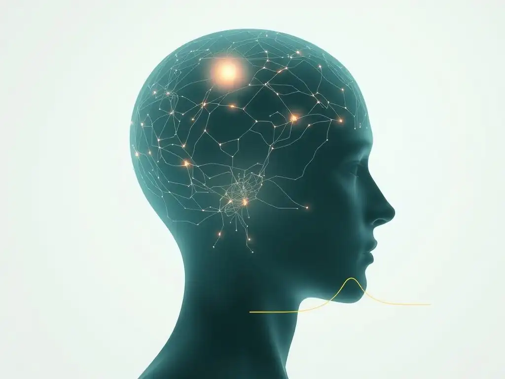
Despite its many potential benefits, qEEG is not without limitations and requires careful consideration in its application. Like any assessment tool, qEEG findings must be interpreted within the broader clinical context and should not be used in isolation for diagnosis. Several factors can influence qEEG results, and the technology itself has inherent limitations regarding the source localization of electrical activity.
It is crucial for clinicians and patients to understand that qEEG provides correlational data – it identifies patterns of brain activity that are associated with certain conditions, but it does not definitively cause them. The relationship between specific qEEG patterns and complex psychiatric symptoms is still an active area of research, and findings can vary across studies and populations. Therefore, qEEG is best viewed as a complementary tool within a comprehensive assessment battery.
Factors Affecting qEEG Results
Several factors can influence qEEG results, potentially leading to inaccurate or misleading findings if not properly controlled. These include:
- Artifacts: Muscle tension, eye movements, blinking, poor electrode contact, and electrical interference from external sources can introduce noise into the EEG recording, distorting the brain activity signals. Rigorous artifact detection and removal procedures are essential.
- Medication: Psychotropic medications can significantly alter brain electrical activity patterns. It is crucial to account for a patient's medication status when interpreting qEEG results, ideally conducting assessments when the patient is not on medication or comparing findings to normative data adjusted for medication effects, if available.
- State: The patient's state during the recording (e.g., alert, drowsy, anxious) can influence brainwave patterns. Standardized recording protocols (e.g., eyes closed vs. eyes open) are used, but individual variability can still exist.
- Technical Factors: The quality of the EEG equipment, the expertise of the technician performing the recording, and the software used for analysis can all impact the reliability and validity of the qEEG results.
The Importance of Clinical Context
Interpreting qEEG findings requires integrating the quantitative data with the individual's full clinical picture. This includes their symptoms, medical history, psychiatric history, family history, psychosocial factors, and results from other assessments (e.g., psychological evaluations, neurocognitive testing). A qEEG report should not be read in isolation but used to inform and refine the clinical understanding of the patient.
For example, an elevated frontal theta/beta ratio might be observed in a qEEG. While this pattern is often associated with ADHD, it can also be seen in other conditions or even in individuals without any clinical diagnosis. Therefore, the finding must be considered alongside the patient's reported symptoms of inattention, impulsivity, and hyperactivity, as well as their performance on cognitive tests. The clinical context provides the necessary framework for determining the relevance and significance of the qEEG findings in relation to the patient's mental health challenges.
qEEG as a Complementary Tool
Given its limitations and the factors that can influence results, qEEG is best utilized as a complementary tool within a comprehensive mental health assessment. It provides valuable objective data on brain function that can support clinical hypotheses, help differentiate between conditions, and guide treatment planning, but it does not replace the need for a thorough clinical interview, behavioral observations, and other diagnostic procedures.
Think of qEEG as adding another layer to the assessment puzzle. It offers insights into the biological substrate of symptoms, but these insights must be integrated with psychological, social, and environmental factors. A comprehensive assessment utilizing qEEG for mental health assessment alongside traditional methods offers the most robust approach to understanding complex mental health presentations and developing effective, personalized treatment strategies.
The Future of qEEG in Mental Health
The field of qEEG is continuously evolving, driven by advancements in technology, data analysis techniques, and a growing understanding of brain-behavior relationships. As research progresses and technology becomes more sophisticated, the utility and accessibility of qEEG for mental health assessment are expected to increase. Future developments hold promise for even greater precision, integration with other brain imaging modalities, and broader application in personalized mental healthcare.
The increasing availability of larger normative databases and more advanced algorithms for artifact correction and analysis are improving the reliability and validity of qEEG findings. Furthermore, research is moving towards identifying more specific and reliable qEEG biomarkers for different subtypes and stages of mental health conditions, which could revolutionize how these disorders are diagnosed and treated.
Advancements in qEEG Technology
Technological advancements are making qEEG systems more portable, affordable, and user-friendly. High-density EEG systems with a greater number of electrodes (up to 256 channels) are becoming more common, providing higher spatial resolution and a more detailed picture of brain activity. Wireless and dry electrode systems are also being developed, making the recording process more comfortable and less time-consuming.
Improvements in signal processing algorithms are enhancing the ability to remove artifacts and extract cleaner brain activity data. Machine learning and artificial intelligence techniques are being applied to qEEG analysis to identify complex patterns and predict treatment response with greater accuracy. These technological leaps are expanding the capabilities of qEEG and making it more practical for routine clinical use and large-scale research studies, further solidifying the role of qEEG for mental health assessment in the digital age.
Integration of qEEG with Other Brain Imaging Techniques
One of the most exciting areas of development is the integration of qEEG with other brain imaging techniques, such as functional Magnetic Resonance Imaging (fMRI) and Positron Emission Tomography (PET). While qEEG excels at capturing the timing (temporal resolution) of brain activity, fMRI and PET provide excellent spatial resolution, showing where activity is occurring in the brain. Combining these modalities can provide a more comprehensive understanding of brain function, capturing both the timing and location of neural events.
For example, simultaneous EEG-fMRI recordings allow researchers to correlate changes in brainwave patterns captured by EEG with changes in blood flow measured by fMRI, offering a richer dataset for investigating neural networks involved in mental health conditions. This multimodal approach holds significant potential for advancing our understanding of the complex interplay between brain structure, function, and mental health symptoms, enhancing the power of brain mapping techniques.
Expanding qEEG for Personalized Mental Healthcare
The future of qEEG lies in its potential to contribute significantly to personalized mental healthcare. As research identifies more specific qEEG patterns associated with different treatment responses, clinicians may be able to use qEEG data to predict which interventions are most likely to be effective for a particular individual. This could lead to more efficient and successful treatment pathways, reducing the trial-and-error often involved in finding the right therapy or medication.
Furthermore, longitudinal qEEG assessments could provide objective markers for tracking the progression of conditions or the impact of lifestyle interventions (e.g., exercise, mindfulness) on brain function. This would empower individuals and clinicians to make data-informed decisions about ongoing care. The vision is that qEEG for mental health assessment will become a standard tool in the clinician's arsenal, helping to tailor interventions to the unique brain profile of each individual.
qEEG and Brain Imaging
qEEG is a form of brain imaging, specifically functional brain imaging, as it measures brain activity rather than structure. However, it differs significantly from other functional imaging techniques like fMRI and PET in terms of what it measures and its spatiotemporal resolution. While qEEG measures the electrical activity of neurons with very high temporal resolution (milliseconds), fMRI measures blood flow changes (BOLD signal) as an indirect indicator of neural activity with good spatial resolution but lower temporal resolution (seconds), and PET measures metabolic activity or neurotransmitter binding with good spatial resolution but poor temporal resolution (minutes).
Understanding the strengths and weaknesses of each modality is key to their effective use, particularly when considering qEEG for mental health assessment alongside other techniques. The ability to combine the high temporal precision of qEEG with the excellent spatial localization of fMRI or PET offers a powerful approach to investigating the neural basis of mental health conditions, providing a more complete picture than any single technique alone.
Integrating qEEG with fMRI and PET
Integrating qEEG with fMRI and PET scans can provide a more complete picture of brain function in mental health. Simultaneous EEG-fMRI recordings allow researchers to correlate electrical brain activity (measured by EEG) with changes in blood flow (measured by fMRI) in real-time. This helps to understand how rapid electrical events relate to activity in specific brain regions and networks. For example, researchers might investigate how specific patterns of alpha or theta activity measured by EEG correlate with activity in limbic structures or prefrontal cortex measured by fMRI during emotional tasks.
Similarly, combining qEEG with PET allows researchers to relate patterns of electrical activity to metabolic rates or neurotransmitter system function in specific brain areas. For instance, a study might explore how altered dopamine receptor availability measured by PET correlates with specific qEEG patterns in individuals with schizophrenia or ADHD. These integrated approaches provide richer datasets for understanding the complex neurobiological underpinnings of mental health disorders, advancing the field of brain mapping in psychiatry.
Advancing Mental Health Research
The integration of qEEG with other brain imaging techniques is significantly advancing mental health research. By combining the strengths of different modalities, researchers can investigate brain function at multiple levels simultaneously, from rapid electrical oscillations to metabolic activity and structural connectivity. This multimodal approach allows for a more nuanced understanding of the complex neural circuits involved in psychiatric disorders.
For example, research using combined EEG-fMRI has provided insights into the altered connectivity within resting-state networks in depression, while studies combining EEG and PET have explored the relationship between neurotransmitter systems and specific brainwave abnormalities in conditions like Parkinson's disease with associated mood symptoms. These integrated studies are crucial for identifying reliable biomarkers, understanding disease mechanisms, and developing more targeted interventions for mental health conditions, highlighting the vital role of advanced brain imaging, including qEEG for mental health assessment, in pushing the boundaries of neuroscience and clinical practice.
Conclusion: Leveraging qEEG for Better Mental Health Outcomes
Quantitative EEG (qEEG) offers a powerful, non-invasive method for assessing brain function by quantifying and analyzing electrical brain activity. By transforming raw EEG data into objective metrics and topographical maps, qEEG provides insights into neural patterns that may be associated with various mental health conditions, including depression, anxiety, ADHD, PTSD, schizophrenia, and bipolar disorder. This objective data complements traditional clinical evaluations, contributing to a more comprehensive understanding of an individual's mental state.
While qEEG is not a standalone diagnostic tool and requires careful interpretation within the full clinical context, its potential benefits are significant. It can enhance diagnostic accuracy by helping to differentiate between conditions with overlapping symptoms, guide the development of personalized treatment plans by identifying specific neural targets, and objectively monitor treatment response. As technology advances and research further validates specific qEEG patterns, the utility of qEEG for mental health assessment is expected to grow, paving the way for more data-driven, personalized, and effective mental healthcare interventions aimed at improving outcomes for individuals facing mental health challenges.
Frequently Asked Questions
What is a qEEG and how is it used in mental health assessment?
A qEEG, or Quantitative Electroencephalogram, is a non-invasive brain imaging technique that measures electrical activity in the brain. It's used in mental health assessment to identify patterns of brain activity that may be associated with various mental health conditions. This provides objective data to supplement clinical evaluations.
How does a qEEG differ from a standard EEG?
While both EEGs measure brain electrical activity, a qEEG uses computer analysis to quantify and compare the data against a normative database. Standard EEGs are typically interpreted visually by a trained professional, whereas qEEGs provide statistical analysis and brain maps.
What mental health conditions can a qEEG potentially help diagnose or assess?
qEEGs have been used to assess conditions like ADHD, anxiety, depression, PTSD, and other disorders related to brain function. It can help identify underlying neurological imbalances that may contribute to these conditions. However, it's crucial to remember it's just one piece of the diagnostic puzzle.
Is a qEEG a painful or invasive procedure?
No, a qEEG is non-invasive and painless. Electrodes are attached to the scalp using a conductive gel to record brain activity. There's no electrical stimulation involved, only recording.
What are the limitations of using qEEG for mental health assessment?
qEEG results should be interpreted with caution, as they are not a standalone diagnostic tool. They should be used in conjunction with clinical interviews, psychological testing, and other relevant information. Further research is needed to fully understand the clinical utility of qEEG in mental health.
Final Reflection: Unlocking the Brain's Electrical Language for Mental Health
The ability to objectively measure brainwave activity using qEEG is revolutionizing mental health assessment, offering a powerful new lens through which to understand and treat a wide range of conditions. This non-invasive technology moves beyond subjective reporting, providing clinicians with quantifiable data to inform diagnoses and personalize treatment plans, ultimately leading to improved patient outcomes.
The impact of qEEG on the field of mental health is profound and continues to grow. Its ability to identify subtle neurological imbalances often missed by traditional methods offers hope for earlier intervention and more effective therapies. This technology is not just about diagnosis; it's about fostering a deeper understanding of the intricate relationship between brain function and mental well-being. The brain is the most complex organ in the body, and understanding its electrical activity is crucial to understanding mental health.
This simple statement underscores the transformative potential of qEEG.
Consider the implications of this technology for your own life, or the lives of those you care about. Are you curious about the underlying neurological patterns contributing to your mental health? Could qEEG offer a more precise understanding of your challenges and guide you toward more effective treatment strategies? Take the time to explore the possibilities of qEEG. Learn more, ask questions, and advocate for its wider adoption in mental health care. The future of mental health assessment is here, and it's powered by the brain's own electrical language.
Unlock Your Brain's Potential: Take Charge of Your Mental Wellbeing
Thanks for diving into the fascinating world of quantitative EEG (qEEG)! You've now learned how this powerful tool can provide valuable insights into brain function and mental health. Ready to take the next step?
- Schedule a qEEG assessment with a qualified professional: This is the most direct way to apply what you've learned and gain personalized insights into your brain's activity.
- Explore online resources and learn more about qEEG's role in different mental health conditions: Deepen your understanding by researching further – you might find surprising connections to your own experiences.
- Share this article with someone who might benefit: Help spread awareness and empower others to explore this life-changing technology.
What specific questions do you have about utilizing qEEG to improve your brain health and mental wellbeing? Let's discuss!