
Brain Imaging in Forensic Psychiatry: Applications and Limitations

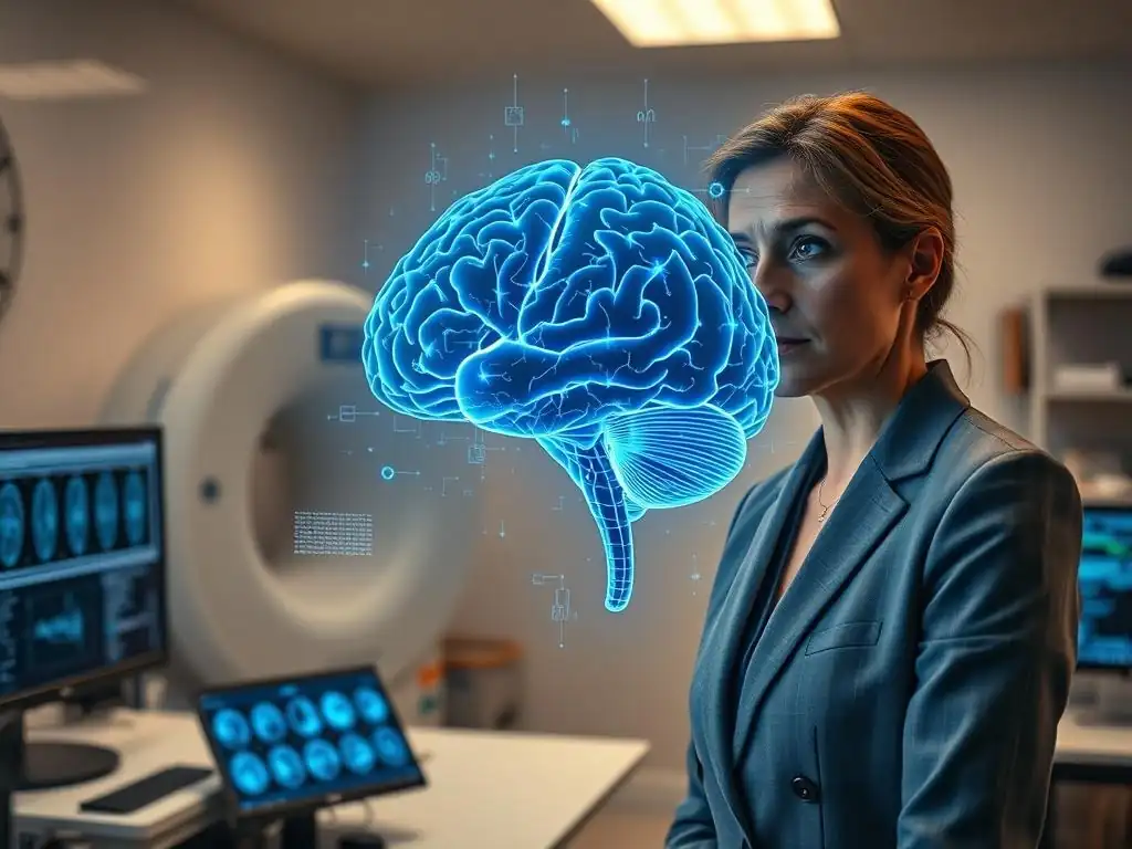
Welcome to Mind Care Guide, a compassionate space dedicated to understanding and nurturing your mental well-being. Ever wondered how technology is helping us unravel the complexities of the mind in even the most challenging legal contexts? Delve into our exploration of " Forensic psychiatry brain imaging" and discover its fascinating applications and limitations within the field, as we explore the Neuroscience of Mental Health. Prepare to have your perspective broadened and your curiosity piqued as we uncover the power of advanced imaging techniques in understanding the human brain.
- Introduction to Forensic Psychiatry Brain Imaging
- Applications of Brain Imaging in Forensic Psychiatry
- Types of Brain Imaging Techniques Used in Forensic Settings
- Limitations of Brain Imaging in Forensic Psychiatry
- The Future of Brain Imaging in Forensic Psychiatry
- Conclusion: Balancing Potential and Practicality of Brain Imaging in Forensic Psychiatry
-
Frequently Asked Questions
- What is the role of brain imaging in forensic psychiatry?
- What types of brain imaging techniques are used in forensic psychiatry?
- How can brain imaging contribute to legal proceedings?
- What are the limitations of using brain imaging in forensic psychiatry?
- How does brain imaging assist with the diagnosis of mental illness in forensic settings?
- Final Reflection: The Evolving Landscape of Forensic Psychiatry and Brain Imaging
- Unlock the Mysteries of the Mind: Your Next Steps in Forensic Psychiatry
Introduction to Forensic Psychiatry Brain Imaging
Understanding the Role of Neuroimaging in Forensic Mental Health
Forensic psychiatry, the intersection of mental health and the legal system, often grapples with complex questions about an individual's state of mind at the time of an alleged offense or their capacity to participate in legal proceedings. Traditionally, assessments rely heavily on clinical interviews, behavioral observations, and psychological testing. However, advancements in neuroscience, particularly brain imaging techniques, are introducing new dimensions to these evaluations. Forensic psychiatry brain imaging offers a potential window into the structural integrity and functional activity of the brain, which may provide objective data relevant to diagnoses, cognitive abilities, and potentially, behaviors pertinent to legal questions.
The application of neuroimaging in forensic settings is still evolving and subject to significant debate. While techniques like MRI and fMRI can reveal brain features associated with various conditions, translating these findings into definitive legal conclusions is challenging. The field of forensic psychiatry brain imaging aims to explore how these technologies can enhance our understanding of the neural underpinnings of behaviors relevant to crime and competency, while also acknowledging the inherent limitations and ethical pitfalls involved in using such complex data in courtrooms and legal decision-making processes.
The Growing Importance of Brain Scans in Legal Contexts
In recent decades, brain imaging evidence has appeared with increasing frequency in legal cases, particularly in the United States. Lawyers and forensic psychiatrists are exploring how structural and functional brain scans might corroborate clinical findings, explain behavior, or even potentially influence perceptions of culpability or competency. This trend reflects both the rapid progress in neuroimaging technology and a growing public fascination with the brain as the seat of behavior. The integration of forensic psychiatry brain imaging into legal discourse is forcing a critical examination of how neuroscience can inform legal principles and practices.
The allure of seemingly objective brain data is powerful in a legal system often reliant on subjective interpretation of behavior and testimony. For example, evidence of a frontal lobe injury revealed by an MRI scan might be presented to support an argument that a defendant's impulse control was impaired. However, the scientific community remains cautious about the direct causal link between specific brain findings and complex behaviors like criminal actions or legal capacity. The increasing presence of forensic psychiatry brain imaging in court necessitates careful consideration of its scientific validity, interpretive challenges, and potential for misuse or overstatement.
Applications of Brain Imaging in Forensic Psychiatry
Assessing Criminal Responsibility with Neuroimaging Techniques
One of the most challenging areas in forensic psychiatry is assessing criminal responsibility, which often involves evaluating a defendant's mental state at the time of the offense – specifically, their capacity to understand the nature and quality of their actions or that their actions were wrong. Traditional methods rely on retrospective clinical evaluation. Forensic psychiatry brain imaging explores whether neuroimaging can provide objective data to support or refute such assessments, though this remains highly controversial and generally not accepted as definitive proof in legal settings.
Research has attempted to correlate structural or functional brain abnormalities with behaviors relevant to criminal responsibility, such as impulse control deficits, impaired judgment, or lack of empathy. For instance, studies using structural MRI have identified differences in prefrontal cortex volume in some individuals with impulsive violent behavior compared to control groups, with effect sizes often being small to moderate.
Similarly, functional MRI (fMRI) studies have explored neural responses during tasks related to moral reasoning or emotional processing in individuals with certain diagnoses like psychopathy, though these findings are group-level averages and cannot reliably predict individual behavior or mental state at a specific past time.
Using fMRI to Evaluate Intent and Cognitive Function
Functional Magnetic Resonance Imaging (fMRI) measures brain activity by detecting changes in blood flow. In forensic psychiatry, fMRI has been investigated for its potential to shed light on cognitive functions relevant to intent or mens rea – the mental state element of a crime. The theory is that certain brain activation patterns might correlate with specific cognitive processes like planning, decision-making, or understanding consequences. However, directly linking fMRI signals recorded in a scanner to a person's intent or cognitive state during a past event is fraught with methodological and theoretical difficulties.
While fMRI can show where and when brain activity occurs during specific tasks in the scanner, translating this to an individual's mental state during a real-world event is a huge inferential leap. For example, research might show that individuals with certain conditions exhibit atypical activation in areas like the prefrontal cortex or amygdala when performing tasks related to executive function or emotional regulation.
However, this does not mean a specific fMRI pattern can prove or disprove that a defendant intended to commit a crime or understood their actions were wrong at the time it happened. The complexity of behavior, the influence of external factors, and the inherent variability in brain function across individuals make such direct interpretations unreliable for legal purposes.
Structural MRI for Identifying Brain Abnormalities Relevant to Criminal Behavior
Structural Magnetic Resonance Imaging (MRI) provides detailed images of the brain's anatomy, allowing forensic psychiatrists to identify structural abnormalities such as lesions, tumors, atrophy, or developmental anomalies. The presence of such abnormalities, particularly in areas known to be involved in executive functions, emotional regulation, or impulse control (like the frontal or temporal lobes), might be presented as potential contributing factors to a defendant's behavior. For instance, a history of severe traumatic brain injury (TBI) clearly visible on an MRI could be relevant in explaining behavioral changes that preceded criminal conduct.
However, establishing a direct causal link between a structural abnormality and a specific criminal act is complex. Many individuals with significant brain abnormalities do not engage in criminal behavior, and many who do commit crimes show no discernible structural differences on standard MRI scans.
Furthermore, even when an abnormality exists, determining how it specifically impacted the individual's capacity or mental state at the time of the offense requires careful clinical correlation, not just the imaging finding itself. For example, while a large study might show that individuals with frontal lobe lesions have a statistically higher propensity for aggression, this finding cannot definitively explain why one specific individual with a frontal lesion committed a specific aggressive act.
Evaluating Mental Competency Through Brain Imaging
Mental competency in the legal sense refers to an individual's capacity to understand legal proceedings and assist in their own defense (fitness to stand trial), or their capacity to make decisions about their medical care, finances, etc. (decisional capacity). Traditional competency evaluations rely on clinical interviews and standardized psychological tests assessing cognitive functions like memory, understanding, and reasoning. Forensic psychiatry brain imaging is being explored as a potential adjunct to these evaluations, offering objective data about brain structures and functions underlying these cognitive capacities.
For example, neuroimaging might be used to support a diagnosis of a condition known to impair cognitive function, such as severe dementia or the effects of a significant stroke. While the imaging itself doesn't directly measure competency, it can provide evidence for the presence and extent of neurological conditions that are highly correlated with impaired capacity. The challenge lies in translating group-level correlations between brain features and cognitive deficits into an assessment of a specific individual's functional capacity in a legal context at a particular point in time.
Brain Imaging and the Assessment of Fitness to Stand Trial
Fitness to stand trial requires a defendant to have a factual and rational understanding of the legal process and the ability to consult with their attorney with a reasonable degree of rational understanding. These capacities rely on intact cognitive functions such as memory, attention, reasoning, and communication. Forensic psychiatry brain imaging could potentially provide objective evidence regarding the structural or functional integrity of brain regions critical for these functions.
For instance, a defendant with a documented history of stroke causing extensive damage to language processing areas (verified by structural MRI) might have an objective basis for impaired communication, relevant to their ability to assist counsel. Similarly, functional imaging could, in principle, explore brain activity during tasks related to legal decision-making, though this application is highly experimental.
Current practice does not rely on brain imaging alone to determine fitness; it remains a clinical and legal determination informed by all available evidence, including clinical examination, history, and behavioral observation, with imaging serving only as potential supporting data regarding underlying neurological conditions.
Identifying Cognitive Impairments Affecting Legal Capacities
Legal capacities, such as the ability to manage one's affairs, make medical decisions, or understand a contract, all depend on a range of cognitive functions, including memory, executive function, judgment, and insight. Neuroimaging techniques in forensic psychiatry can help identify and characterize brain pathology or dysfunction that might underlie severe impairments in these areas. Conditions like advanced Alzheimer's disease, severe intellectual disability, or major neurocognitive disorder due to TBI often present with clear structural changes on MRI (e.g., atrophy) and potentially altered function measurable by fMRI or PET.
For example, a PET scan showing significantly reduced metabolic activity in the frontal lobes might support a clinical finding of impaired executive function severe enough to compromise financial decision-making capacity in an elderly individual. However, it's crucial to note that brain imaging provides information about the brain, not a direct measure of legal capacity.
Legal capacity is a functional assessment based on observed behavior and performance on specific tasks relevant to the capacity in question, interpreted within a legal framework. Brain imaging serves only to potentially explain or corroborate the cause or basis of observed cognitive deficits, not to quantify the legal capacity itself.
Detecting Malingering and Deception Using Brain Imaging
Malingering, the intentional production of false or grossly exaggerated physical or psychological symptoms, is a significant challenge in forensic evaluations. Defendants or plaintiffs may feign illness or cognitive deficits to avoid responsibility or gain advantage. Traditional methods for detecting malingering rely on inconsistencies in self-report, performance on symptom validity tests, and discrepancies between reported symptoms and objective findings. Forensic psychiatry brain imaging has been explored as a potential novel tool to detect deception or feigning, particularly using fMRI.
The premise behind using fMRI for lie detection is that lying is cognitively more demanding than telling the truth and involves greater activation in certain brain regions, such as the prefrontal cortex and anterior cingulate cortex, associated with conflict monitoring and cognitive control. Studies have shown that fMRI can achieve accuracies above chance in controlled laboratory settings when participants are instructed to lie about simple facts. However, the leap from a controlled lab setting to detecting malingering or deception in a complex forensic context is enormous and currently insurmountable for practical, reliable application.
fMRI-Based Lie Detection in Forensic Settings
Research into fMRI-based lie detection for forensic purposes has generated considerable interest but also significant skepticism. While laboratory studies report accuracies ranging from roughly 70% to over 90% under specific conditions, these studies typically involve simple, instructed lies in healthy volunteers, a scenario far removed from the high-stakes, emotionally charged environment of a forensic evaluation involving complex past events.
Factors like countermeasures (strategies to deliberately alter brain activity), individual variability in brain function, the impact of mental illness, substance use, or emotional states on brain activity, and the lack of a single, universal "lie signature" in the brain severely limit the reliability and validity of this technology for real-world forensic applications.
Furthermore, the interpretation of fMRI data is complex and subject to methodological choices (e.g., how data is processed and analyzed). A signal difference in a particular brain region might be due to deception, but it could also be related to anxiety, attempts to recall details, or other cognitive processes.
Currently, no fMRI technique for lie detection has been scientifically validated for use in legal settings, and major scientific and legal bodies have expressed strong reservations about its reliability and admissibility in court. The use of forensic psychiatry brain imaging for lie detection remains firmly in the realm of research, not established clinical or forensic practice.
Ethical Considerations in Using Brain Scans for Deception Detection
The prospect of using brain imaging to detect deception raises profound ethical questions. If fMRI lie detection were ever deemed sufficiently reliable (a significant 'if'), its use could potentially infringe upon fundamental rights, such as the right against self-incrimination. Could individuals be compelled to undergo brain scans? How would the results be interpreted, and what margin of error would be acceptable in legal proceedings?
Moreover, the technology is not foolproof and could yield false positives or negatives, potentially leading to miscarriages of justice. There are concerns about the potential for coercion, the privacy of thoughts and cognitive processes accessed via brain activity, and the potential for bias in the interpretation of complex neuroimaging data. The empathetic tone required in mental health discussions underscores the need for extreme caution when considering technologies that probe the brain in ways that could be perceived as invasive or potentially harmful to individuals' rights and dignity, especially within the high-stakes environment of forensic psychiatry brain imaging applications.
Predicting Future Risk of Violence with Neuroimaging
Forensic psychiatry is often tasked with assessing the likelihood that an individual will engage in future violent behavior, particularly in contexts like sentencing, parole decisions, or civil commitment proceedings. This is inherently a probabilistic assessment based on known risk factors (e.g., history of violence, substance abuse, certain mental illnesses). Neuroimaging research is exploring whether brain features can serve as additional risk markers, potentially improving the accuracy of violence risk assessments.
Studies have investigated structural and functional differences in individuals with a history of violence or aggression compared to non-violent populations. For example, some research suggests that reduced gray matter volume or decreased functional connectivity in areas of the prefrontal cortex involved in impulse control and decision-making might be associated with increased risk of violence. Similarly, atypical activity in the amygdala (involved in processing fear and emotion) has been explored. However, these findings are correlations observed at the group level and are influenced by numerous confounding factors (e.g., trauma history, environmental stressors).
Identifying Neural Correlates of Aggression and Impulsivity
Research using fMRI, PET, and structural MRI has identified several brain regions and networks potentially involved in aggression and impulsivity, behaviors often relevant to violent crime. These include the prefrontal cortex (involved in executive control, planning, and inhibiting inappropriate behavior), the amygdala (involved in processing emotional stimuli, particularly fear and anger), the hippocampus (involved in memory and contextual processing), and circuits connecting these areas. For example, some studies using fMRI have observed diminished activation in the ventral prefrontal cortex during tasks requiring impulse inhibition in individuals with a history of impulsive aggression.
Structural studies using MRI have reported findings such as reduced gray matter volume in the orbitofrontal cortex or anterior cingulate cortex in some populations with aggressive or antisocial behavior. However, these findings are not universal, often have small effect sizes, and are subject to variations based on the specific population studied (e.g., individuals with specific diagnoses like antisocial personality disorder, incarcerated populations, individuals with TBI). Identifying a definitive "neural correlate" that reliably predicts aggression across all individuals remains elusive, and the relationship between brain structure/function and complex behaviors is far from a simple one-to-one mapping.
The Predictive Power of Brain Imaging in Forensic Risk Assessment
Despite the identification of potential neural correlates, the predictive power of brain imaging for future violence risk in forensic psychiatry is currently very limited. Risk assessment instruments rely on a combination of static (unchangeable, like criminal history) and dynamic (changeable, like substance use) risk factors, which have been validated over decades of research. While neuroimaging might eventually contribute to these assessments by providing data on underlying biological vulnerabilities, it cannot currently predict whether a specific individual will commit a violent act in the future with any clinically meaningful accuracy.
The probabilistic nature of risk assessment means we are predicting the likelihood of an event occurring within a population or group with certain characteristics, not predicting a specific individual's future actions definitively. Brain imaging findings are, at best, potential risk factors among many others.
A 2013 study, for instance, suggested that reduced activity in the anterior cingulate cortex during an impulse control task predicted recidivism in a group of incarcerated individuals, but the predictive power was modest, and the findings require replication and validation in diverse populations. Using brain imaging alone or giving it undue weight in high-stakes predictions of future violence is scientifically premature and ethically questionable in forensic psychiatry brain imaging applications.
Types of Brain Imaging Techniques Used in Forensic Settings
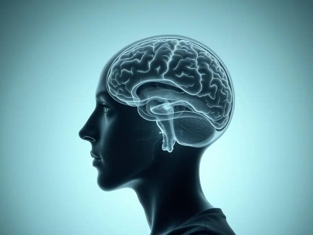
Several different brain imaging modalities are relevant to forensic psychiatry, each providing unique information about the brain's structure or function. The choice of technique depends on the specific question being asked (e.g., is there structural damage? Is a specific brain area functioning abnormally during a task?). Understanding the principles behind these techniques is crucial for interpreting forensic psychiatry brain imaging data.
These techniques broadly fall into categories of structural imaging (showing anatomy) and functional imaging (showing activity or metabolism). While each technique has its strengths, they also have limitations regarding spatial and temporal resolution, cost, and clinical availability, all of which impact their utility and admissibility in forensic contexts.
Structural Imaging: MRI and CT Scans
Structural imaging techniques like Magnetic Resonance Imaging (MRI) and Computed Tomography (CT) provide detailed images of the brain's physical structure. They are used to identify abnormalities in brain anatomy, such as tumors, lesions, areas of atrophy, or the effects of trauma. These techniques are well-established in clinical medicine and are often the first line of imaging used when there is suspicion of underlying neurological pathology relevant to a forensic case.
MRI uses strong magnetic fields and radio waves to create detailed cross-sectional images of brain tissue, offering excellent resolution and differentiation between gray matter, white matter, and cerebrospinal fluid. CT scans use X-rays from multiple angles to create cross-sectional images; they are faster and more readily available than MRI, particularly useful in acute settings (like immediately following head trauma), but provide less detailed images of soft tissue compared to MRI. Both can reveal gross structural damage that might be relevant to behavioral changes or cognitive impairments in forensic psychiatry brain imaging.
Understanding Anatomical Brain Differences
Structural MRI allows for detailed examination of brain anatomy, enabling the identification of differences in volume, shape, or integrity of specific brain structures. For example, studies using structural MRI have compared groups of individuals with certain psychiatric disorders or behavioral profiles (e.g., individuals with schizophrenia, psychopathy, or a history of TBI) to control groups to identify potential anatomical correlates. Findings might include reduced gray matter volume in specific cortical areas, abnormalities in white matter tracts (connective pathways) visible with techniques like Diffusion Tensor Imaging (DTI), or enlarged ventricles.
While these studies can reveal statistically significant anatomical differences on average between groups, there is considerable overlap between groups, and these findings cannot reliably diagnose a condition or explain an individual's behavior. For instance, while some studies show reduced prefrontal cortex volume in groups of individuals with antisocial personality disorder, many individuals with this disorder do not show this finding, and many individuals without the disorder do. In forensic psychiatry brain imaging, structural findings are typically used to support or refute the presence of significant neurological damage or disease, rather than to directly explain complex behaviors or mental states.
Identifying Traumatic Brain Injury and its Legal Implications
Traumatic Brain Injury (TBI), ranging from mild concussion to severe penetrating injury, can cause significant structural damage to the brain, particularly in frontal and temporal lobes, which are critical for executive function, emotional regulation, and social behavior. Structural MRI and CT are standard tools for identifying the presence and extent of TBI. Findings like contusions, hemorrhages, diffuse axonal injury (often visualized with DTI, a form of MRI), or atrophy in specific areas can provide objective evidence of brain damage.
In forensic contexts, evidence of TBI identified through structural imaging is often relevant because TBI can lead to lasting cognitive deficits (e.g., memory problems, impaired executive function) and behavioral changes (e.g., increased impulsivity, irritability, aggression). This evidence might be used to support arguments regarding a defendant's mental state at the time of an offense, their competency to stand trial, or the extent of injury in civil litigation.
For example, a CT scan showing a significant frontal lobe contusion following a severe head injury could provide an anatomical basis for observed changes in personality and behavior that become relevant in a legal case involving forensic psychiatry brain imaging.
Functional Imaging: fMRI, PET, and SPECT
Functional imaging techniques measure brain activity or metabolism rather than just structure. These techniques provide insight into how different brain regions are working, how they interact, and how brain activity might differ in individuals with certain conditions or during specific tasks. In forensic psychiatry, functional imaging is explored for its potential to reveal neural correlates of mental states or cognitive processes relevant to legal questions, though its application is even more debated than structural imaging.
Functional MRI (fMRI) measures changes in blood oxygenation level (BOLD signal) as an indirect indicator of neural activity. Positron Emission Tomography (PET) and Single-Photon Emission Computed Tomography (SPECT) measure metabolic activity or blood flow by detecting radioactive tracers injected into the bloodstream. These tracers can be designed to target specific neurotransmitter systems or glucose metabolism, providing insight into regional brain function.
Measuring Brain Activity and Neural Networks
fMRI is widely used in research to map brain activity during cognitive or emotional tasks. By presenting stimuli or asking participants to perform tasks (e.g., decision-making, emotional processing, impulse control tasks) while in the scanner, researchers can observe which brain areas become more or less active. This allows for the study of neural networks involved in various functions relevant to forensic psychiatry, such as those underlying empathy, moral reasoning, risk-taking, or response inhibition.
For example, fMRI studies have shown that individuals with psychopathy may exhibit reduced activity in the amygdala and ventromedial prefrontal cortex when processing emotional stimuli compared to control groups. PET and SPECT can measure baseline metabolic rates in different brain regions, which can be altered in various neurological and psychiatric conditions.
A PET scan might show reduced glucose metabolism in the frontal lobes of individuals with severe dementia, correlating with their cognitive decline. These techniques provide data on brain function, but translating these findings to an individual's mental state or behavior outside the scanner remains a major challenge in forensic psychiatry brain imaging.
Assessing the Impact of Mental Disorders on Brain Function
Many mental disorders are associated with alterations in brain function and connectivity. Functional imaging can provide objective evidence supporting the presence of such alterations, potentially corroborating a clinical diagnosis that is relevant to a forensic issue. For instance, fMRI studies in individuals with schizophrenia have revealed disrupted connectivity in brain networks involved in cognitive control and reality monitoring. PET studies in depression have shown altered activity in limbic and prefrontal circuits.
While these functional imaging findings can provide a biological context for mental illness, they are typically group-level findings with considerable individual variability. A functional scan showing altered activity patterns consistent with research findings in a population with a specific disorder does not definitively prove that the individual has that disorder or that their behavior at a specific past time was solely caused by that alteration.
The complexity of mental disorders, the influence of medication, substance use, and environmental factors on brain function, and the lack of diagnostic specificity for most functional imaging findings limit their direct application as standalone evidence in forensic psychiatry brain imaging evaluations.
Electroencephalography (EEG) in Forensic Psychiatry
Electroencephalography (EEG) measures electrical activity in the brain using electrodes placed on the scalp. It primarily reflects the summed activity of large populations of neurons. EEG is a relatively old and inexpensive neurophysiological technique compared to fMRI or PET, with high temporal resolution (it can detect changes in brain activity milliseconds after they occur) but poor spatial resolution (it's difficult to pinpoint exactly where in the brain the activity originates).
In clinical settings, EEG is primarily used to diagnose seizure disorders and evaluate other conditions affecting brain electrical activity. In forensic psychiatry, EEG has limited direct application for most questions of responsibility or competency, but it can be useful in specific circumstances, such as evaluating for seizure disorders that might cause altered states of consciousness relevant to an offense, or assessing general brain dysfunction.
Identifying Brainwave Patterns Associated with Mental Illness
While EEG is not a primary diagnostic tool for most psychiatric disorders, some research has explored associations between specific EEG patterns (e.g., altered alpha, theta, or delta wave activity; reduced coherence between brain regions) and certain conditions like schizophrenia, ADHD, or antisocial personality disorder. Some studies have investigated "P300" event-related potentials (ERPs), a type of EEG response associated with attention and memory, in forensic contexts, but these applications are experimental.
For example, some early research explored using P300 responses in a "guilty knowledge test" paradigm, attempting to detect if a suspect recognizes details related to a crime, suggesting they have "guilty knowledge." However, like fMRI lie detection, this application of EEG is controversial, lacks sufficient validation for forensic use, and is susceptible to countermeasures and confounding factors. Currently, EEG findings are rarely used as standalone evidence in forensic psychiatry brain imaging contexts regarding specific mental states or behaviors, though they can contribute to an overall picture of neurological health.
Detecting Seizure Activity and Cognitive Dysfunction
One of the more established uses of EEG relevant to forensic psychiatry is the detection of seizure disorders or other conditions causing generalized brain dysfunction. Certain types of seizures, particularly complex partial or absence seizures, can result in periods of altered consciousness, confusion, or automatic behaviors during which an individual may not be fully aware of their actions or may behave in uncharacteristic ways. An abnormal EEG showing epileptic activity can provide objective evidence supporting a claim that a defendant was experiencing a seizure at the time of an alleged offense.
Similarly, diffuse slowing of brain waves on an EEG can indicate generalized brain dysfunction, potentially related to metabolic issues, intoxication, or widespread neurological disease. While not specific, such findings can contribute to an overall clinical picture when assessing cognitive impairment or altered mental status relevant to competency or responsibility. However, it is crucial to remember that a normal EEG does not rule out all forms of brain dysfunction, nor does an abnormal EEG definitively explain a specific complex behavior like a criminal act.
Limitations of Brain Imaging in Forensic Psychiatry
Despite the promise and growing interest in forensic psychiatry brain imaging, its application in legal contexts faces significant limitations. These challenges stem from the complexity of the brain-behavior relationship, methodological issues in neuroimaging research, the inherent difficulties in interpreting group-level data for individual cases, and profound ethical and legal hurdles. Overlooking these limitations is crucial for avoiding the misuse or overstatement of neuroimaging evidence in court.
The brain is an incredibly complex organ, and behavior is the product of intricate interactions between brain function, genetics, environment, experience, and immediate context. Reducing complex legal concepts like criminal responsibility or competency to simple correlates on a brain scan is scientifically unsound and ethically problematic.
Methodological Challenges in Neuroimaging Research
Neuroimaging research, particularly studies attempting to link brain features to complex behaviors or mental states, is subject to numerous methodological challenges. These include issues related to sample size, the variability of brain structure and function across individuals and populations, the difficulty of controlling for confounding factors (such as medication, substance use, comorbidities, educational background, scanner differences), and the complexities of data acquisition and analysis.
Interpreting functional imaging data (fMRI, PET, SPECT) is particularly challenging. The BOLD signal measured by fMRI is an indirect measure of neural activity and can be influenced by physiological factors unrelated to the task at hand. PET and SPECT require the injection of radioactive tracers, limiting their repeated use. All modalities are susceptible to artifacts (e.g., head motion in MRI/fMRI, metallic implants) that can distort results. Ensuring reproducibility and reliability of findings across different studies and scanning sites remains an ongoing effort in the field of forensic psychiatry brain imaging research.
Sample Size and Statistical Power in Forensic Brain Imaging Studies
Many neuroimaging studies, especially those investigating specific forensic populations (e.g., individuals acquitted by reason of insanity, defendants found incompetent to stand trial), are limited by small sample sizes. Studying these populations is inherently difficult due to access issues and privacy concerns. Small sample sizes reduce the statistical power of a study, making it harder to detect true effects and increasing the risk of both false positives (finding a statistically significant difference that doesn't exist in the real population) and false negatives (failing to detect a true difference).
Findings from underpowered studies are less likely to be replicable. When results from small studies showing brain differences in a specific forensic group are presented in court, there is a risk that these preliminary, potentially underpowered findings will be misinterpreted as definitive evidence applicable to the individual defendant. Rigorous forensic psychiatry brain imaging research requires larger, well-characterized samples to ensure findings are robust and generalizable, a significant challenge given the nature of forensic populations.
Addressing Artifacts and Noise in Brain Scan Data
Brain imaging data is inherently noisy and susceptible to various artifacts that can mimic or obscure real biological signals. Head motion during scanning is a major source of artifacts, particularly problematic with children, individuals with certain mental illnesses (e.g., psychosis), or those experiencing anxiety or discomfort in the scanner. Even small movements can significantly distort fMRI signals or structural measurements.
Other potential artifacts include scanner hardware issues, physiological noise (like breathing and heartbeat affecting the BOLD signal), and susceptibility artifacts (distortions caused by differences in magnetic properties within the head, such as near air cavities or metal implants). Advanced data processing techniques are used to minimize the impact of noise and artifacts, but they cannot eliminate them entirely. Interpreting forensic psychiatry brain imaging data requires expertise not only in neuroanatomy and function but also in understanding the limitations of the imaging process and the potential for misleading results due to noise or artifacts.
Interpreting Brain Imaging Results in a Legal Context
Perhaps the most significant limitation of forensic psychiatry brain imaging lies in the challenge of interpreting complex neuroscientific data within the framework of legal questions. Legal standards like "criminal responsibility," "intent," or "competency" are behavioral and legal constructs, not direct measures of brain states. Translating brain imaging findings into conclusions relevant to these legal standards requires making significant inferential leaps that are often not scientifically warranted.
The brain is plastic and constantly changing, influenced by myriad factors throughout life. A scan taken at one point in time may not accurately reflect the brain's state at a different, legally relevant time (e.g., the time of an offense). Furthermore, group-level statistical differences found in research studies do not reliably apply to any single individual. Presenting complex statistical maps or colorful brain images in court risks oversimplifying the science and potentially misleading judges and juries.
The Complexity of Linking Brain Activity to Behavior
Linking specific patterns of brain activity or structure to complex human behavior, particularly behaviors relevant to criminal actions or legal capacity, is extraordinarily difficult. Many different brain regions and networks interact in complex ways to produce any given behavior. A single brain abnormality or functional difference is rarely the sole cause of a complex behavior like violence or impaired judgment.
For example, while reduced activity in the prefrontal cortex might be associated with poor impulse control in some individuals, poor impulse control can also result from environmental factors, upbringing, substance use, or other psychological issues, even in individuals with seemingly typical brain scans. Conversely, many individuals with documented prefrontal abnormalities do not exhibit criminal behavior. The relationship is probabilistic, not deterministic. Using forensic psychiatry brain imaging to argue for a direct causal link between a brain finding and a specific criminal act or lack of capacity is generally scientifically unsupported and overly reductionistic.
Avoiding Neurological Determinism and Reductionism
A major risk in presenting brain imaging evidence in court is the potential for neurological determinism – the idea that behavior is solely determined by brain structure and function, negating free will or agency. This reductionistic view oversimplifies the complex interplay of biological, psychological, and social factors that contribute to human behavior. Presenting a brain scan as the "cause" of a crime ignores the multitude of other influences at play.
Judges and juries, often lacking expertise in neuroscience, may be unduly influenced by seemingly objective and technical brain images, potentially giving them more weight than is scientifically warranted. This can lead to the "CSI effect," where jurors expect definitive scientific evidence like brain scans, even when it's not appropriate or reliable. Forensic psychiatry brain imaging must be presented with extreme caution, emphasizing that findings are potential correlates or contributors among many factors, not deterministic causes of behavior. The empathetic tone requires acknowledging the individual's full context, not just their brain scan.
Ethical and Legal Considerations in Forensic Neuroimaging
The use of brain imaging in forensic settings raises significant ethical and legal challenges. These include issues of privacy and confidentiality of sensitive brain data, the potential for coercion (e.g., pressuring defendants to undergo scans), the risk of discrimination based on brain findings, and the fundamental question of the admissibility and appropriate use of neuroimaging evidence in court.
The legal system is designed to protect individual rights and ensure fair trials. Introducing complex, potentially misunderstood, and often non-definitive neuroimaging data requires careful consideration of how it aligns with legal principles and safeguards. The debate surrounding forensic psychiatry brain imaging highlights the tension between the potential for new scientific insights and the need to uphold justice and protect individual liberties.
Privacy and Confidentiality of Brain Scan Data
Brain scans contain highly personal and potentially revealing information about an individual's health, cognitive abilities, and potentially even predispositions. This data is highly sensitive and requires robust protection. In forensic contexts, brain imaging data may be obtained as part of an evaluation ordered by the court or requested by defense or prosecution. Questions arise regarding who owns this data, who has access to it, how it is stored, and under what circumstances it can be shared.
Unlike traditional medical records, brain imaging data could potentially be used in unforeseen ways in the future as neuroscience advances, raising concerns about long-term privacy. For example, could forensic psychiatry brain imaging data collected for one case be used later in a different legal matter, or even for non-legal purposes like employment screening or insurance? Establishing clear guidelines for the collection, storage, access, and use of forensic neuroimaging data is essential to protect individuals' privacy rights.
The Admissibility of Brain Imaging Evidence in Court
The admissibility of scientific evidence in court is governed by legal standards (e.g., Daubert standard in US federal courts, Frye standard in some state courts) that assess the reliability and relevance of the evidence. For neuroimaging evidence, this means demonstrating that the technique is scientifically valid, the theory underlying the application is reliable, the error rate is known, and the findings are generally accepted within the relevant scientific community.
Currently, most applications of forensic psychiatry brain imaging, particularly those attempting to link specific brain findings directly to complex behaviors like criminal intent or future dangerousness, have not met these standards for reliability and general acceptance in the scientific community. While structural imaging showing gross pathology (like a large tumor or stroke) is often admissible as evidence of a potential underlying issue, functional imaging used to interpret mental states or detect deception is generally viewed with skepticism by courts and often excluded due to lack of validation, reliability concerns, and potential to mislead.
The legal system is appropriately cautious about admitting scientific evidence that may appear more definitive than it is.
Cost and Accessibility of Brain Imaging Technologies
Brain imaging technologies like MRI, fMRI, and PET are expensive to acquire and operate. The cost of performing a single scan can range from several hundred to several thousand dollars, depending on the modality and location. This presents a significant practical limitation in forensic settings, particularly within public defender systems or for indigent defendants.
The high cost means that access to potentially relevant brain imaging evaluations may not be equitable. Defendants with financial resources may be able to commission comprehensive neuroimaging evaluations, while those without may not, potentially creating disparities in the evidence presented in court. This raises concerns about fairness and equal access to justice within the application of forensic psychiatry brain imaging.
The Economic Burden of Neuroimaging in Forensic Settings
Incorporating advanced neuroimaging into forensic evaluations adds substantial costs to the legal process. Beyond the cost of the scan itself, there are expenses related to expert interpretation by qualified neuroradiologists and forensic psychiatrists, potential adversarial experts reviewing the data, and the time and resources required for attorneys to understand and present the complex evidence.
For example, a comprehensive forensic psychiatric evaluation incorporating neuroimaging might cost tens of thousands of dollars, a figure far beyond the resources available in many court-appointed cases. This economic burden limits the routine use of forensic psychiatry brain imaging and raises questions about whether its potential benefits justify the considerable expense, especially given the current limitations in interpreting the findings for legal purposes.
Ensuring Equitable Access to Brain Imaging for All Defendants
The disparity in access to neuroimaging based on socioeconomic status is a significant ethical concern in the legal system. If brain imaging evidence becomes increasingly influential in determining outcomes related to criminal responsibility, competency, or sentencing, then unequal access to these technologies could undermine the principle of equal justice under the law.
Efforts to ensure equitable access would require significant investment in public defender resources or the establishment of court-funded neuroimaging services specifically for indigent defendants. Without such measures, the potential for forensic psychiatry brain imaging to become a tool that primarily benefits those who can afford it remains a significant challenge to its fair implementation in legal practice.
The Future of Brain Imaging in Forensic Psychiatry
Despite the current limitations, research in forensic psychiatry brain imaging continues to advance rapidly. Technological improvements are leading to higher resolution images and more sophisticated ways to analyze brain data. Future developments may enhance the ability to identify subtle brain differences or functional patterns associated with conditions relevant to forensic issues.
However, the scientific community remains appropriately cautious. The future utility of brain imaging in forensic psychiatry will depend not only on technological advancements but also on rigorous research validating specific applications, developing standardized protocols for data acquisition and analysis in forensic contexts, and establishing clear guidelines for interpretation and presentation of findings in court.
Advancements in Neuroimaging Technologies
Advancements in scanner hardware, pulse sequences, and data acquisition techniques are leading to higher spatial and temporal resolution in MRI and fMRI. This means future scans may be able to visualize smaller brain structures or capture faster changes in brain activity with greater detail. Development of ultra-high field strength MRI scanners (e.g., 7T and higher) offers the potential for even finer anatomical detail and functional specificity, although these are currently primarily research tools.
Improvements in diffusion imaging (like DTI) are enhancing the ability to map white matter tracts and assess the integrity of brain connectivity, which is crucial for understanding how different brain regions communicate. Developments in PET tracers may allow for more specific targeting of neurochemical systems relevant to psychiatric disorders and behavior. These technological strides offer the potential for more nuanced insights into brain structure and function relevant to forensic psychiatry brain imaging.
High-Resolution Imaging and Advanced Data Analysis Techniques
Higher resolution imaging techniques provide more detailed anatomical information and potentially more precise localization of functional activity. This could, in principle, improve the ability to detect subtle structural abnormalities or map functional networks more accurately. Alongside hardware improvements, sophisticated data analysis techniques are being developed, including machine learning and artificial intelligence approaches.
These advanced analytical methods aim to identify complex patterns in large datasets of brain imaging data that might be predictive of certain conditions or behaviors. While promising in research settings, applying these methods to individual cases in forensic psychiatry brain imaging is still in its infancy. Machine learning models trained on group data may not generalize well to individuals, and the "black box" nature of some AI algorithms can make it difficult to explain why a particular prediction or classification is made, posing challenges for legal admissibility and interpretation.
Developing More Sensitive and Specific Biomarkers of Mental Illness
A key goal of neuroimaging research in mental health is the identification of reliable biomarkers – objective biological indicators of a disease state or condition. For most psychiatric disorders relevant to forensic psychiatry (e.g., schizophrenia, bipolar disorder, antisocial personality disorder), there are currently no definitive neuroimaging biomarkers that can diagnose the condition in an individual with the certainty required for legal settings.
Future research aims to identify more sensitive and specific neuroimaging measures (structural, functional, or connectivity-based) that can reliably distinguish individuals with certain conditions or behavioral profiles relevant to forensic questions. This would require large-scale, longitudinal studies. Even if such biomarkers are found, the challenge of linking them to a specific past mental state or legal capacity will remain. The development of reliable forensic psychiatry brain imaging biomarkers is a long-term goal, not a present reality for most applications.
Integrating Brain Imaging with Other Forensic Assessments
Perhaps the most realistic future for brain imaging in forensic psychiatry is not as a standalone diagnostic or predictive tool, but as one component within a comprehensive forensic evaluation. Neuroimaging findings could provide additional data points that, when considered alongside clinical interviews, behavioral observations, psychological testing, historical information, and collateral reports, contribute to a more complete understanding of an individual.
In this integrated model, brain imaging would not be used to determine legal outcomes directly but to inform the clinical opinion of the forensic psychiatrist regarding the presence and potential impact of underlying neurological or psychiatric conditions relevant to legal questions. This approach acknowledges the limitations of neuroimaging while leveraging its potential to provide objective data about brain structure and function.
Combining Neuroimaging with Clinical Interviews and Psychological Testing
A comprehensive forensic psychiatric evaluation traditionally involves thorough clinical interviews to gather history and assess mental state, and psychological testing to evaluate cognitive function, personality, and symptom validity. Integrating neuroimaging means using scan findings to potentially corroborate or help interpret information gathered through these traditional methods.
For example, if a defendant reports significant memory problems during interviews and performs poorly on neuropsychological tests measuring memory, an MRI scan showing atrophy in the hippocampus (a brain structure critical for memory) could provide objective support for the clinical and test findings. This integrated approach leverages the strengths of each method while mitigating the weaknesses of relying on any single source of information in forensic psychiatry brain imaging evaluations.
Creating Comprehensive Forensic Psychiatric Evaluations
Ultimately, the future of forensic psychiatry brain imaging lies in its inclusion as one valuable, but not determinative, piece of evidence in comprehensive forensic evaluations. Such evaluations synthesize information from multiple sources – clinical, historical, behavioral, psychological, and potentially neurobiological – to formulate an expert opinion relevant to the specific legal question at hand (e.g., criminal responsibility, competency).
A comprehensive report would present neuroimaging findings within the context of the individual's full history, clinical presentation, and performance on other assessments. It would carefully explain what the imaging shows and, crucially, what it doesn't show, avoiding overstatement or deterministic claims. This integrated approach ensures that forensic psychiatry brain imaging serves to enrich, rather than replace, the complex clinical and legal reasoning required in forensic mental health assessments.
Conclusion: Balancing Potential and Practicality of Brain Imaging in Forensic Psychiatry
The application of brain imaging in forensic psychiatry holds both exciting potential and significant challenges. Techniques like MRI, fMRI, PET, and SPECT offer unprecedented views into the living brain, providing objective data about structure and function that can be relevant to understanding underlying neurological and psychiatric conditions. This information can potentially inform assessments of criminal responsibility, competency, and risk, complementing traditional clinical methods.
However, the scientific and legal communities must approach forensic psychiatry brain imaging with caution and a clear understanding of its current limitations. Translating complex brain data into definitive conclusions about past mental states, legal capacity, or future behavior is fraught with scientific uncertainty and ethical pitfalls. Methodological challenges in research, the difficulty of interpreting group-level findings for individual cases, and the risk of oversimplification or misuse in court require careful consideration.
Summarizing the Benefits and Drawbacks of Neuroimaging in Legal Contexts
The potential benefits of forensic psychiatry brain imaging include providing objective evidence of brain pathology or dysfunction, potentially corroborating clinical findings, and advancing our scientific understanding of the brain's relationship to behavior relevant to the legal system. In specific cases, such as documenting severe TBI or neurological disease, imaging can offer critical support for clinical opinions regarding cognitive or behavioral changes.
The drawbacks are substantial: the current inability to reliably link specific brain findings to complex legal constructs like intent or responsibility; the risk of misinterpretation and undue influence on judges and juries; significant ethical concerns regarding privacy, consent, and potential coercion; legal hurdles regarding admissibility and reliability; and the practical limitations of high cost and unequal access. The field of forensic psychiatry brain imaging must constantly balance the potential insights offered by technology against these significant limitations and risks.
Emphasizing the Need for Responsible and Ethical Use of Brain Imaging in Forensic Mental Health
Given the power and complexity of brain imaging data, responsible and ethical use is paramount in forensic psychiatry. This requires adherence to rigorous scientific standards, ensuring that neuroimaging evidence presented in court is based on validated techniques and interpreted accurately, avoiding overstatement of its meaning or implications for legal outcomes.
Forensic psychiatrists and legal professionals must be educated about both the capabilities and, crucially, the limitations of forensic psychiatry brain imaging. Guidelines are needed for the appropriate acquisition, analysis, interpretation, and presentation of neuroimaging data in legal settings. Ultimately, brain imaging should be used, if at all, as an adjunct to comprehensive clinical and legal evaluations, approached with scientific humility and a deep respect for the complexities of the human mind and the principles of justice. The empathetic approach reminds us that behind every scan is an individual whose rights and dignity must be protected throughout the legal process.
Frequently Asked Questions
What is the role of brain imaging in forensic psychiatry?
Brain imaging in forensic psychiatry aims to objectively assess brain structure and function, potentially offering insights into an individual's mental state at the time of an offense and informing legal proceedings. It is used to supplement other clinical assessments.
What types of brain imaging techniques are used in forensic psychiatry?
Common techniques include MRI, which provides detailed anatomical images, and fMRI, which measures brain activity. PET scans and SPECT scans are also used in specific cases to assess brain metabolism and neurotransmitter activity.
How can brain imaging contribute to legal proceedings?
Brain imaging findings might be used to support claims of diminished capacity, assess competency to stand trial, or evaluate the potential impact of brain injuries on behavior. However, its use is controversial and subject to legal scrutiny.
What are the limitations of using brain imaging in forensic psychiatry?
Brain imaging findings are not always definitive and should be interpreted cautiously. Correlation does not equal causation, and brain scans cannot definitively prove intent or predict future behavior. Ethical considerations are also paramount.
How does brain imaging assist with the diagnosis of mental illness in forensic settings?
While not a standalone diagnostic tool, brain imaging can help in the differential diagnosis of mental disorders, particularly when combined with clinical evaluations. It can rule out organic brain disorders that might mimic psychiatric conditions and inform treatment strategies.
Final Reflection: The Evolving Landscape of Forensic Psychiatry and Brain Imaging
The intersection of forensic psychiatry and brain imaging represents a critical frontier in understanding and addressing complex legal and mental health issues. This technology offers unprecedented potential to illuminate the neural underpinnings of criminal behavior and mental illness, moving beyond simple diagnostic categories to a more nuanced understanding of individual cases. However, the responsible and ethical application of this powerful tool remains paramount.
The impact of brain imaging on forensic psychiatry is profound and far-reaching, challenging long-held assumptions about culpability and responsibility. Its influence extends beyond the courtroom, shaping how we conceptualize mental illness and its relationship to criminal behavior within society. The ethical considerations surrounding privacy, bias in interpretation, and the potential for misuse are ongoing conversations that require careful attention. The brain is not a lie detector; it is a complex organ whose activity can be influenced by a multitude of factors.
This quote highlights the need for cautious interpretation and a multi-faceted approach to forensic evaluation, integrating brain imaging data with clinical assessments and other relevant information.
The future of forensic psychiatry hinges on our ability to harness the power of brain imaging responsibly and ethically. Consider the implications of this technology for yourself: How can we ensure that its application promotes justice and fairness, while protecting individual rights and avoiding misinterpretations? You have a role to play in this ongoing conversation. Engage with the ethical debates, advocate for responsible research and application, and demand transparency in the use of this powerful tool. The future of forensic psychiatry, and indeed, the future of justice, depends on it.
Unlock the Mysteries of the Mind: Your Next Steps in Forensic Psychiatry
Thanks for exploring the fascinating world of brain imaging in forensic psychiatry with us! You've now gained valuable insights into its applications and limitations. Ready to delve deeper?
- Dive into a related case study: Explore real-world examples of how brain imaging has impacted forensic psychiatric evaluations. Consider the ethical and practical implications of such evidence. This will deepen your understanding of the complexities involved.
- Share your thoughts: What are your key takeaways from this article? What questions do you still have about the ethical considerations of brain imaging in forensic contexts? Let's discuss in the comments below!
- Challenge your perspective: Consider how the limitations discussed might influence future advancements in forensic neuroimaging. What innovations do you envision in the next decade?
What groundbreaking applications of brain imaging in forensic psychiatry do you predict will shape the field in the future?