
The Power of Pictures: Communicating Brain Imaging Findings in Mental Health


Welcome to Mind Care Guide, a sanctuary for understanding and nurturing your mental well-being. Feeling overwhelmed by the complexities of mental health? You're not alone. In this guide, we delve into the fascinating intersection of neuroscience and communication, specifically focusing on brain imaging mental health communication. Ever wondered how these powerful visuals can help us understand and communicate about mental health conditions? Let's unlock the secrets together and explore how a picture truly can be worth a thousand words. Intrigued? Read on to discover the power of pictures in shaping our understanding of the brain and mental health.
The Power of Pictures: Communicating Brain Imaging Findings in Mental Health
- Introduction: Bridging the Gap in Brain Imaging Mental Health Communication
- Understanding Brain Imaging Modalities for Mental Health Professionals
- Challenges in Communicating Complex Brain Imaging Data
- Strategies for Effective Brain Imaging Mental Health Communication
- Case Studies: Successful Examples of Brain Imaging Communication in Mental Health
- The Future of Brain Imaging Mental Health Communication
- Conclusion: Empowering Understanding Through Effective Brain Imaging Communication
-
Frequently Asked Questions
- How does brain imaging enhance communication in mental health?
- What types of brain imaging are typically used in mental health communication?
- How can brain imaging findings be communicated effectively to individuals without a medical background?
- What are the ethical considerations when using brain imaging to communicate about mental health?
- How does brain imaging contribute to destigmatizing mental health conditions?
- Final Reflection: Visualizing the Mind, Bridging the Gap
- Unlock the Stories Within: Visualizing Mental Health
Introduction: Bridging the Gap in Brain Imaging Mental Health Communication
Why Effective Communication of Brain Imaging is Crucial for Mental Health Understanding
Effective brain imaging mental health communication is paramount because it translates complex scientific discoveries into understandable insights for diverse audiences. For individuals navigating mental health challenges, understanding the potential biological underpinnings revealed by techniques like fMRI or PET can reduce stigma and foster a sense of agency. When findings about altered brain connectivity in depression or metabolic changes in schizophrenia are clearly explained, it helps demystify conditions often perceived solely through behavioral symptoms. This clarity can empower patients to engage more actively in their treatment plans and discussions with clinicians.
Moreover, clear communication of brain imaging findings is vital for advancing public understanding and support for mental health research. When the public can grasp how scientists are using sophisticated tools to uncover the biological bases of conditions like anxiety or PTSD, it builds trust and highlights the importance of continued investment in the field.
For instance, studies showing atypical brain responses to fear stimuli in individuals with anxiety disorders, when communicated effectively, can demonstrate that these are not simply "in their head" but are associated with measurable differences in brain function. This level of understanding facilitates empathy and can shift societal perspectives towards mental illness.
The Role of Visualizations in Mental Health Research Dissemination
Visualizations are indispensable tools in brain imaging mental health communication, transforming raw data into interpretable images and graphics. Brain scans themselves are powerful visual representations, depicting activity levels, structural differences, or connectivity patterns. However, these raw images often require expert interpretation and further processing to highlight relevant findings. Researchers employ techniques like statistical parametric mapping (SPM) to create heat maps overlaid on anatomical brain images, visually indicating areas of significant difference between groups (e.g., patients vs. controls) or conditions (e.g., during a cognitive task).
Beyond direct brain images, visualizations include graphs, charts, and diagrams that illustrate relationships between brain activity and behavior, changes over time, or network interactions. For example, a graph might show how connectivity between the prefrontal cortex and the amygdala is reduced in individuals with certain mood disorders compared to healthy controls, correlating with symptom severity.
Effective visualizations simplify complex statistical results, making them accessible to clinicians who need to understand the latest research, policymakers deciding on funding priorities, and the general public seeking to learn more about mental health conditions. They are the essential bridge in translating the abstract language of neuroscience into compelling and understandable narratives.
Understanding Brain Imaging Modalities for Mental Health Professionals
Overview of Common Brain Imaging Techniques Used in Mental Health Research
Brain imaging techniques offer non-invasive ways to peer inside the living brain, providing invaluable data for understanding the neural correlates of mental health conditions. Each modality captures different aspects of brain function or structure, contributing unique pieces to the complex puzzle of mental illness. Researchers often utilize a combination of these techniques to gain a more comprehensive picture, studying everything from the brain's electrical rhythms to its metabolic activity and structural integrity.
Selecting the appropriate brain imaging technique depends heavily on the specific research question being asked. If the goal is to understand which brain regions are active during a particular emotional task in individuals with PTSD, fMRI is likely the choice. If researchers want to study the rapid electrical responses of the brain during sleep in patients with schizophrenia, EEG would be more suitable. Investigating the distribution of neurotransmitter receptors in addiction might call for PET scanning. Understanding the strengths and limitations of each method is fundamental for accurate interpretation and, subsequently, effective brain imaging mental health communication.
Functional Magnetic Resonance Imaging (fMRI) and Mental Health Applications
Functional Magnetic Resonance Imaging (fMRI) measures brain activity by detecting changes in blood flow associated with neural activity. When a brain region is more active, it requires more oxygen, leading to changes in the ratio of oxygenated to deoxygenated hemoglobin, which affects the magnetic signal. This blood-oxygen-level-dependent (BOLD) signal provides an indirect measure of neural activity. fMRI has been widely used to study the neural circuits involved in various mental health conditions, identifying altered activation patterns in response to stimuli or during rest.
In mental health research, fMRI has revealed crucial insights into conditions like depression, anxiety disorders, and schizophrenia. For instance, studies have consistently shown altered activity in the amygdala, a region involved in processing emotions, in individuals with anxiety disorders when presented with fearful faces. Research in depression using fMRI has highlighted dysregulation in brain networks associated with mood regulation, reward processing, and self-referential thought, such as the default mode network and the salience network. These findings contribute significantly to our biological understanding of these complex conditions.
Electroencephalography (EEG) and its Role in Mental Disorder Diagnosis
Electroencephalography (EEG) measures the electrical activity of the brain using electrodes placed on the scalp. It provides excellent temporal resolution, meaning it can capture rapid changes in brain activity occurring in milliseconds. EEG is particularly useful for studying brain states (like sleep and wakefulness), diagnosing seizure disorders, and investigating event-related potentials (ERPs), which are averaged EEG responses time-locked to specific stimuli.
In the context of mental health, EEG and ERP studies have identified various abnormalities associated with different disorders. For example, studies in schizophrenia have shown altered patterns of gamma-band oscillations, believed to be involved in cognitive functions like perception and memory binding.
Research on attention-deficit/hyperactivity disorder (ADHD) using ERPs has revealed differences in components related to attention and inhibitory control, such as reduced P300 amplitude during Go/No-Go tasks, reflecting difficulties in response inhibition. While not typically used as a standalone diagnostic tool for most mental disorders, EEG provides valuable insights into the dynamic electrical processes underlying cognitive and emotional dysregulation.
Positron Emission Tomography (PET) Scans: Insights into Brain Chemistry and Mental Illness
Positron Emission Tomography (PET) is a nuclear imaging technique that uses small amounts of radioactive tracers to measure metabolic activity, blood flow, or the distribution of neurochemicals in the brain. Tracers are designed to bind to specific molecules, such as glucose (to measure metabolism) or receptors for neurotransmitters like dopamine or serotonin. By tracking the decay of the tracer, PET scans provide quantitative data on these biological processes.
PET scanning has been instrumental in understanding the neurochemical basis of mental illnesses. For instance, studies using PET have demonstrated altered dopamine receptor availability in individuals with schizophrenia, particularly in the striatum, which is implicated in psychosis. Research in depression has used PET to investigate serotonin transporter binding, finding reduced availability in certain brain regions, supporting the role of serotonin system dysfunction. PET also plays a role in addiction research, showing how drugs of abuse impact dopamine release and receptor levels. These chemical insights complement the functional and structural information provided by other imaging modalities.
Magnetic Resonance Spectroscopy (MRS): Analyzing Brain Metabolism in Mental Health
Magnetic Resonance Spectroscopy (MRS) is a non-invasive technique derived from MRI that provides information about the concentration of specific metabolites in a localized region of the brain. Instead of creating images based on water protons, MRS detects signals from other molecules containing hydrogen atoms, such as N-acetylaspartate (NAA), creatine, choline, glutamate, and GABA. These metabolites are involved in energy metabolism, neuronal integrity, and neurotransmission.
MRS offers unique insights into the biochemical environment of the brain in mental health conditions. For example, reduced levels of NAA, often considered a marker of neuronal health, have been reported in certain brain regions in disorders like schizophrenia and bipolar disorder. Alterations in glutamate and GABA levels, the brain's primary excitatory and inhibitory neurotransmitters, respectively, have also been observed in various conditions, including depression and anxiety. MRS provides a window into the metabolic and neurochemical state of specific brain areas, offering valuable biochemical data that complements the structural and functional information from other brain imaging techniques.
Challenges in Communicating Complex Brain Imaging Data
The Complexity of Brain Imaging Data and its Interpretation
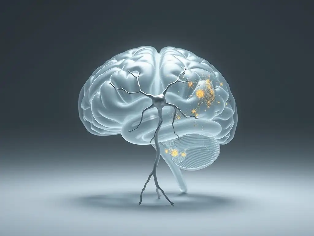
Brain imaging data is inherently complex, involving high-dimensional datasets that require sophisticated analysis techniques. A single fMRI scan can generate millions of data points representing activity across thousands of brain voxels (3D pixels) over time. Interpreting these vast datasets necessitates advanced statistical methods, computational models, and a deep understanding of neuroanatomy and function. Findings often represent statistical probabilities across groups, not deterministic statements about an individual brain, which is a critical nuance often lost in simplified communication.
Furthermore, brain imaging studies are subject to various sources of variability, including individual differences, scanner parameters, and analysis choices. Reconciling findings across different studies, which may use slightly different tasks, populations, or analytical pipelines, can be challenging even for experts. Communicating these inherent complexities – the statistical nature of findings, the influence of methodology, and the probabilistic rather than deterministic insights – presents a significant hurdle in effective brain imaging mental health communication to non-experts.
Avoiding Misinterpretations and Over-Simplifications of Brain Scans in Mental Health
A significant challenge in brain imaging mental health communication is the risk of misinterpretation and over-simplification. Brain scans can appear deceptively simple, often presented as colorful images highlighting specific brain regions. This visual directness can lead to the misconception that a scan provides a straightforward "diagnosis" or a definitive "picture" of a mental illness, akin to an X-ray showing a broken bone. In reality, current brain imaging techniques in mental health research provide insights into group-level differences or correlations with symptoms, rather than individual diagnostic markers.
The media and public often gravitate towards sensationalized headlines like "Brain scan shows cause of depression," which contribute to this over-simplification. For instance, while fMRI studies show altered activity in certain brain areas in depressed individuals, this doesn't mean the scan causes depression or that a single scan can definitively diagnose it in an individual.
Effective brain imaging mental health communication must actively counteract this tendency, emphasizing that these techniques reveal associations and potential mechanisms, not simple causes or individual diagnoses. It requires careful framing to manage expectations and ensure accurate understanding of what brain imaging can and cannot currently do for mental health diagnosis and treatment.
Ethical Considerations in Presenting Brain Imaging Results to Patients and the Public
Presenting brain imaging results, particularly to patients or the general public, raises important ethical considerations. Given the potential for misinterpretation, there is a risk that individuals might feel their condition is solely biological and fixed, diminishing the perceived value of psychological interventions or lifestyle changes. Conversely, patients might feel stigmatized if their scan is presented as showing a "broken" brain, or feel a false sense of certainty about their prognosis based on preliminary research findings.
Responsible brain imaging mental health communication requires transparency about the limitations of the technology and the research findings. For patients, results should be discussed within the broader context of their symptoms, history, and treatment options, emphasizing that scans provide information, not a complete explanation or cure. For the public, it's crucial to avoid deterministic language and highlight that mental health is influenced by complex interactions between biological, psychological, and social factors. Ethical communication prioritizes accuracy, avoids hype, and respects the individual's autonomy and well-being, ensuring that brain imaging information is empowering rather than misleading or harmful.
Strategies for Effective Brain Imaging Mental Health Communication
Visual Aids: Leveraging Images, Graphs, and Videos for Clarity
Visual aids are fundamental to effective brain imaging mental health communication, transforming complex data into more digestible formats. Static images of brain regions with overlaid statistical maps, like those generated from fMRI or PET data, are powerful for showing where differences or activity occur. However, these need careful annotation and explanation to be understood. Graphs can illustrate quantitative findings, such as correlations between brain activity and symptom severity, or changes in brain metrics over time or with treatment.
Dynamic visualizations, such as short animations or videos, can be particularly effective for explaining complex processes, like neural network interactions or the flow of information between brain regions. For example, visualizing altered connectivity patterns in conditions like autism or schizophrenia can be more intuitive through animated diagrams than static images. The key is to ensure these visuals are not just aesthetically pleasing but are accurate, clearly labeled, and accompanied by accessible explanations that reinforce the core message without oversimplification, thereby improving brain imaging mental health communication.
Designing Accessible and Understandable Brain Imaging Visualizations
Designing accessible visualizations for brain imaging mental health communication requires careful consideration of the target audience and the complexity of the information. For a general audience or patients, visualizations should minimize jargon, use intuitive color schemes, and focus on conveying the main message clearly. For example, instead of showing a raw statistical map, one might show a simplified diagram highlighting key brain regions and explaining their general function before showing how activity or structure differs in a specific condition.
It is crucial to provide context and scale. Showing a brain region abnormality is more meaningful if the visualization also shows where that region is located within the whole brain and briefly explains its typical role. Using annotations, legends, and accompanying text that explains what the colors, shapes, and patterns represent is essential. The goal is to make the visual information immediately interpretable, allowing the viewer to grasp the core finding without needing extensive prior knowledge of neuroscience or brain imaging techniques.
Using Color Palettes Effectively in Brain Imaging Representation
The choice of color palette significantly impacts how brain imaging data is perceived and understood. In many brain imaging visualizations, color is used to represent statistical significance, magnitude of effect, or activity levels. For instance, a common approach is to use a gradient from cool colors (like blue) to warm colors (like red or yellow) to indicate increasing levels of activity or statistical strength. However, careful consideration is needed to avoid misleading representations.
It is important to use color palettes that are perceptually uniform, meaning that changes in color correspond linearly to changes in the data they represent. Sequential color palettes are often best for showing a range from low to high values, while diverging palettes are useful for showing deviations from a central point (e.g., differences from a control group, with positive and negative differences represented by different hues).
Additionally, using colorblind-friendly palettes is crucial to ensure accessibility. Consistent use of color schemes across multiple visualizations within an article or presentation helps build familiarity and aids interpretation, supporting effective brain imaging mental health communication.
Narrative Storytelling: Connecting Brain Imaging Findings to Real-World Experiences
Narrative storytelling is a powerful strategy for making complex brain imaging findings relatable and understandable, particularly in the context of mental health. Instead of just presenting data and statistics, connecting the findings to the lived experiences of individuals with mental health conditions can create a compelling narrative. This involves explaining how altered brain activity or structure might relate to the symptoms a person experiences in their daily life.
For example, explaining that reduced connectivity in a specific brain circuit (shown by fMRI) might manifest as difficulty regulating emotions or impaired decision-making (reported by patients) builds a bridge between the abstract neuroscience and the tangible reality of the illness. Sharing anonymized vignettes or case examples (with permission and care) that illustrate how brain imaging research is contributing to understanding why certain symptoms occur can resonate deeply with audiences.
This human-centered approach makes brain imaging mental health communication more empathetic and impactful, moving beyond just showing pictures of the brain to explaining what those pictures might mean for a person's life.
Tailoring Communication to Different Audiences: Patients, Clinicians, and the General Public
Effective brain imaging mental health communication requires tailoring the content, language, and level of detail to the specific audience. What is appropriate for a fellow researcher is vastly different from what is needed for a patient or a member of the general public. Failing to adapt can lead to confusion, disengagement, or misinterpretation.
For patients, the focus should be on how research findings relate to their condition, potential treatment approaches, and reducing stigma, using clear, non-technical language. For clinicians, the communication needs to be more detailed, explaining methodologies, statistical significance, and implications for clinical practice or future research directions. For the general public, the goal is often to raise awareness, explain the value of research, and provide a basic understanding of how the brain is involved in mental health, using engaging visuals and relatable analogies.
Communicating Brain Imaging Results to Patients with Mental Health Conditions
Communicating brain imaging results to patients with mental health conditions requires sensitivity, clarity, and a focus on empowerment. It's crucial to explain what the scans show in the context of their individual situation, while managing expectations about the diagnostic or prognostic value of the imaging. For instance, explaining that a scan showing atypical activity in a fear circuit is consistent with their anxiety symptoms helps validate their experience, but it must be clear that this isn't the only cause and doesn't replace clinical assessment.
Using simple language, avoiding jargon, and employing clear visual aids that are easy to interpret are essential. Patients should be given ample opportunity to ask questions and express their concerns. The communication should emphasize that brain imaging research is contributing to a better understanding of their condition, potentially leading to improved treatments in the future, rather than presenting the scan as a definitive answer or label for their current state. This empathetic approach in brain imaging mental health communication helps patients feel informed and respected.
Communicating Brain Imaging Findings to Clinicians and Researchers
Communicating brain imaging findings to clinicians and researchers requires a higher level of technical detail and precision. For clinicians, the focus should be on the clinical relevance of the findings: how they might inform diagnosis (in the future), treatment selection, or prognosis, while acknowledging the current limitations of using imaging in individual clinical practice. Data should be presented with appropriate statistical rigor, discussing sample sizes, effect sizes, and limitations.
For fellow researchers, communication involves presenting methodology in detail, including acquisition parameters, preprocessing steps, and analytical approaches. Discussion should include how findings relate to previous literature, potential confounds, and directions for future research. This level of detail is necessary for replication, critical evaluation, and building cumulative knowledge within the field. Presentations often include complex graphs, statistical maps, and network diagrams, assuming a baseline understanding of neuroscience and imaging principles.
Communicating Brain Imaging Discoveries to the General Public and Media
Communicating brain imaging discoveries to the general public and media requires striking a balance between accuracy and accessibility, while avoiding hype. The goal is to inform, educate, and garner support for mental health research without overstating findings or promoting neuro-essentialist views. Analogies can be helpful, but they must be used carefully to avoid misrepresentation.
Key strategies include focusing on the "so what?" – explaining why the finding matters for understanding mental health. Visuals should be engaging but simplified, often using diagrams or illustrative graphics rather than raw scan data.
Collaboration with science communicators, journalists, or public relations professionals is often beneficial to ensure the message is clear, accurate, and appropriately framed for mass consumption. For instance, instead of stating "fMRI shows decreased activity in the subgenual cingulate cortex in depression," a public-friendly message might be "Research using brain scans suggests that a brain area involved in regulating emotions shows reduced activity in people with depression, helping us understand the biological basis of low mood."
Case Studies: Successful Examples of Brain Imaging Communication in Mental Health
Case Study 1: The MQ Mental Health Science Campaign: Visualizing Depression
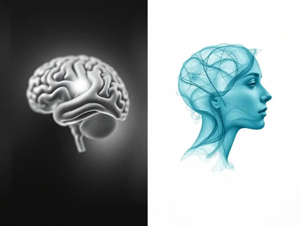
MQ: Transforming Mental Health, a leading mental health research charity, has launched campaigns aimed at explaining complex mental health science to the public. One approach involves using accessible visuals and narratives to communicate findings related to conditions like depression. Instead of relying solely on abstract graphs, they often use simplified brain diagrams or illustrations that metaphorically represent altered brain function.
For example, in explaining the neuroscience of depression, MQ might use graphics depicting communication pathways in the brain as broken or weakened bridges, correlating this visual to the symptoms of impaired emotional regulation or motivation. While not showing raw scan data, these visuals are informed by brain imaging research showing reduced connectivity or altered activity in specific circuits. This approach focuses on explaining the implication of the brain science in a relatable way, effectively bridging the gap between research findings and public understanding of mental health conditions, demonstrating successful brain imaging mental health communication.
Case Study 2: Neuroimaging Training Program for Psychiatrists at UCLA
Recognizing the importance of integrating neuroscience into clinical practice, institutions like UCLA have developed training programs specifically for psychiatrists on neuroimaging. These programs are designed to improve clinicians' ability to understand, interpret, and apply brain imaging findings reported in research literature. A key component of such programs is teaching effective brain imaging mental health communication tailored for the clinical setting.
Participants learn about the principles and limitations of various imaging modalities (fMRI, PET, EEG, etc. ), how to critically evaluate research papers presenting imaging data, and most importantly, how to discuss potential implications of neuroimaging research with patients. The curriculum often includes workshops on visualizing data, interpreting statistical maps, and framing findings in a way that is relevant to clinical symptoms and treatment decisions, while avoiding deterministic language. This training equips clinicians to be better communicators of brain imaging science to their patients and colleagues.
A classic example of communicating specific brain imaging findings is research published in high-impact journals like Biological Psychiatry. A study examining amygdala reactivity to social stimuli in individuals with social anxiety disorder using fMRI serves as a good illustration. The study might report findings showing significantly increased amygdala activation in the social anxiety group compared to controls when viewing images of faces.
Communication of these findings, initially within the scientific community, involves detailed methods sections, statistical results (e.g., t-scores, p-values, effect sizes), and precise anatomical locations of activation clusters. For broader communication, press releases or summaries might highlight the key finding – that the brain's "fear center" shows heightened responses in social anxiety – and explain how this relates to the core symptom of intense fear in social situations. While the journal article provides the rigorous scientific evidence, the summaries translate the core message for a wider audience, illustrating different levels of brain imaging mental health communication.
The Future of Brain Imaging Mental Health Communication
Emerging Technologies for Visualizing Brain Data
The future of brain imaging mental health communication will be significantly shaped by emerging visualization technologies. Advances in computational power and graphics are enabling increasingly sophisticated and interactive ways to represent complex brain data. This includes tools for visualizing brain networks in 3D, allowing users to explore connectivity patterns and information flow in a more intuitive way than static diagrams.
Virtual and augmented reality (VR/AR) hold immense potential for creating immersive experiences that allow users to "walk through" a brain or interact with neural circuits. Imagine a patient or student being able to visualize their own brain scan data or explore a model of a neural pathway implicated in their condition. While still in early stages, these technologies could revolutionize how brain imaging data is taught, discussed, and understood, making brain imaging mental health communication more engaging and potentially more impactful.
The Importance of Collaboration Between Researchers, Clinicians, and Communication Experts
Improving brain imaging mental health communication hinges on enhanced collaboration between the scientists who generate the data, the clinicians who work with patients, and professional science communicators. Researchers possess the deep understanding of the findings and their nuances but may lack the skills or time to translate this complexity for non-expert audiences. Clinicians understand the patient perspective and what information is most relevant and sensitive.
Science communicators, including writers, designers, and multimedia specialists, have the expertise in crafting clear, engaging, and accessible messages for specific audiences. Bringing these groups together from the outset of research projects or communication initiatives can ensure that findings are not only scientifically sound but also packaged in a way that is understandable, ethical, and relevant to those who need it most. This collaborative approach is crucial for effective brain imaging mental health communication.
Future Directions in Improving Brain Imaging Communication for Better Mental Health Outcomes
Future efforts to improve brain imaging mental health communication should focus on several key areas to enhance mental health outcomes. Firstly, developing standardized guidelines and best practices for communicating brain imaging findings to different audiences is crucial to minimize misinterpretation and ensure ethical presentation. This includes recommendations on language, visuals, and contextual information.
Secondly, investing in training for researchers and clinicians on effective communication techniques, including the use of visual aids and narrative, is essential. Just as important is involving patients and the public in the communication process, perhaps through co-design of materials or feedback sessions, to ensure the information is relevant and understandable from their perspective. Ultimately, better brain imaging mental health communication can foster greater understanding, reduce stigma, inform treatment decisions, and build support for the ongoing research necessary to improve mental health care globally.
Conclusion: Empowering Understanding Through Effective Brain Imaging Communication
Key Takeaways: Improving Brain Imaging Communication in Mental Health
Effective brain imaging mental health communication is not merely about presenting data; it's about building bridges of understanding between complex neuroscience and the real-world experiences of those affected by mental health conditions. Key strategies involve leveraging clear and accessible visual aids, employing narrative storytelling to connect findings to lived experiences, and rigorously tailoring communication to specific audiences – patients, clinicians, and the general public.
Successfully navigating the challenges of data complexity, potential misinterpretations, and ethical considerations requires careful planning and execution. By prioritizing accuracy, empathy, and clarity, while utilizing appropriate tools and collaborations, we can ensure that the powerful insights gained from brain imaging research contribute meaningfully to reducing stigma, improving treatment approaches, and fostering a more informed and compassionate society regarding mental health.
Resources for Learning More About Brain Imaging and Mental Health
For those interested in delving deeper into the fascinating world of brain imaging and its role in understanding mental health, numerous resources are available. University websites and research institution pages often feature public summaries of their latest findings, explaining complex studies in simpler terms. Organizations dedicated to mental health research, such as MQ: Transforming Mental Health or the Brain & Behavior Research Foundation, provide accessible articles and reports on neuroimaging discoveries.
Scientific journals like Biological Psychiatry or JAMA Psychiatry publish the primary research, though these require a higher level of technical understanding. Educational platforms and online courses offer introductions to neuroscience and brain imaging techniques. Engaging with these resources, critically evaluating the information, and seeking clarification when needed are excellent ways to enhance one's understanding of how brain imaging is shedding light on the complexities of mental health.
Frequently Asked Questions
How does brain imaging enhance communication in mental health?
Brain imaging provides visual evidence of brain activity and structure, making complex mental health conditions more tangible and understandable for patients, families, and the public. This can lead to improved patient engagement and reduced stigma.
What types of brain imaging are typically used in mental health communication?
Common techniques include fMRI (functional Magnetic Resonance Imaging) showing brain activity, MRI (Magnetic Resonance Imaging) visualizing brain structure, and PET (Positron Emission Tomography) providing information about brain function using radioactive tracers. Each provides unique insights into brain health.
How can brain imaging findings be communicated effectively to individuals without a medical background?
Use clear, non-technical language and avoid jargon. Focus on the key findings and their implications, using visual aids like simplified diagrams and analogies to explain complex concepts.
What are the ethical considerations when using brain imaging to communicate about mental health?
It's crucial to avoid overstating the certainty or implications of brain imaging results. Emphasize that brain scans are just one piece of the puzzle in mental health diagnosis and treatment, and ensure patient privacy and confidentiality.
How does brain imaging contribute to destigmatizing mental health conditions?
By demonstrating the biological basis of many mental illnesses, brain imaging can help reduce the perception that mental health conditions are simply due to personal failings or character flaws. This can foster greater understanding and empathy.
Final Reflection: Visualizing the Mind, Bridging the Gap
The effective communication of brain imaging findings in mental health is not merely a scientific endeavor; it's a crucial bridge between complex research and the lived experiences of individuals and communities. Misunderstandings surrounding brain scans can lead to stigmatization and hinder access to appropriate care. Accurate and empathetic communication is paramount to fostering hope and driving progress in mental health treatment.
The power of visual communication in this field is undeniable. Images, when presented thoughtfully, can demystify the brain and its complexities, fostering a sense of understanding and reducing fear. A picture is worth a thousand words,
and in the context of mental health, those words can be the difference between isolation and connection, between despair and hope. The ongoing challenge lies in ensuring that these images are not only scientifically accurate but also presented in a way that is accessible and relatable to a broad audience, avoiding the perpetuation of harmful stereotypes.
Consider the impact of these images. How can we, as individuals and as a society, ensure that the power of brain imaging is used responsibly and ethically? You have the power to advocate for clearer, more compassionate communication surrounding mental health. Engage with research, question assumptions, and demand accessible information. By understanding the brain, we can better understand ourselves and each other, paving the way for a more empathetic and supportive future for all.
Unlock the Stories Within: Visualizing Mental Health
Thanks for taking the time to explore the powerful world of brain imaging and its role in understanding mental health! We hope you gained a clearer understanding of how visuals can communicate complex findings effectively. Ready to take the next step?
- Craft your own compelling visual: Try creating a simple infographic summarizing a key mental health finding—it's a great way to solidify your understanding!
- Share this article with a colleague or friend: Let's spark a conversation about improving communication in mental health research.
- Leave a comment below: What surprising insights did you gain from visualizing brain imaging data, and how can we use this knowledge more effectively?
Ready to delve deeper into the world of visual communication in mental health?