
The Role of Glial Cells: More Than Just Support for Neurons and Mood

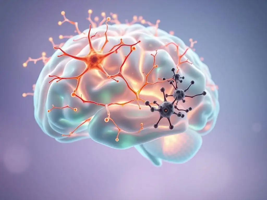
Welcome to Mind Care Guide, where we believe understanding is the first step towards healing.
Are you aware that the secret to better mental well-being might lie in the unsung heroes of your brain? What if the key to unlocking improved mood and resilience wasn't just about neurons, but about the intricate network of support surrounding them? Dive in to explore the fascinating role of glial cells mental health support and discover how these often-overlooked cells are revolutionizing our understanding of mood disorders and overall mental wellness. We're here to guide you on this journey of discovery.
The Role of Glial Cells: More Than Just Support for Neurons and Mood
- Introduction: Understanding Glial Cells and Mental Wellbeing
- Glial Cells: Types and Functions in the Brain
- Glial Cells and Neuropsychology of Mood Disorders
- Glial Cells, Brain Chemistry, and Mental Health Support
- How Glial Cells Contribute to Mental Resilience
- Glial Cell Imaging and Research Advancements
- Future Directions: Glial Cells as Therapeutic Targets for Mental Health
- Conclusion: Glial Cells – Essential Players in Mental Wellbeing
-
Frequently Asked Questions
- What are glial cells and what is their primary function in the brain?
- How do glial cells contribute to mental health?
- What is the connection between glial cell dysfunction and mental health disorders?
- Can glial cells be targeted therapeutically for mental health support?
- Where can I find more information and resources for mental health support related to glial cells?
- Final Reflection: Unveiling the Unsung Heroes of Mental Well-being
- Unlock Your Brain's Hidden Potential: Discover the Power of Glial Cells
Introduction: Understanding Glial Cells and Mental Wellbeing
Glial Cells: Beyond Neuronal Support
For decades, neurons were considered the undisputed stars of the brain, solely responsible for processing information and transmitting signals. Glial cells, often referred to simply as "glia," were relegated to a secondary role, primarily seen as structural support or "glue" holding neurons together. This view significantly underestimated their complexity and vital functions. Modern neuroscience has revealed that glial cells are active participants in virtually all aspects of brain function, from development and plasticity to maintaining the delicate balance required for healthy cognitive and emotional states.
This paradigm shift highlights that a comprehensive understanding of brain function, and consequently mental health, must include the intricate activities of glial cells. They are not merely passive bystanders but dynamic entities that influence neuronal activity, regulate the brain's environment, and play crucial roles in the immune response within the central nervous system. Exploring the diverse functions of glia is essential for appreciating their profound impact on our mental wellbeing and developing effective strategies for mental health support.
The Emerging Role of Glia in Brain Function and Mental Health
Recent research has increasingly implicated glial cells in the pathophysiology of various mental health conditions, including mood disorders, anxiety disorders, and neurodevelopmental conditions. Their ability to communicate with neurons and other glial cells, respond to environmental changes, and modulate synaptic transmission positions them as critical players in brain circuitry and function. Disruptions in glial cell activity or numbers are now recognized as potential contributors to the complex origins of mental illness.
This growing body of evidence underscores the importance of studying glial cells for mental health support. For instance, changes in astrocyte morphology or microglial activation states have been observed in post-mortem brain tissue from individuals who suffered from major depressive disorder or bipolar disorder. Understanding these cellular alterations provides new avenues for research into the underlying mechanisms of these conditions and offers potential targets for novel therapeutic interventions aimed at restoring healthy glial function to provide better mental health support.
Glial Cells: Types and Functions in the Brain
Astrocytes: Regulating the Neuronal Environment and Mood
Astrocytes are the most abundant type of glial cell in the brain, characterized by their star-like shape. They perform a multitude of essential functions, including maintaining the blood-brain barrier, regulating cerebral blood flow, and controlling the chemical environment around neurons. A critical role of astrocytes is the uptake and metabolism of neurotransmitters like glutamate and GABA at synapses, which is vital for regulating synaptic transmission and preventing excitotoxicity. They form close associations with synapses, creating what is known as the "tripartite synapse," where communication involves the pre-synaptic neuron, the post-synaptic neuron, and the surrounding astrocyte.
Beyond their direct support roles, astrocytes are increasingly recognized for their active participation in neuronal signaling and plasticity. They can release gliotransmitters, such as D-serine and ATP, which can modulate synaptic activity. Dysfunction in astrocytes has been linked to various neurological and psychiatric disorders.
For example, studies have shown reduced density or altered morphology of astrocytes in certain brain regions, like the prefrontal cortex and hippocampus, in individuals with major depressive disorder. These changes can impair their ability to regulate glutamate levels or provide metabolic support to neurons, potentially contributing to mood dysregulation. Understanding these astrocyte-specific changes is key to developing targeted glial cells mental health support strategies.
Oligodendrocytes: Myelination and its Impact on Mental Processes
Oligodendrocytes are glial cells found in the central nervous system (CNS) whose primary function is to produce myelin, a fatty sheath that wraps around neuronal axons. Myelin acts as an insulator, allowing electrical signals (action potentials) to propagate much faster along the axon through a process called saltatory conduction. This efficient transmission is crucial for coordinating complex brain functions, including cognitive processes and emotional regulation. In the peripheral nervous system, Schwann cells perform a similar myelination role.
Disruptions in myelination or damage to oligodendrocytes can impair neuronal communication speed and efficiency, potentially contributing to symptoms seen in various mental health conditions. For instance, studies using diffusion tensor imaging (DTI) have suggested alterations in white matter integrity, indicative of potential myelination issues, in individuals with conditions like schizophrenia and bipolar disorder.
While the direct link between oligodendrocyte dysfunction and mood disorders is still an active area of research, it is plausible that impaired myelination could affect the connectivity and function of neural circuits involved in mood regulation. Supporting the health and function of oligodendrocytes is another facet of comprehensive glial cells mental health support.
Microglia: Immune Defense and Neuroinflammation in Mental Disorders
Microglia are the primary immune cells of the central nervous system, acting as the brain's resident macrophages. In their resting state, they constantly survey the brain environment, extending and retracting processes to monitor for pathogens, tissue damage, or abnormal cellular debris. When they detect a threat or injury, microglia become activated, changing their morphology and releasing inflammatory molecules like cytokines. This immune response is crucial for protecting the brain, but chronic or dysregulated microglial activation can lead to neuroinflammation, which is increasingly implicated in the pathology of various mental health disorders.
Research has found evidence of increased microglial activation in certain brain regions in individuals with major depression, anxiety disorders, and obsessive-compulsive disorder. For example, PET imaging studies using ligands that bind to the translocator protein (TSPO), a marker of microglial activation, have shown elevated binding in areas like the prefrontal cortex and anterior cingulate cortex in depressed patients compared to healthy controls.
This suggests that neuroinflammation driven by activated microglia may contribute to the symptoms of mood and anxiety disorders. Modulating microglial activity to reduce excessive neuroinflammation is a promising area for developing novel glial cells mental health support strategies.
Ependymal Cells: Cerebrospinal Fluid and Brain Homeostasis
Ependymal cells are specialized epithelial cells that line the ventricles of the brain and the central canal of the spinal cord. These cells have cilia that beat rhythmically, helping to circulate cerebrospinal fluid (CSF). They also form the choroid plexus, where CSF is produced. CSF plays a critical role in brain homeostasis by providing mechanical protection, removing metabolic waste products, and transporting nutrients and signaling molecules throughout the CNS. Ependymal cells also act as a barrier between the CSF and the brain parenchyma, regulating the passage of substances.
While less directly studied in the context of mood disorders compared to astrocytes, oligodendrocytes, and microglia, the maintenance of a stable brain environment by ependymal cells is fundamental to overall neuronal and glial health. Disruptions in CSF flow or composition, potentially influenced by ependymal cell function, could indirectly impact the delicate balance required for healthy brain function and mood regulation.
Furthermore, ependymal cells have been shown to act as neural stem cell niches in certain areas, contributing to neurogenesis, a process implicated in the recovery from depression. Supporting overall brain homeostasis, partly facilitated by ependymal cells, contributes to the broader context of glial cells mental health support.
Glial Cells and Neuropsychology of Mood Disorders
Glial Dysfunction in Depression: Investigating the Connection
Major Depressive Disorder (MDD) is a complex condition, and research increasingly points to glial cell dysfunction as a significant contributing factor, not just a consequence. Studies examining post-mortem brain tissue from individuals with MDD have consistently reported reductions in the density and function of astrocytes and oligodendrocytes in key mood-regulating areas such as the prefrontal cortex, hippocampus, and amygdala.
For example, a meta-analysis of multiple studies indicated a significant decrease in the number of glial cells (both astrocytes and oligodendrocytes) in the prefrontal cortex of depressed patients compared to non-depressed controls, sometimes showing reductions of 20% or more in specific layers.
Furthermore, microglial activation and associated neuroinflammation are frequently observed in depression. Elevated levels of pro-inflammatory cytokines like IL-1β, IL-6, and TNF-α have been found in the brains and peripheral blood of depressed individuals. These inflammatory molecules, released by activated microglia, can interfere with neurotransmitter systems, disrupt synaptic plasticity, and impair neurogenesis, all of which are implicated in the pathophysiology of depression. Understanding these specific glial abnormalities provides crucial insights into the biological underpinnings of depression and highlights glial cells as prime targets for developing novel mental health support strategies.
Bipolar Disorder: Glial Cell Abnormalities and Mood Swings
Bipolar Disorder (BD), characterized by dramatic shifts in mood, energy, and activity levels, also shows clear links to glial cell pathology. Similar to depression, post-mortem studies in individuals with BD have revealed significant reductions in glial cell numbers, particularly astrocytes and oligodendrocytes, in regions such as the prefrontal cortex, anterior cingulate cortex, and amygdala. Some studies report glial cell deficits ranging from 15% to 30% in specific cortical layers of BD patients compared to controls. These deficits are thought to contribute to impaired neuronal function and connectivity within mood-regulating circuits.
Microglial activation and neuroinflammation are also implicated in BD, although the pattern might differ between manic and depressive episodes. Studies using PET imaging or post-mortem tissue analysis have shown increased markers of microglial activation in certain brain areas during mood episodes. The fluctuating nature of BD symptoms suggests that glial function might be dynamically altered throughout the illness course. Understanding the specific types of glial dysfunction and inflammatory processes involved in BD is crucial for developing targeted glial cells mental health support interventions that can stabilize mood and improve long-term outcomes.
The Role of Glia in Anxiety Disorders: New Research Insights
Anxiety disorders, including generalized anxiety disorder (GAD), panic disorder, and post-traumatic stress disorder (PTSD), are also increasingly linked to glial cell dysfunction. Research suggests that altered glial activity, particularly involving astrocytes and microglia, may contribute to the heightened fear and anxiety responses characteristic of these conditions. For instance, studies in animal models of anxiety have shown changes in astrocyte morphology and function in brain regions like the amygdala and hippocampus, areas critical for processing fear and memory.
Neuroinflammation, driven by activated microglia, appears to play a role in anxiety as well. Stress, a major risk factor for anxiety disorders, is known to activate microglia and induce the release of inflammatory cytokines in the brain. These cytokines can influence neuronal excitability and synaptic plasticity in fear circuits, potentially exacerbating anxiety symptoms.
Understanding how glial cells contribute to the dysregulation of these circuits is a key area of current research. Developing therapies that modulate glial activity to reduce neuroinflammation or restore proper astrocyte function could offer new avenues for providing effective glial cells mental health support for individuals struggling with anxiety.
Glial Cells, Brain Chemistry, and Mental Health Support
Glial-Neuron Interactions: The Key to Understanding Brain Chemistry
The traditional view of brain chemistry focused primarily on the synthesis, release, and reception of neurotransmitters by neurons. However, it is now clear that glial cells, particularly astrocytes, are intimately involved in regulating neurotransmitter levels and synaptic function. Astrocytes surround synapses and possess transporters that remove neurotransmitters like glutamate and GABA from the synaptic cleft, preventing excitotoxicity and ensuring proper signal termination. They also store and release glutamate and its precursor, glutamine, which is recycled back to neurons for neurotransmitter synthesis.
This dynamic interaction between neurons and glia, often referred to as the "gliotransmission" concept, highlights that glial cells actively modulate neuronal communication and brain chemistry. Disruptions in these interactions, such as impaired glutamate uptake by astrocytes or altered release of gliotransmitters, can lead to imbalances in excitatory and inhibitory signaling, potentially contributing to neuropsychiatric symptoms. Understanding these complex glial-neuron interactions is fundamental to deciphering the neurobiological basis of mental illness and identifying new targets for glial cells mental health support.
Glutamate, GABA, and Glial Cells: The Tripartite Synapse
The tripartite synapse concept emphasizes the functional unit formed by the pre-synaptic terminal, the post-synaptic terminal, and the surrounding perisynaptic astrocyte. Within this structure, astrocytes actively participate in neurotransmission, particularly involving the major excitatory neurotransmitter glutamate and the major inhibitory neurotransmitter GABA. Astrocytes express glutamate transporters (like EAAT1 and EAAT2) and GABA transporters (like GAT-3) that rapidly clear these neurotransmitters from the synaptic cleft, regulating their availability and preventing spillover to neighboring synapses.
Furthermore, astrocytes are involved in the glutamate-glutamine cycle, where glutamate released by neurons is taken up by astrocytes, converted to glutamine, and then transported back to neurons for re-synthesis into glutamate or GABA. This cycle is essential for maintaining adequate neurotransmitter pools. Dysregulation of astrocyte transporters or the glutamate-glutamine cycle has been implicated in various neurological and psychiatric conditions, including mood disorders. For example, reduced expression of EAAT2 in certain brain regions has been observed in individuals with depression. Targeting these glial mechanisms offers a potential strategy for restoring neurotransmitter balance and providing glial cells mental health support.
Neuroinflammation and Glial Activation: Implications for Mood
Neuroinflammation, characterized by the activation of microglia and astrocytes and the release of inflammatory mediators within the brain, is increasingly recognized as a significant contributor to mood disorders. While a necessary protective response to injury or infection, chronic or excessive neuroinflammation can disrupt neuronal function, impair synaptic plasticity, and even lead to neuronal damage, all of which are relevant to the pathology of depression and bipolar disorder. Activated microglia release pro-inflammatory cytokines (e.g., IL-1β, TNF-α, IL-6) and chemokines, which can directly affect neuronal excitability and neurotransmitter metabolism.
Astrocytes also participate in the inflammatory response by releasing inflammatory mediators, although they can also release anti-inflammatory factors. In chronic inflammation, however, reactive astrocytes can contribute to neurotoxicity. The interplay between activated microglia and reactive astrocytes creates a pro-inflammatory environment that can negatively impact mood circuits. Studies have shown that systemically administered inflammatory challenges can induce depressive-like symptoms in both humans and animal models. Understanding the specific pathways involved in glial activation and neuroinflammation in mental illness is vital for developing anti-inflammatory strategies as a form of glial cells mental health support.
How Glial Cells Contribute to Mental Resilience
Glial Plasticity and Brain Adaptability
Glial cells are not static entities; they exhibit significant plasticity, meaning their morphology, function, and even number can change in response to various stimuli, including environmental experiences, stress, and therapeutic interventions. This glial plasticity is crucial for overall brain adaptability and resilience. For instance, astrocytes can alter their morphology and coverage of synapses in response to changes in neuronal activity, potentially modulating synaptic strength and plasticity. Oligodendrocytes can undergo myelination or demyelination processes in response to learning or injury, affecting the speed of neural circuit communication.
Microglia also exhibit plasticity, shifting between different activation states ranging from pro-inflammatory to anti-inflammatory and pro-resolving phenotypes. Their ability to adapt and change function is essential for maintaining brain health and responding to challenges. Stress, for example, can induce changes in glial morphology and function in stress-sensitive brain regions like the hippocampus and prefrontal cortex. Understanding and harnessing this intrinsic glial plasticity represents a promising avenue for enhancing brain resilience and developing novel glial cells mental health support strategies that help individuals cope with stress and recover from mental health challenges.
The Potential of Glial-Targeted Therapies for Mental Health
Given the growing evidence linking glial dysfunction and neuroinflammation to mental illness, targeting glial cells directly has emerged as a promising therapeutic strategy. Unlike traditional psychotropic medications that primarily target neuronal receptors or neurotransmitter reuptake, glial-targeted therapies aim to restore normal glial function, reduce neuroinflammation, or promote glial-mediated neuroprotection. These approaches could offer novel ways to address the underlying pathology of mental disorders, potentially leading to more effective and longer-lasting treatment outcomes.
Potential glial-targeted therapies include:
- Agents that modulate microglial activation states (e.g., inhibiting pro-inflammatory pathways or promoting anti-inflammatory phenotypes).
- Drugs that enhance astrocyte function, such as improving glutamate uptake or restoring metabolic support.
- Strategies to promote remyelination by oligodendrocytes.
- Compounds that interfere with glial-neuron signaling pathways that are dysregulated in disease.
Developing such targeted interventions requires a deep understanding of the specific glial cell types and pathways involved in different mental health conditions, paving the way for more personalized and effective glial cells mental health support.
Minocycline: Targeting Microglia for Depression Treatment
Minocycline, a tetracycline antibiotic, is one example of a compound that has been investigated for its potential to target glial cells, particularly microglia, in the context of mental health conditions like depression. While primarily known for its antibacterial properties, minocycline also possesses anti-inflammatory and neuroprotective effects. Research suggests that minocycline can cross the blood-brain barrier and modulate microglial activation, reducing the release of pro-inflammatory cytokines.
Preclinical studies in animal models of depression have shown that minocycline can reduce depressive-like behaviors and markers of neuroinflammation in the brain. Clinical trials investigating minocycline as an adjunctive treatment for major depressive disorder have yielded mixed results, with some studies showing a modest improvement in depressive symptoms, particularly in individuals with evidence of inflammation, while others have not found a significant effect.
Despite variable clinical outcomes, the exploration of minocycline highlights the concept of targeting glial cells, specifically microglia and neuroinflammation, as a viable strategy for providing glial cells mental health support and underscores the need for further research into more potent and specific glial-modulating agents.
Glial Cell Imaging and Research Advancements
Advancements in Glial Cell Research Methods
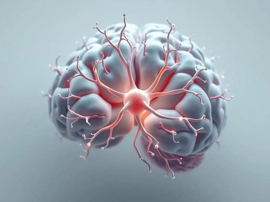
Understanding the complex roles of glial cells in brain health and disease has been greatly advanced by the development of sophisticated research techniques. Beyond traditional histology and immunohistochemistry, which allow for the visualization of glial cell morphology and markers in fixed tissue, live-cell imaging techniques now permit researchers to observe glial activity in real-time in living brains, particularly in animal models. Techniques like two-photon microscopy enable the visualization of calcium signaling within astrocytes or the dynamic movements of microglial processes in response to stimuli.
Genetic tools, such as transgenic mice with fluorescently labeled glial cells or inducible gene expression systems specific to different glial subtypes (e.g., using Cre-lox recombination with specific promoters like GFAP for astrocytes or IBA1 for microglia), allow for selective manipulation or study of individual glial populations. Single-cell RNA sequencing has revolutionized our ability to characterize the diverse subtypes of glial cells and understand how their gene expression profiles change in different brain states or diseases. These powerful tools are essential for uncovering the specific contributions of glial cells to mental health and developing targeted glial cells mental health support strategies.
Positron Emission Tomography (PET) Imaging of Microglial Activation
Positron Emission Tomography (PET) imaging is a non-invasive neuroimaging technique that allows researchers and clinicians to visualize and quantify specific molecular targets in the living human brain. For studying microglial activation, PET ligands that bind to the translocator protein (TSPO) have been particularly valuable. TSPO is an outer mitochondrial membrane protein that is upregulated in activated microglia and reactive astrocytes. While not exclusively expressed by microglia, TSPO binding is widely used as a proxy marker for neuroinflammation and glial activation in vivo.
Studies using TSPO-PET imaging have provided crucial evidence for increased glial activation in the brains of individuals with various mental health conditions, including major depressive disorder, bipolar disorder, schizophrenia, and anxiety disorders. For example, research has shown elevated TSPO binding in regions like the prefrontal cortex, anterior cingulate cortex, and insula in depressed patients compared to healthy controls. These findings provide in-vivo support for the role of neuroinflammation in the pathophysiology of mood disorders and reinforce the importance of considering glial cells in the search for effective mental health support.
Understanding Glial Cells in Different Brain Regions
The functions and characteristics of glial cells can vary significantly depending on their location within the brain. Glia in the cortex may have different properties and roles compared to glia in the hippocampus, amygdala, or cerebellum. For example, astrocyte morphology and gene expression profiles differ between brain regions. Microglia also exhibit regional heterogeneity in their baseline states and responses to stimuli. This regional specialization means that glial dysfunction in one area might contribute to specific symptoms, while dysfunction in another area could manifest differently.
For instance, glial deficits in the prefrontal cortex might primarily impact executive function and mood regulation, while changes in the hippocampus could affect memory formation and stress response, both of which are relevant to mental health conditions. Research is increasingly focused on understanding the unique roles of glial cells within specific brain circuits implicated in mood and anxiety. This detailed, region-specific approach is crucial for accurately pinpointing the source of glial pathology in different disorders and developing highly targeted and effective glial cells mental health support interventions.
Future Directions: Glial Cells as Therapeutic Targets for Mental Health
Developing Novel Glial-Targeted Medications
The growing understanding of the critical roles glial cells play in mental health is driving significant interest in developing novel pharmacological agents that specifically target glial function. Unlike current psychotropic drugs that primarily act on neuronal systems, these new medications aim to correct underlying glial abnormalities, such as excessive microglial activation, impaired astrocyte support, or myelin deficits. This represents a major shift in psychopharmacology, moving beyond the neuron-centric view.
Research pipelines are exploring various approaches, including small molecules that modulate specific glial receptors or signaling pathways, antibodies that target inflammatory cytokines released by glia, and gene therapies aimed at restoring normal glial protein expression. For instance, compounds that inhibit the NLRP3 inflammasome, a key complex involved in microglial activation and cytokine release, are being investigated for their potential in treating neuroinflammation-associated mood disorders. Developing safe and effective glial-targeted medications holds immense promise for providing more precise and potentially curative glial cells mental health support.
The Promise of Personalized Mental Healthcare Based on Glial Function
Recognizing that glial pathology can vary between individuals and across different mental health conditions opens the door to personalized medicine approaches. Future mental healthcare might involve assessing an individual's specific glial profile – perhaps through advanced neuroimaging like TSPO-PET or analysis of biomarkers indicative of glial activation or dysfunction in CSF or blood – to tailor treatment strategies. For example, if a patient's depression is strongly linked to evidence of neuroinflammation, a glial-targeted anti-inflammatory therapy might be prioritized.
This personalized approach could move beyond the trial-and-error method often used in current psychiatric treatment. By identifying the specific glial mechanisms contributing to an individual's symptoms, clinicians could select therapies most likely to be effective for that person. This requires continued research to develop reliable biomarkers of glial function and dysfunction and to understand how different glial pathologies manifest clinically. The potential for personalized glial cells mental health support based on an individual's unique biological signature is a significant step towards more effective and compassionate care.
Investigating Glial-Specific Gene Expression in Mental Illness
Genomic studies are providing valuable insights into the molecular mechanisms underlying glial dysfunction in mental illness. By analyzing gene expression profiles in specific glial cell populations isolated from post-mortem brain tissue or using single-cell sequencing techniques, researchers can identify genes that are dysregulated in conditions like depression, bipolar disorder, and schizophrenia. For example, studies have identified altered expression of genes related to inflammatory pathways, neurotransmitter metabolism, and cellular structure specifically within astrocytes or microglia from individuals with these disorders.
Identifying these glial-specific genetic changes can reveal the molecular pathways that go awry and serve as potential targets for intervention. For instance, if a specific transporter protein gene in astrocytes is consistently downregulated in depression, therapies could be designed to upregulate its expression or enhance its function. Furthermore, understanding the genetic basis of glial dysfunction could help identify individuals at higher risk for developing certain mental illnesses. Continued research into glial-specific gene expression is fundamental to unraveling the complex etiology of mental illness and developing highly specific glial cells mental health support strategies.
Conclusion: Glial Cells – Essential Players in Mental Wellbeing
Recognizing the Crucial Role of Glial Cells for Mental Health Support
The era of viewing glial cells as mere support structures for neurons is over. Research over the past few decades has unequivocally demonstrated their dynamic and essential roles in every aspect of brain function, including the complex processes that underpin mood, cognition, and behavior. Glial cells – astrocytes, oligodendrocytes, microglia, and ependymal cells – are active participants in neuronal signaling, synaptic plasticity, neuroinflammation, and maintaining brain homeostasis. Their proper function is absolutely critical for maintaining mental wellbeing, and their dysfunction is increasingly recognized as a key contributor to the development and progression of various mental health conditions.
From regulating neurotransmitter levels at the tripartite synapse to myelinating axons for efficient communication and acting as the brain's immune defenders, glial cells provide foundational support that is indispensable for healthy neuronal activity. Understanding their diverse roles and interdependencies is vital for a complete picture of brain health. Recognizing their crucial involvement paves the way for developing more comprehensive and effective strategies for glial cells mental health support, moving beyond neuron-centric approaches to address the full complexity of the brain.
The Importance of Further Research on Glial Cells and Mood
Despite significant progress, our understanding of the intricate relationship between glial cells and mood disorders is still evolving. Further research is essential to fully elucidate the specific mechanisms by which different glial cell types contribute to the pathophysiology of conditions like depression, bipolar disorder, and anxiety. Key areas for future investigation include:
- Mapping the heterogeneity of glial cells across different brain regions and their specific roles in distinct neural circuits.
- Understanding how environmental factors (e.g., stress, diet, gut microbiome) interact with genetic predispositions to influence glial function and susceptibility to mental illness.
- Developing more specific and reliable biomarkers of glial dysfunction and neuroinflammation that can be used in clinical settings.
- Conducting rigorous clinical trials to test the efficacy and safety of novel glial-targeted therapies.
Continued investment in glial cell research is paramount for unlocking new therapeutic opportunities and improving the lives of individuals affected by mental health challenges.
Glial cells represent a promising new frontier for understanding and treating mental illness.
The shift in focus towards glial cells has opened up exciting new possibilities for the future of mental healthcare. By recognizing glia not just as support staff but as active, dynamic players, researchers are gaining unprecedented insights into the root causes of mood disorders and other psychiatric conditions. This knowledge is directly translating into the development of innovative therapeutic strategies aimed at restoring healthy glial function and mitigating neuroinflammation.
Targeting glial cells offers the potential for novel treatments that address distinct pathological pathways, possibly leading to greater efficacy, fewer side effects, and even the potential for disease modification rather than just symptom management. As research continues to reveal the complex interplay between glial cells, neurons, and the brain environment, the promise of glial cells as a frontier for understanding and treating mental illness grows stronger, offering hope for significant advancements in providing effective mental health support.
Frequently Asked Questions
What are glial cells and what is their primary function in the brain?
Glial cells are brain cells that were previously considered just support cells for neurons. However, they have many functions, including providing nutrients and physical support to neurons.
How do glial cells contribute to mental health?
Glial cells are now understood to play a crucial role in regulating neurotransmitter levels and synaptic plasticity, both of which are vital for mental well-being and cognitive function.
What is the connection between glial cell dysfunction and mental health disorders?
Dysfunctional glial cells have been linked to various mental health disorders, including depression, anxiety, and schizophrenia, highlighting their importance in maintaining brain health.
Can glial cells be targeted therapeutically for mental health support?
Research is exploring therapeutic strategies that target glial cells to improve mental health. This includes developing drugs that modulate glial cell activity and reduce inflammation in the brain.
Mind Care Guide offers a comprehensive guide to mental well-being, providing information, support, and resources related to various mental health challenges, including those potentially involving glial cells.
Final Reflection: Unveiling the Unsung Heroes of Mental Well-being
The intricate dance between glial cells and neuronal function is no longer a niche scientific topic; it's a crucial piece of the puzzle in understanding and treating mental health disorders. The implications of glial cell dysfunction in conditions like depression, anxiety, and schizophrenia are becoming increasingly clear, highlighting the urgent need for further research and innovative therapeutic approaches that target these often-overlooked cells.
The more we learn about glial cells, the more profound our understanding of the brain's complexity becomes, and the more we realize the limitations of solely focusing on neurons in mental health research. This shift in perspective is not merely an academic exercise; it has the potential to revolutionize how we diagnose, treat, and ultimately prevent mental illness. The brain is not just a collection of neurons; it's a complex ecosystem where glial cells play a vital, and often underestimated, role.
This quote encapsulates the core message of this article, urging us to move beyond a simplistic neuron-centric view.
Consider the implications of this research for your own life. Are you, or someone you know, struggling with a mental health condition? Understanding the role of glial cells offers a new lens through which to view these challenges, fostering empathy and hope.
Take a moment to reflect on the interconnectedness of the brain's components and the potential for future breakthroughs in glial cell-targeted therapies. Let this knowledge inspire you to advocate for increased research funding and to support initiatives that promote mental health awareness and understanding. The future of mental health care may well depend on our ability to fully appreciate the unsung heroes within our brains – the glial cells.
Thanks for taking the time to explore the fascinating world of glial cells! You've now gained a valuable understanding of their crucial role in brain health and mood regulation. Ready to delve deeper and share this knowledge?
- Dive into further research: Explore reputable scientific journals and websites to uncover more about specific glial cell types and their functions. Consider focusing on a particular area that intrigued you most.
- Share your newfound knowledge: Discuss this article with friends, family, or colleagues – it's a great conversation starter and could help others understand the complexity of the brain.
- Challenge your understanding: Can you think of specific lifestyle choices that might positively or negatively impact glial cell function? Share your thoughts in the comments below!
Ready to unlock the secrets of your brain? Let's discuss!