
Seeing is Believing: Brain Scans Show LongTerm CBT Effects on Depression


Welcome to Mind Care Guide, your sanctuary for understanding and nurturing mental well-being. Feeling lost in the complexities of depression? What if you could actually see the positive impact of therapy on your brain? Delve into our article, " Seeing is Believing: Brain Scans Show LongTerm CBT Effects on Depression," and discover how cutting-edge neuroscience is revealing the tangible benefits of Cognitive Behavioral Therapy. Curious to learn more about the fascinating link between CBT, depression, and brain scans? Let's explore this groundbreaking research together.
Seeing is Believing: Brain Scans Show LongTerm CBT Effects on Depression
- Introduction: Unveiling the Neuroscience of CBT and Depression
- CBT Depression Brain Scans: A Breakthrough in Mental Health Research
- How Brain Scans Reveal CBT's Long-Lasting Effects on Depression
- Brain Scan Evidence: Demonstrating the Power of CBT for Depression
- Long-Term Benefits of CBT: Supported by Brain Scan Research
- Comparing CBT Brain Scan Results with Other Depression Treatments
- CBT for Depression: Beyond Symptom Relief - A Brain-Based Approach
- Future Directions in CBT and Brain Scan Research for Depression
- Conclusion: The Future of Mental Health is in the Brain
-
Frequently Asked Questions
- How do brain scans support the effectiveness of CBT for depression?
- What specific brain changes might be seen after long-term CBT for depression?
- Is a brain scan necessary to determine if CBT is working for depression?
- Are there limitations to using brain scans to assess CBT's impact on depression?
- Why is it important to understand the brain's response to CBT for depression?
- Final Reflection: The Enduring Power of the Mind's Resilience
- Unlock Your Brain's Potential: Take Charge of Your Mental Well-being
Introduction: Unveiling the Neuroscience of CBT and Depression
Depression is a complex mental health challenge affecting millions globally. While therapeutic interventions like Cognitive Behavioral Therapy (CBT) have long demonstrated clinical effectiveness in alleviating symptoms, the underlying mechanisms in the brain have been a subject of intense scientific inquiry. Understanding how CBT works at a neural level provides crucial insights, not only validating the therapy's efficacy but also paving the way for more targeted and personalized treatments. The integration of neuroscience with psychotherapy is transforming our understanding of mental well-being.
This article explores the compelling evidence from brain imaging studies that illustrates the profound and lasting impact of CBT on the brain structure and function in individuals experiencing depression. By examining how brain scans reveal changes correlated with therapeutic progress, we gain a deeper appreciation for the power of cognitive and behavioral strategies in fostering neural plasticity and recovery. This neurobiological perspective offers a tangible glimpse into the brain's capacity to heal and adapt through focused therapeutic intervention.
Understanding the Impact of Cognitive Behavioral Therapy (CBT) on the Depressed Brain
Cognitive Behavioral Therapy operates on the principle that our thoughts, feelings, and behaviors are interconnected. In the context of depression, CBT helps individuals identify and challenge negative thought patterns (cognitive distortions) and unhelpful behaviors that contribute to their symptoms. The therapy provides tools and strategies to replace these with more balanced thinking and constructive actions.
From a neurobiological standpoint, the persistent negative rumination and emotional dysregulation characteristic of depression are associated with specific patterns of activity and connectivity in various brain regions. CBT aims to modify these patterns.
Studies utilizing techniques like functional Magnetic Resonance Imaging (fMRI) suggest that successful CBT treatment is associated with altered activity in neural circuits involved in emotional regulation, cognitive control, and self-referential processing. For instance, regions often hyperactive in depression, such as the amygdala (involved in processing fear and emotion), may show decreased reactivity following CBT, while regions involved in executive function, like parts of the prefrontal cortex, may show increased engagement.
The Role of Brain Imaging in Studying Depression Treatment Outcomes
Brain imaging techniques have revolutionized our ability to study the neural underpinnings of mental health conditions and their response to treatment. Methods such as fMRI, PET (Positron Emission Tomography), and structural MRI allow researchers to visualize brain activity, connectivity, and structure in living individuals.
These techniques provide objective data points that complement self-report measures and clinical observations of symptom change. By comparing brain scans taken before and after a course of therapy, researchers can identify neural correlates of clinical improvement. For example, a study might show that a reduction in depressive symptoms after CBT is associated with a specific change in the functional connectivity between two brain areas.
This allows scientists to move beyond simply observing that a therapy works and begin to understand how it works at the level of neural circuits. This is particularly powerful for understanding the long-term effects of CBT on depression, as brain scans can potentially reveal persistent changes that underpin sustained recovery and reduced relapse risk.
CBT Depression Brain Scans: A Breakthrough in Mental Health Research
The advent of advanced neuroimaging has provided compelling evidence for the efficacy of CBT, moving beyond subjective reports to reveal objective changes in the brain. Research using CBT depression brain scans has become a significant area of study, offering insights into the neural mechanisms of recovery. These studies often involve scanning participants with depression before they begin CBT, during treatment, and after completion, sometimes even following up months or years later.
This neuroscientific approach helps validate CBT as a treatment that doesn't just manage symptoms but appears to facilitate fundamental shifts in brain function. For example, early studies from the 2000s began to show that successful CBT was associated with metabolic changes in specific brain regions, measured by PET scans, similar in some ways to changes seen with antidepressant medication but often involving different patterns of activity in certain areas like the prefrontal cortex and limbic system.
More recent fMRI studies have focused on connectivity patterns, revealing how CBT may help restore healthier communication between brain networks involved in mood and cognition.
Exploring the Connection Between CBT, Depression, and Neural Activity
Depression is characterized by dysregulation in several key brain networks, including those involved in processing emotions, self-referential thought, and executive control. Brain imaging research consistently shows altered activity and connectivity in these networks in individuals with depression compared to healthy controls. For instance, the default mode network (DMN), often involved in self-referential thinking and rumination, can be hyperactive or aberrantly connected in depression.
CBT aims to modify the cognitive and behavioral patterns associated with these neural abnormalities. Studies using CBT depression brain scans have demonstrated that successful therapy is associated with a normalization of activity in these networks. For example, research might show decreased activity in the DMN during rumination tasks or increased engagement of cognitive control networks (like the frontoparietal network) when participants are asked to regulate their emotions or challenge negative thoughts. These findings suggest that CBT doesn't just teach coping skills; it actively helps individuals rewire their brain's responses to emotional and cognitive challenges.
The Significance of Long-Term Studies in Evaluating CBT Effectiveness
While short-term studies can show immediate changes following a course of CBT, long-term follow-up studies using brain scans are crucial for understanding sustained recovery and the prevention of relapse. Depression is often a recurrent illness, and the ability of a treatment to provide lasting benefits is a critical measure of its success.
Longitudinal brain imaging research on CBT for depression has provided promising results. Some studies have shown that neural changes observed immediately after successful CBT treatment persist for months or even years in individuals who maintain their recovery. For example, one study following participants for two years after CBT found that sustained remission was associated with continued normalized activity in certain prefrontal regions involved in emotional regulation.
These findings suggest that CBT may induce enduring neurobiological changes that support resilience against future depressive episodes. This long-term perspective using CBT depression brain scans reinforces the value of therapy not just as an acute treatment but as an investment in long-term mental health.
How Brain Scans Reveal CBT's Long-Lasting Effects on Depression
Brain scans offer a window into the neural changes that underlie recovery from depression through CBT. They can reveal both functional changes (how different brain areas are active and connected) and structural changes (alterations in the volume or integrity of brain tissue). By comparing scans taken before and after therapy, researchers can identify the neural pathways and regions most affected by CBT.
These neuroimaging findings provide objective support for the cognitive and behavioral shifts that patients experience. For instance, as a patient learns to challenge negative thoughts and engage in more positive activities, brain scans may reveal corresponding shifts in activity patterns in regions associated with self-evaluation, reward processing, and emotional reactivity. The specificity of these changes provides clues about the core mechanisms through which CBT exerts its therapeutic effects, highlighting its ability to promote adaptive neuroplasticity.
Specific Brain Regions Affected by Depression and Targeted by CBT
Several brain regions are consistently implicated in the pathology of depression. These areas form interconnected networks involved in mood regulation, stress response, decision-making, and cognitive processing. CBT's therapeutic strategies are thought to directly or indirectly influence the activity and connectivity within these critical regions.
Understanding which brain areas are most responsive to CBT provides valuable information for tailoring treatment and potentially predicting who might benefit most. Research using CBT depression brain scans has focused particularly on the limbic system, prefrontal cortex, and hippocampus, given their well-established roles in emotion, cognition, and stress response, respectively. Changes in the interplay between these regions are thought to be central to the therapeutic effects of CBT.
The Amygdala: Regulating Emotions and CBT's Influence
The amygdala, located deep within the temporal lobe, is a key component of the brain's limbic system and plays a central role in processing emotions, particularly fear and threat detection. In depression, the amygdala often shows increased activity and reactivity to negative stimuli. This heightened sensitivity can contribute to the pervasive feelings of sadness, anxiety, and hopelessness experienced by individuals with depression.
Brain scan studies on CBT for depression have frequently observed changes in amygdala activity following successful treatment. Research often shows a decrease in amygdala reactivity to negative emotional cues after a course of CBT. For example, fMRI scans might reveal that participants' amygdala responses to images of sad or fearful faces are significantly reduced compared to their responses before therapy.
This suggests that CBT helps individuals process emotional information more adaptively, reducing the brain's exaggerated response to perceived threats or negative situations. This observed dampening of amygdala hypervigilance is a significant neural correlate of improved emotional regulation facilitated by therapy.
The Prefrontal Cortex: Cognitive Control and CBT-Induced Changes
The prefrontal cortex (PFC), located at the front of the brain, is critical for higher-level cognitive functions, including executive control, decision-making, working memory, and regulating emotions. In depression, there is often reduced activity or connectivity in certain areas of the PFC, particularly those involved in cognitive control and reappraisal of negative emotions. This can impair an individual's ability to manage negative thoughts, focus attention, and make decisions.
CBT explicitly targets cognitive processes, teaching individuals to identify and challenge negative automatic thoughts. Brain imaging research suggests that successful CBT is associated with increased activity and improved connectivity within prefrontal regions.
For instance, studies using CBT depression brain scans have shown increased engagement of the dorsolateral prefrontal cortex (dlPFC), an area involved in cognitive control and problem-solving, and the ventromedial prefrontal cortex (vmPFC), which plays a role in regulating emotional responses and evaluating rewards. These findings indicate that CBT strengthens the brain's capacity for cognitive control, enabling individuals to better manage their thoughts and emotions, and providing a neural basis for the development of more adaptive coping strategies.
The Hippocampus: Memory Formation and CBT's Impact on Neuroplasticity
The hippocampus, another structure within the limbic system, is crucial for learning, memory formation, and regulating the stress response. Chronic stress, often associated with depression, can negatively impact the hippocampus, sometimes leading to reduced volume and impaired neurogenesis (the creation of new neurons). This can contribute to cognitive symptoms of depression, such as problems with concentration and memory.
Emerging evidence from brain scan studies suggests that successful CBT may have a positive impact on the hippocampus. While findings are still developing, some research indicates that effective CBT treatment can be associated with changes in hippocampal volume or activity patterns, potentially reflecting improved neuroplasticity.
For example, studies have explored whether the stress reduction and cognitive restructuring techniques taught in CBT can mitigate the negative effects of stress on the hippocampus. While more research is needed, these findings hint at the possibility that CBT not only addresses the symptoms of depression but may also support the brain's capacity for repair and growth, particularly in regions vulnerable to stress-induced damage, offering a potential mechanism for its long-lasting benefits.
Brain Scan Evidence: Demonstrating the Power of CBT for Depression
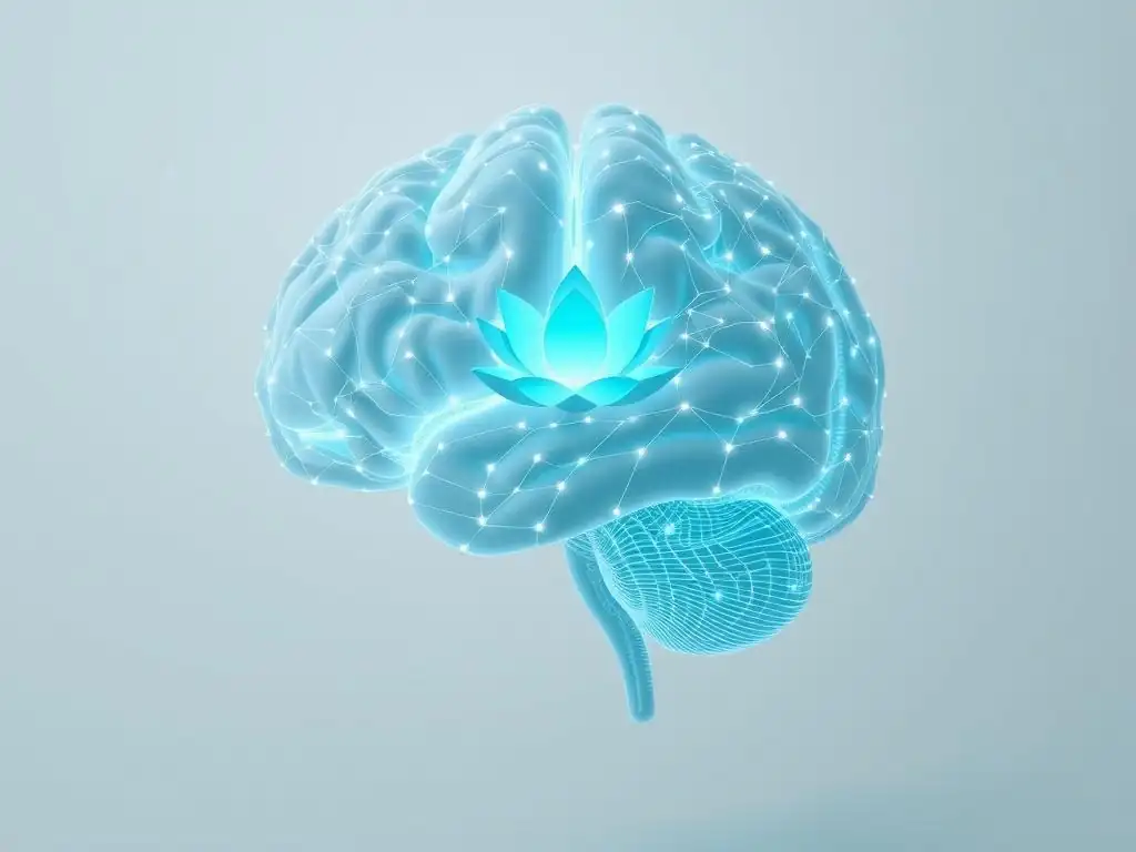
Analyzing brain scans before and after a course of CBT for depression provides compelling visual evidence of the therapy's impact. These studies typically involve a control group (either healthy individuals or those receiving a different treatment) to ensure that observed changes are specific to CBT and not simply due to the passage of time or non-specific therapeutic factors.
The consistent findings across numerous studies using different neuroimaging techniques highlight specific patterns of neural change associated with clinical improvement. These changes are often observed in the very brain circuits known to be dysregulated in depression, providing strong support for the idea that CBT directly influences the underlying neurobiology of the disorder. The power of CBT depression brain scans lies in their ability to objectively demonstrate that therapy instigates measurable changes within the brain, offering a biological basis for its effectiveness.
Analyzing Brain Scan Images Before and After CBT Treatment
The process of analyzing brain scans in CBT research involves sophisticated techniques. Researchers compare baseline scans (taken before therapy) with follow-up scans (taken after therapy completion or during follow-up periods). This comparison can reveal changes in regional brain activity (e.g., how much a specific area "lights up" during a task or at rest), connectivity between brain regions (how well different areas communicate with each other), and sometimes structural changes (like gray matter volume).
For example, a study might use fMRI to measure brain activity while participants view emotional images. Before CBT, individuals with depression might show exaggerated activity in the amygdala and reduced activity in parts of the prefrontal cortex when viewing negative images. After successful CBT, the analysis of post-treatment scans might show a significant reduction in amygdala reactivity and an increase in prefrontal engagement during the same task.
These patterns of change are then correlated with the degree of symptom improvement reported by participants, providing a powerful link between neural changes and clinical outcomes. The analysis of CBT depression brain scans thus provides objective markers of therapeutic success.
Identifying Structural and Functional Changes in the Brain Post-CBT
Brain imaging studies have identified both functional and, in some cases, structural changes in the brains of individuals with depression following successful CBT. Functional changes, often observed with fMRI or PET, include shifts in activity levels in specific regions or altered patterns of communication between brain networks. For instance, studies have shown reduced functional connectivity within the default mode network (associated with rumination) and increased connectivity between the PFC and limbic regions (suggesting better top-down control of emotions).
Structural changes, typically assessed with structural MRI, are less consistently reported but are an area of ongoing research. Some studies have explored whether CBT can influence gray matter volume in regions like the hippocampus or prefrontal cortex, which can be reduced in chronic depression. While findings on structural changes are more varied than those on functional changes, the consistent observation of functional reorganization through CBT depression brain scans strongly supports the idea that therapy facilitates adaptive neuroplasticity, altering how the brain processes information and regulates mood on a fundamental level.
Long-Term Benefits of CBT: Supported by Brain Scan Research
One of the most significant advantages of CBT for depression is its potential for long-term benefits, including sustained symptom relief and a reduced risk of relapse. Brain scan research provides valuable insights into the neural basis of these enduring effects. Unlike treatments that may only manage symptoms acutely, CBT appears to equip individuals with cognitive and behavioral tools that foster lasting changes in brain function.
Studies that track participants over extended periods using brain imaging techniques have shown that the neural changes observed immediately after successful CBT can persist. These sustained neural alterations are thought to underpin the individual's improved ability to cope with stress, manage negative thoughts, and maintain a more positive emotional state, thereby reducing vulnerability to future depressive episodes. The data from CBT depression brain scans supports the notion that therapy builds resilience at a neurobiological level.
Sustained Improvements in Mood and Cognitive Functioning
Long-term follow-up studies consistently show that a significant proportion of individuals who respond to CBT for depression maintain their clinical gains over time. Brain scan research provides a potential explanation for this sustained improvement. Studies have indicated that neural changes associated with successful CBT, such as normalized activity in emotion regulation circuits and enhanced engagement of cognitive control regions, can still be observed months or even years after therapy has ended.
For example, a study might find that individuals who remain in remission after CBT show sustained increased connectivity between areas of the prefrontal cortex and the amygdala, suggesting continued effective top-down control over emotional responses. Furthermore, improvements in cognitive functions often impaired by depression, such as attention and executive function, are also supported by enduring positive changes in prefrontal activity observed in CBT depression brain scans. These findings suggest that CBT facilitates lasting functional reorganization in the brain that supports both sustained mood stability and improved cognitive performance.
Reduced Risk of Relapse: Neural Correlates of Long-Term CBT Efficacy
Relapse is a major challenge in treating depression. The ability of CBT to significantly reduce relapse rates compared to some other interventions is a key strength. Brain scan research is beginning to uncover the neural mechanisms that contribute to this reduced vulnerability. It is hypothesized that the enduring neural changes induced by CBT build resilience against future stressors and negative events that might otherwise trigger a return of symptoms.
Studies investigating the neural correlates of relapse prevention after CBT have focused on the stability of post-treatment brain changes. Research suggests that individuals who maintain the positive neural adaptations achieved through CBT, such as sustained healthy patterns of activity in prefrontal-limbic circuits, are less likely to experience a relapse.
For instance, some studies have found that a return of depressive symptoms is associated with a reversal of the neural changes observed after successful treatment, such as a return to heightened amygdala reactivity or reduced prefrontal control. This suggests that the long-term efficacy of CBT is linked to the persistence of favorable changes in brain function, providing a neurobiological foundation for its role in preventing future episodes, a finding powerfully illustrated by long-term CBT depression brain scans.
Comparing CBT Brain Scan Results with Other Depression Treatments
Understanding the specific neural pathways influenced by different depression treatments is crucial for optimizing care. Brain imaging studies have not only illuminated how CBT works but have also allowed for comparisons of its neural effects with those of other common treatments, most notably antidepressant medications. These comparisons reveal that while different treatments can lead to similar clinical outcomes (symptom reduction), they may achieve these results through partially distinct or overlapping neural mechanisms.
This comparative approach using brain scans helps researchers and clinicians understand the unique strengths and potential synergies of different therapeutic modalities. For example, some studies have shown that while both medication and CBT can reduce activity in overactive limbic regions, they might have different effects on specific prefrontal areas or connectivity patterns. These distinctions can inform decisions about which treatment might be most suitable for an individual based on their specific symptom profile and potential underlying neural patterns, as revealed by brain imaging.
CBT vs. Medication: A Neuroimaging Perspective
Both Cognitive Behavioral Therapy and antidepressant medications (such as SSRIs) are effective treatments for depression, and brain imaging studies have investigated their respective impacts on the brain. While both can lead to clinical improvement and normalization of activity in certain brain regions, the pattern of these changes often differs.
Brain scan research comparing CBT and medication has sometimes shown that CBT is associated with changes primarily in prefrontal cortical regions involved in cognitive control and emotional regulation, suggesting a "top-down" influence where cognitive restructuring impacts emotional processing. In contrast, medication is sometimes associated with more pronounced changes in limbic areas like the amygdala or hippocampus, suggesting a more "bottom-up" modulation of emotional reactivity.
For example, one meta-analysis of neuroimaging studies found that while both treatments reduced amygdala activity, CBT was more consistently associated with increased activity in the dorsolateral prefrontal cortex. These findings from CBT depression brain scans highlight that while the clinical outcome may be similar, the brain's journey to recovery can differ depending on the treatment path taken, potentially influencing long-term outcomes and relapse vulnerability.
Combining CBT with Other Therapies: Investigating Synergistic Brain Changes
Given that depression is multifaceted, combining different therapeutic approaches is often beneficial. Research is increasingly exploring the effects of combining CBT with other therapies, such as medication or other forms of psychotherapy, and using brain imaging to understand the synergistic effects on the brain.
Studies investigating combined treatments using brain scans are still emerging but hold significant promise. The hypothesis is that combining therapies might target different aspects of the brain's dysfunction in depression, leading to more comprehensive or robust neural normalization than either treatment alone.
For example, combining medication's potential to quickly modulate neurotransmitter systems and affect basic emotional reactivity with CBT's ability to build cognitive control and coping skills might lead to a more complete restoration of healthy brain function. While specific findings on synergistic brain changes are complex and require further research, the use of CBT depression brain scans in combination therapy studies is a vital step towards understanding how best to integrate treatments for optimal outcomes.
CBT for Depression: Beyond Symptom Relief - A Brain-Based Approach
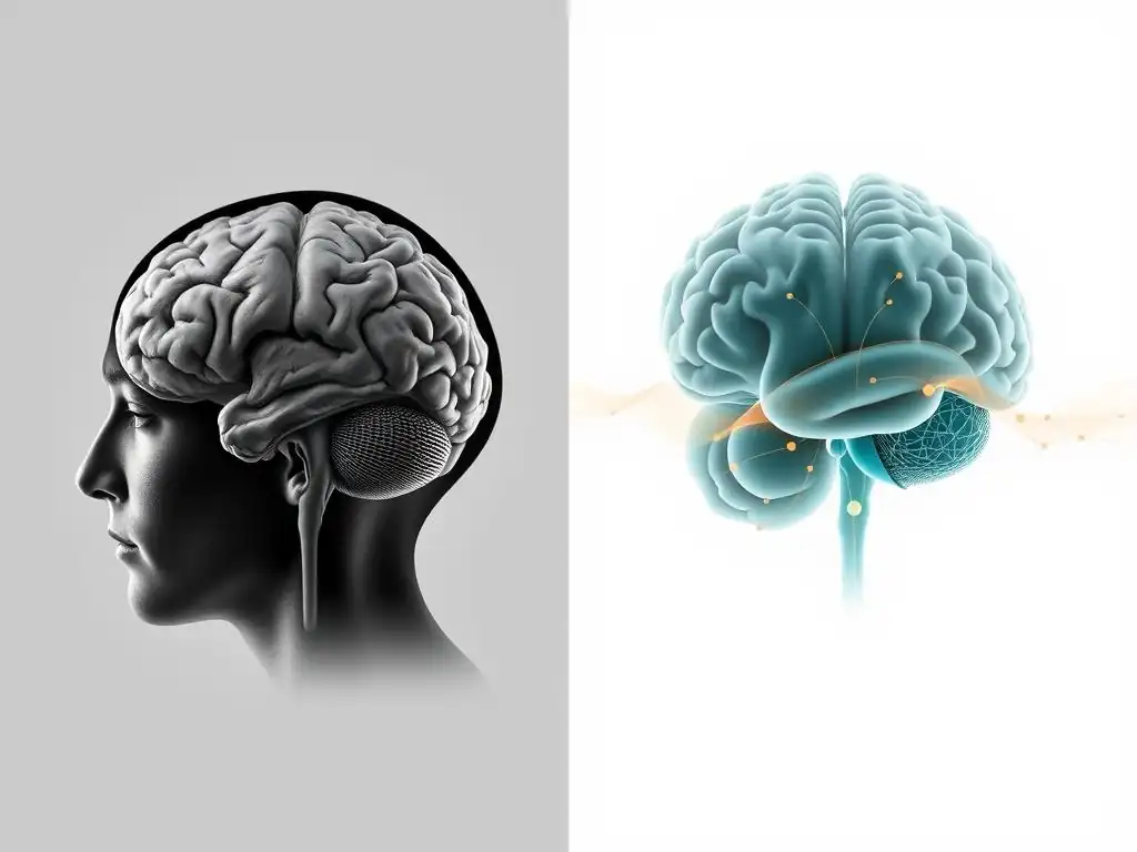
Viewing CBT through the lens of neuroscience reveals that its impact extends beyond merely alleviating depressive symptoms. It appears to facilitate fundamental changes in brain function and structure, promoting neuroplasticity and building resilience. This brain-based understanding elevates CBT from a set of coping techniques to a powerful intervention that helps the brain rewire itself for healthier emotional and cognitive processing.
The insights gained from CBT depression brain scans are transforming how we understand the mechanisms of therapeutic change. They demonstrate that engaging in cognitive restructuring and behavioral activation exercises isn't just about changing thoughts and actions; it's about actively training the brain to respond differently to internal and external stimuli. This perspective underscores the active role of the individual in their recovery process and highlights the brain's remarkable capacity for adaptation.
Understanding the Underlying Mechanisms of CBT Through Brain Imaging
Brain imaging provides objective evidence for the proposed psychological mechanisms of CBT. For instance, CBT posits that challenging negative thoughts reduces their impact on mood. Brain scans can show this by demonstrating decreased activity in brain regions associated with negative self-referential processing (like parts of the medial prefrontal cortex) when individuals engage in cognitive reappraisal after CBT.
Similarly, behavioral activation, a core component of CBT that encourages engaging in rewarding activities, is thought to counteract the anhedonia (loss of pleasure) characteristic of depression. Brain imaging can investigate this by examining activity in reward circuits (such as the striatum) in response to positive stimuli before and after behavioral activation interventions within CBT. By correlating specific therapeutic techniques with observable brain changes, CBT depression brain scans help researchers refine models of how therapy works and identify which components are most effective at a neural level.
Promoting Neuroplasticity and Resilience Through Cognitive Behavioral Therapy
Neuroplasticity, the brain's ability to reorganize itself by forming new neural connections throughout life, is a key concept in understanding recovery from depression. Chronic depression is associated with reduced neuroplasticity in certain brain regions. CBT appears to be a powerful tool for promoting adaptive neuroplasticity.
By repeatedly practicing new ways of thinking and behaving, individuals undergoing CBT are essentially engaging in a form of brain training. This practice strengthens neural pathways associated with healthier cognitive and emotional processing and weakens those associated with depressive patterns. Brain scan studies provide evidence for this, showing changes in gray matter density, white matter connectivity, and functional network organization following successful CBT.
These neuroplastic changes are thought to build resilience, equipping the brain with enhanced capacity to cope with stress and preventing a return to depressive states. The evidence from CBT depression brain scans supports the idea that therapy fosters lasting brain changes that protect against future episodes.
Future Directions in CBT and Brain Scan Research for Depression
The field of CBT and brain scan research is rapidly evolving. While significant progress has been made in understanding the general neural effects of CBT, future research aims to refine this understanding and translate it into more personalized and effective clinical practice.
One major area of focus is using brain imaging data to predict individual responses to CBT. Not everyone benefits from the same treatment, and identifying biomarkers in the brain that predict who is most likely to respond to CBT could revolutionize treatment selection. Furthermore, integrating brain imaging with other technologies, such as neurofeedback, holds potential for enhancing the delivery and effectiveness of CBT. The continued exploration using CBT depression brain scans promises exciting advancements in mental healthcare.
Advancing Personalized CBT Treatment Strategies Using Brain Imaging Data
Currently, treatment selection for depression often involves trial and error. Brain imaging data offers the potential to move towards a more personalized approach. Researchers are exploring whether specific patterns of brain activity or connectivity observed in baseline scans can predict an individual's likelihood of responding to CBT versus other treatments like medication.
For example, some preliminary research suggests that individuals with a certain pattern of connectivity within the default mode network might be particularly good candidates for CBT. If replicated, such findings could allow clinicians to use brain scans as a tool to guide treatment decisions, directing individuals to the therapy most likely to be effective for their specific neurobiological profile. This advancement, driven by insights from CBT depression brain scans, could significantly reduce the time and distress associated with finding the right treatment, leading to faster recovery and better outcomes.
Exploring the Potential of Neurofeedback to Enhance CBT Outcomes
Neurofeedback is a technique that allows individuals to see their own brain activity in real-time and learn to regulate it. Combining neurofeedback with CBT is an exciting area of future research. The idea is that by providing individuals with direct feedback on the activity of brain regions involved in emotional regulation or cognitive control, they might be able to learn to modulate these areas more effectively, potentially enhancing the skills taught in CBT.
For example, an individual might receive neurofeedback on their amygdala activity while processing emotional stimuli, learning to reduce its reactivity. This learned self-regulation could complement the cognitive restructuring and behavioral techniques of CBT. While still largely experimental, the integration of neurofeedback, informed by findings from CBT depression brain scans, represents a promising avenue for developing more potent and targeted therapeutic interventions for depression, leveraging the power of brain imaging to directly support the process of neural change.
Conclusion: The Future of Mental Health is in the Brain
The evidence from brain scan research strongly supports the profound impact of Cognitive Behavioral Therapy on the brain in individuals with depression. These studies provide objective, visual proof that CBT is not just a superficial coping mechanism but a powerful intervention that facilitates significant and lasting changes in neural activity, connectivity, and potentially even structure.
By revealing how CBT normalizes activity in key brain regions like the amygdala and prefrontal cortex and promotes adaptive neuroplasticity, brain imaging enhances our understanding of why this therapy is so effective. The findings from CBT depression brain scans validate the lived experience of recovery and open up exciting possibilities for personalized treatment approaches and enhanced therapeutic techniques. As research continues to advance, the insights gained from studying the brain will undoubtedly play an increasingly central role in our efforts to understand, treat, and ultimately prevent mental health challenges like depression.
CBT Depression Brain Scans: A Promising Avenue for Understanding and Treating Depression
The field of CBT depression brain scans represents a critical intersection of psychology and neuroscience. It provides a compelling narrative supported by objective data: that talking therapy can change the brain. This evidence is invaluable for reducing stigma around mental health issues and encouraging individuals to seek treatment, knowing that effective therapies like CBT are grounded in measurable biological changes.
Future research using brain imaging will likely continue to refine our understanding of the specific neural signatures of different depression subtypes and how they respond to CBT. This will pave the way for even more precise and effective interventions. The promise of using brain scans to personalize therapy and potentially even predict treatment response highlights the transformative potential of this research for the future of mental healthcare. The journey towards understanding and treating depression is increasingly becoming a journey through the brain, guided by the insights from neuroimaging.
Mind Care Guide: Your Resource for Evidence-Based Mental Health Information
At Mind Care Guide, we are committed to providing accessible, evidence-based information on mental health and well-being. Understanding the science behind effective treatments like CBT, supported by data from studies like those using CBT depression brain scans, is crucial for making informed decisions about your mental health journey.
We strive to translate complex scientific findings into clear and understandable content, empowering you with knowledge about conditions like depression and the therapeutic approaches that can help. The research on CBT and brain changes is a powerful example of how scientific inquiry is advancing our ability to support mental health recovery. Explore our resources to learn more about depression, CBT, and the many pathways to well-being, all grounded in the latest understanding of the mind and brain.
Frequently Asked Questions
How do brain scans support the effectiveness of CBT for depression?
Brain scans can provide visual evidence of how CBT impacts brain activity. Studies show that long-term CBT can lead to observable changes in brain regions associated with mood regulation and emotional processing. This provides tangible proof of CBT's impact.
What specific brain changes might be seen after long-term CBT for depression?
Scans may reveal increased activity in areas related to cognitive control and emotion regulation. Conversely, they might show decreased activity in regions associated with negative emotions and rumination. The amygdala and prefrontal cortex are often key areas of interest.
Is a brain scan necessary to determine if CBT is working for depression?
No, brain scans are not typically required. Clinical improvement, measured through self-reported symptoms and therapist assessments, is the primary indicator. Brain scans are primarily used in research settings to better understand CBT's mechanisms.
Are there limitations to using brain scans to assess CBT's impact on depression?
Yes, brain scans can be expensive and complex to interpret. They also only provide a snapshot in time and may not capture the full dynamic of brain changes. Further, individual variability in brain structure and function exists.
Why is it important to understand the brain's response to CBT for depression?
Understanding the neurological basis of CBT's effectiveness can help refine treatment approaches and develop more targeted interventions. It also helps validate CBT as a biologically based treatment and reduce stigma surrounding mental health. This leads to better and more effective treatments in the future.
Final Reflection: The Enduring Power of the Mind's Resilience
The groundbreaking research linking CBT to observable changes in the brain underscores a powerful truth: our thoughts and behaviors are not merely abstract concepts, but tangible forces shaping our neurological landscape. This isn't just a scientific curiosity; it's a revolutionary shift in our understanding of mental health, offering concrete evidence of the efficacy of CBT and providing hope for millions struggling with depression. The ability to see the positive impact of therapy on the brain itself is a game-changer, moving beyond anecdotal evidence to a demonstrably visible transformation.
The implications of this research extend far beyond the clinical setting. It challenges the stigma surrounding mental illness, offering a visual representation of the brain's remarkable plasticity and capacity for healing. The brain is not a static organ; it is constantly changing and adapting in response to our experiences.
This quote perfectly encapsulates the transformative power of CBT, showing how targeted interventions can literally reshape neural pathways and alleviate the debilitating symptoms of depression. This understanding fosters empathy and encourages a more compassionate approach to mental health, moving away from simplistic explanations and towards a nuanced appreciation of the intricate interplay between mind and brain.
Consider the impact this knowledge has on your own life. Perhaps you've struggled with depression, or know someone who has. This research offers not just hope, but a tangible understanding of the potential for recovery.
Take a moment to reflect on the power of your own mind, its resilience, and its capacity for change. Embrace the possibility of healing, explore the potential of CBT, and remember that the visible changes in the brain are a testament to the enduring strength of the human spirit. Seek support, advocate for yourself or others, and let this knowledge inspire you to cultivate a healthier and more resilient mind.
Unlock Your Brain's Potential: Take Charge of Your Mental Well-being
Thanks for taking the time to learn about the powerful, long-term effects of CBT on depression! You've gained valuable insight into how this therapy can reshape your brain and improve your mental health. Now it's time to put that knowledge into action.
- Schedule a consultation with a CBT therapist: This is the most impactful step towards reclaiming your mental well-being. Finding the right therapist is key, so take your time researching and finding someone you connect with.
- Share this article with someone you care about: Knowledge is power, and spreading awareness about effective depression treatments can make a huge difference in someone's life.
- Explore MindCareGuide.org for more resources: Discover additional articles, tips, and support to help you on your journey towards better mental health.
Ready to begin your journey towards a brighter, healthier future? What's the first step you'll take to prioritize your mental well-being today?