
Understanding Brain Plasticity Through Imaging in Mental Health Recovery

Welcome to Mind Care Guide, a sanctuary dedicated to your mental well-being.
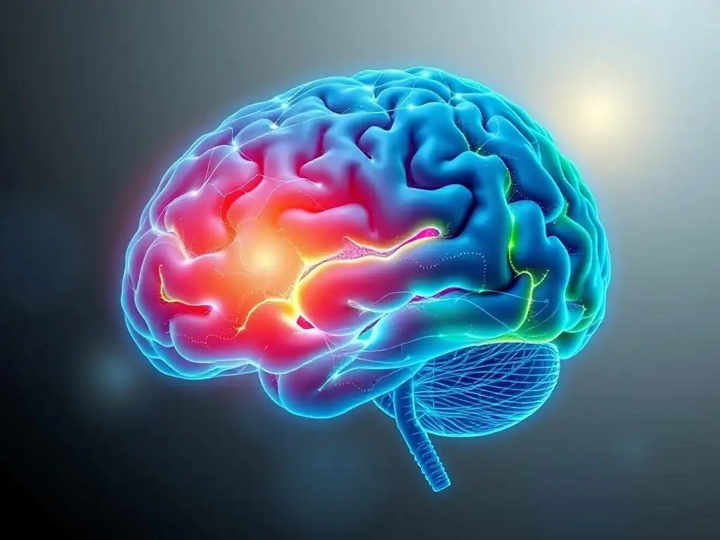
Feeling stuck? Know that your brain has an incredible capacity for change. Delve into Understanding Brain Plasticity Through Imaging in Mental Health Recovery and uncover the fascinating science that proves recovery is possible. Curious about how your brain can reshape itself, even after facing challenges? Let's explore the neuroscience of hope and healing together...
Understanding Brain Plasticity Through Imaging in Mental Health Recovery
- Introduction: Brain Plasticity and Mental Health Recovery
- What is Brain Plasticity? A Comprehensive Overview
- Brain Imaging Techniques for Assessing Brain Plasticity
-
How Brain Imaging Reveals Plastic Changes in Mental Health Conditions
- Brain Plasticity in Depression: Imaging Studies and Findings
- Neuroplasticity in Anxiety Disorders: Insights from Brain Scans
- Brain Imaging and Recovery from Trauma: Understanding PTSD Through Plasticity
- Schizophrenia and Brain Plasticity: Exploring Neural Reorganization
- The Role of Brain Plasticity in Addiction Recovery: Imaging Evidence
- The Link Between Brain Plasticity, Mental Health Treatment, and Imaging
- Limitations and Future Directions in Brain Plasticity Imaging Research
- Conclusion: Harnessing Brain Plasticity for Mental Health Recovery
-
Frequently Asked Questions
- How does brain plasticity relate to mental health recovery?
- Can brain imaging techniques actually show evidence of brain plasticity?
- What are some specific mental health conditions where brain plasticity plays a significant role in recovery?
- What lifestyle factors can enhance brain plasticity and support mental health recovery?
- Are there specific therapeutic approaches that leverage brain plasticity to improve mental health outcomes?
- Final Reflection: The Rewiring of Hope: Brain Plasticity and Mental Health Recovery
- Unlock Your Brain's Potential: Start Your Recovery Journey Today!
Introduction: Brain Plasticity and Mental Health Recovery
The Role of Brain Plasticity in Mental Health Improvement
The human brain is not a static organ; it possesses a remarkable ability to change and adapt throughout life. This capacity, known as brain plasticity or neuroplasticity, is fundamental to learning, memory, and recovery from injury. In the context of mental health, understanding brain plasticity is crucial because it provides a biological basis for improvement and recovery.
It suggests that the neural pathways associated with conditions like depression, anxiety, or trauma are not fixed but can be altered through experiences, therapy, and other interventions. This dynamic nature of the brain offers a hopeful perspective, indicating that positive changes in mental well-being are achievable through targeted efforts that encourage adaptive neuroplasticity.
Scientific research has increasingly focused on how harnessing brain plasticity can support mental health recovery. Studies show that engaging in certain activities, such as mindfulness meditation or cognitive behavioral therapy (CBT), can lead to measurable changes in brain structure and function. For instance, some research indicates that regular meditation practice may increase gray matter density in areas associated with self-awareness and emotional regulation, like the prefrontal cortex and hippocampus.
Similarly, successful therapy for anxiety disorders has been linked to altered connectivity in fear circuits involving the amygdala. These findings underscore the powerful link between our experiences, our brain's ability to change, and the potential for significant improvement in mental health, highlighting the importance of understanding brain plasticity in mental health recovery.
Why Brain Imaging is Crucial for Understanding Plasticity
While behavioral changes provide observable evidence of mental health improvement, brain imaging techniques offer a window into the underlying neural mechanisms driving these changes. Non-invasive methods allow researchers and clinicians to visualize and measure structural and functional alterations in the living brain over time. This is particularly crucial for studying brain plasticity, as it allows us to see how the brain is reorganizing itself in response to interventions or life experiences. Without imaging, our understanding of neuroplasticity would be largely theoretical, based only on behavioral outcomes or post-mortem studies.
Brain imaging provides tangible evidence of the brain's capacity for change, reinforcing the concept of brain plasticity in mental health recovery. Techniques like MRI, fMRI, EEG, MEG, and PET scans can reveal changes in brain volume, connectivity patterns, metabolic activity, and neurotransmitter levels. For example, tracking changes in hippocampal volume using structural MRI has provided insights into recovery from depression or PTSD.
Functional MRI can show how brain networks reconfigure as symptoms improve. This empirical evidence not only validates the principles of neuroplasticity but also helps refine therapeutic approaches by identifying which interventions are most effective at inducing positive brain changes, thus advancing our understanding of how to foster brain plasticity for mental health recovery.
What is Brain Plasticity? A Comprehensive Overview
Defining Brain Plasticity: Neuroplasticity Explained
Brain plasticity, often referred to as neuroplasticity, is the nervous system's ability to change its structure, function, and organization in response to experience. This fundamental property allows the brain to adapt to new situations, learn new skills, and recover from injury or disease. At a cellular level, plasticity involves changes in the strength of connections between neurons (synaptic plasticity), the formation of new synapses (synaptogenesis), and even the birth of new neurons in certain brain regions (neurogenesis). These micro-level changes accumulate to influence the overall macro-level structure and function of brain circuits and networks.
The concept of neuroplasticity challenges the older view that the adult brain is a fixed entity. Instead, it portrays the brain as a dynamic and adaptable organ, constantly being molded by our interactions with the environment, our thoughts, and our behaviors. This adaptability is crucial for all aspects of brain function, from simple motor skills to complex cognitive processes and emotional regulation. Understanding this inherent capacity for change is foundational to appreciating how recovery and improvement are possible in the context of mental health challenges, emphasizing the potential of brain plasticity for mental health recovery.
Types of Brain Plasticity: Structural and Functional Changes
Brain plasticity manifests in two primary forms: structural plasticity and functional plasticity. Structural plasticity refers to changes in the physical structure of the brain. This includes alterations in the size of specific brain regions, the thickness of the cerebral cortex, the density of gray matter (neuronal cell bodies), the integrity of white matter (axonal connections), and the number and complexity of dendrites and synapses. For instance, learning a new language might lead to an increase in gray matter density in language-related areas.
Functional plasticity, on the other hand, refers to changes in the way the brain processes information. This can involve shifts in how specific brain regions are activated during tasks, alterations in the strength or efficiency of connections within neural networks, or even the recruitment of entirely new areas to perform a function previously handled elsewhere. For example, after a stroke, the brain might reroute neural pathways to regain lost motor skills, demonstrating functional reorganization. Both structural and functional plasticity are interconnected and contribute significantly to the brain's ability to adapt and facilitate brain plasticity in mental health recovery.
Factors Influencing Brain Plasticity: Therapy, Medication, Lifestyle
Numerous factors can influence the degree and direction of brain plasticity. Therapeutic interventions are powerful drivers of plastic change. Psychotherapies like Cognitive Behavioral Therapy (CBT), Dialectical Behavior Therapy (DBT), and Eye Movement Desensitization and Reprocessing (EMDR) work by helping individuals learn new thought patterns, emotional regulation skills, and behavioral responses, which in turn can reorganize neural circuits associated with maladaptive patterns. For example, exposure therapy for phobias can lead to reduced activity in the amygdala and increased activity in the prefrontal cortex when encountering the feared stimulus.
Medications also play a role in influencing neuroplasticity, particularly those that affect neurotransmitter systems. Antidepressants, for instance, not only alter levels of neurotransmitters like serotonin and norepinephrine but have also been shown in some studies to promote neurogenesis in the hippocampus and increase synaptic connectivity, potentially facilitating long-term mood regulation. Beyond direct interventions, lifestyle factors significantly impact brain plasticity. Regular physical exercise has been widely demonstrated to promote neurogenesis, particularly in the hippocampus, and improve synaptic function.
Adequate sleep is essential for memory consolidation and synaptic homeostasis. A nutritious diet, rich in omega-3 fatty acids and antioxidants, supports brain health and plasticity. Engaging in intellectually stimulating activities, social interaction, and stress management techniques also foster positive brain changes, all contributing to the potential for brain plasticity in mental health recovery.
Brain Imaging Techniques for Assessing Brain Plasticity

Magnetic Resonance Imaging (MRI) and Brain Plasticity Studies
Magnetic Resonance Imaging (MRI) is one of the most widely used non-invasive techniques for studying the brain and its plasticity. It uses strong magnetic fields and radio waves to generate detailed images of brain structure and function. MRI is versatile, offering different modalities that can capture various aspects of neural tissue and activity, making it invaluable for assessing the dynamic nature of the brain over time. Serial MRI scans taken before and after interventions or over periods of recovery can reveal subtle yet significant changes in brain anatomy and connectivity that reflect neuroplastic processes.
The safety profile of standard MRI (without contrast agents) makes it suitable for repeated scanning, which is essential for longitudinal studies tracking brain plasticity in mental health recovery. Researchers can use MRI to examine differences in brain structure or function between individuals with mental health conditions and healthy controls, and critically, to observe how these differences change as individuals undergo treatment or experience recovery. This capability provides objective evidence of the brain's capacity to adapt and heal, reinforcing the potential for positive outcomes driven by brain plasticity in mental health recovery.
Structural MRI: Examining Brain Volume and Connectivity
Structural MRI provides high-resolution images of brain anatomy, allowing researchers to measure the size and shape of different brain regions and assess the integrity of white matter tracts. Techniques like Voxel-Based Morphometry (VBM) can statistically compare local concentrations of gray matter or white matter across groups or over time within individuals. For example, studies using structural MRI have reported reduced gray matter volume in the hippocampus and prefrontal cortex in individuals with chronic depression, and some research tracks increases in volume in these areas following successful antidepressant treatment or psychotherapy, suggesting structural plasticity.
Diffusion Tensor Imaging (DTI), a type of structural MRI, specifically measures the diffusion of water molecules in white matter, providing insights into the directionality and integrity of nerve fiber bundles. Changes in DTI metrics, such as fractional anisotropy (FA), can indicate alterations in the structure and organization of white matter pathways, reflecting changes in connectivity between brain regions. Observing these changes over time, for instance, in individuals recovering from trauma or addiction, provides concrete evidence of structural neuroplasticity underlying brain plasticity in mental health recovery.
Functional MRI (fMRI): Measuring Brain Activity Changes
Functional MRI (fMRI) measures brain activity by detecting changes in blood flow. When a brain region is more active, it requires more oxygenated blood, and fMRI can capture the resulting changes in the blood oxygen level-dependent (BOLD) signal. By having participants perform tasks or simply rest within the scanner, researchers can identify which brain areas are active and how different areas communicate with each other (functional connectivity). This allows for the study of how brain network activity patterns change in response to stimuli, during cognitive processes, or over time with interventions.
In the context of mental health, fMRI studies have revealed altered activity and connectivity patterns in various conditions. For example, individuals with anxiety disorders often show hyperactivation in the amygdala (a fear processing region) and altered connectivity between the amygdala and prefrontal cortex (involved in regulation). Longitudinal fMRI studies have shown that effective therapy can normalize these activity patterns and connectivity strengths, demonstrating functional plasticity. Tracking these changes provides direct evidence of how the brain's functional organization adapts, illustrating the dynamic nature of brain plasticity in mental health recovery.
Electroencephalography (EEG) and Magnetoencephalography (MEG) for Neuroplasticity Research
Electroencephalography (EEG) and Magnetoencephalography (MEG) are neuroimaging techniques that measure the electrical and magnetic activity produced by neuronal populations in the brain, respectively. Unlike MRI, which typically has higher spatial resolution, EEG and MEG offer excellent temporal resolution, meaning they can track brain activity changes on the order of milliseconds. This allows researchers to study the rapid dynamics of neural processing and observe how brain responses evolve over very short periods, which is valuable for understanding the timing of plastic changes.
These techniques are particularly useful for studying rhythmic brain activity (brainwaves) and event-related potentials/fields (ERPs/ERFs), which are brain responses time-locked to specific events (like presenting a stimulus). Changes in brainwave patterns or ERP/ERF components over time can reflect plastic adaptations in neural processing speed and efficiency. For instance, changes in certain ERP components have been linked to improved cognitive processing following interventions for conditions like ADHD or learning disabilities, providing insights into the functional neuroplasticity supporting brain plasticity in mental health recovery.
EEG: Tracking Brainwave Patterns in Mental Health Recovery
EEG measures electrical activity via electrodes placed on the scalp. It can identify different frequency bands of brainwaves (e.g., delta, theta, alpha, beta, gamma), each associated with different states of consciousness or cognitive processes. Abnormalities in these brainwave patterns are often observed in mental health conditions; for example, altered alpha or theta activity has been noted in depression and anxiety. Longitudinal EEG studies can track how these patterns change with treatment or recovery.
Changes in EEG coherence (a measure of synchronization between different brain regions) can also indicate alterations in functional connectivity, reflecting neuroplastic changes. For instance, studies might look at how coherence between frontal and limbic areas changes as emotional regulation improves with therapy. The relatively low cost and portability of EEG compared to other imaging modalities make it a practical tool for some types of neuroplasticity research, helping to map the dynamic electrical landscape associated with brain plasticity in mental health recovery.
MEG: High-Resolution Mapping of Brain Activity
MEG measures the magnetic fields produced by electrical currents in the brain, offering a higher spatial resolution than EEG, particularly for activity originating from deeper brain structures. Like EEG, it boasts excellent temporal resolution, capturing rapid neural events. MEG is often used to study the dynamics of neural networks and oscillations with greater precision than EEG.
MEG studies can identify changes in the location and strength of neural sources generating activity, as well as alterations in the synchronization of activity between different brain regions. This provides a detailed view of how functional brain networks reorganize over time. For example, MEG could be used to track changes in the processing of auditory stimuli in schizophrenia or alterations in resting-state network dynamics in depression, offering high-resolution insights into the functional neuroplasticity that underpins brain plasticity in mental health recovery.
Positron Emission Tomography (PET) Scans and Brain Plasticity: Assessing Neurotransmitter Activity
Positron Emission Tomography (PET) is a nuclear imaging technique that uses small amounts of radioactive tracers to measure metabolic activity, blood flow, or the distribution of specific molecules, such as neurotransmitter receptors or transporters, in the brain. PET scans are particularly valuable for studying changes in neurochemistry and metabolism that are associated with mental health conditions and may reflect or drive plastic changes.
By using different tracers, PET can provide quantitative data on the density of receptors for neurotransmitters like dopamine, serotonin, or opioids, or measure glucose metabolism (an indicator of overall neuronal activity). Changes in receptor availability or metabolic activity in specific brain regions over time can reflect compensatory mechanisms or adaptations in response to illness or treatment. For instance, PET studies have investigated changes in dopamine receptor density in addiction recovery or alterations in serotonin transporter availability in depression following treatment, offering unique neurochemical insights into the mechanisms supporting brain plasticity in mental health recovery.
How Brain Imaging Reveals Plastic Changes in Mental Health Conditions
Brain Plasticity in Depression: Imaging Studies and Findings
Imaging studies have consistently revealed structural and functional alterations in the brains of individuals with depression, and critically, have tracked how these changes relate to recovery, highlighting the role of brain plasticity in mental health recovery. Structural MRI studies often report reduced gray matter volume in areas like the hippocampus (involved in memory and mood), the prefrontal cortex (involved in executive function and emotional regulation), and the anterior cingulate cortex (involved in emotion and decision-making). For example, some studies suggest a volume reduction in the hippocampus of approximately 9-13% in individuals with recurrent depression compared to healthy controls.
Functional MRI studies in depression frequently show altered activity and connectivity within neural networks, including the default mode network (DMN), which is often hyperactive, and networks involved in cognitive control and reward processing, which may be hypoactive. Successful treatment with antidepressants or psychotherapy has been associated with reversals or improvements in these structural and functional abnormalities. For instance, some fMRI studies have shown a normalization of connectivity between the prefrontal cortex and amygdala after psychotherapy, suggesting a restoration of regulatory control over emotional responses, providing tangible evidence of brain plasticity in mental health recovery.
Neuroplasticity in Anxiety Disorders: Insights from Brain Scans
Brain imaging provides crucial insights into the neural underpinnings of anxiety disorders and how neuroplasticity contributes to recovery. Conditions like Generalized Anxiety Disorder (GAD), Panic Disorder, and Social Anxiety Disorder are often associated with hyperactivity in limbic regions, particularly the amygdala, and altered connectivity with prefrontal regulatory areas. For example, fMRI studies in individuals with social anxiety disorder might show exaggerated amygdala responses to social stimuli compared to healthy controls.
Therapeutic interventions for anxiety, such as CBT or exposure therapy, aim to modify fear responses and improve emotional regulation. Imaging studies have shown that these therapies can induce significant neuroplastic changes. Research using fMRI has demonstrated a decrease in amygdala reactivity and an increase in activity in prefrontal regions (like the ventromedial prefrontal cortex) following successful CBT for phobias or GAD.
Structural changes, such as alterations in gray matter volume in these areas, have also been reported in some longitudinal studies. These findings provide compelling evidence that the brain's fear circuits are not fixed and can be rewired through therapeutic efforts, illustrating the power of brain plasticity in mental health recovery from anxiety.
Brain Imaging and Recovery from Trauma: Understanding PTSD Through Plasticity
Post-Traumatic Stress Disorder (PTSD) is characterized by persistent symptoms following exposure to a traumatic event, and brain imaging has been instrumental in understanding its neural basis and the potential for recovery via brain plasticity. Individuals with PTSD often exhibit a smaller hippocampus, hyperactive amygdala, and hypoactive medial prefrontal cortex compared to trauma-exposed individuals without PTSD. These structural and functional differences are thought to contribute to difficulties with memory, fear regulation, and emotional processing.
Effective treatments for PTSD, such as trauma-focused psychotherapies like EMDR or Prolonged Exposure (PE), aim to process traumatic memories and reduce hyperarousal. Longitudinal imaging studies have tracked brain changes associated with these therapies. For instance, some research has shown increases in hippocampal volume and normalization of amygdala-prefrontal connectivity following successful treatment, suggesting structural and functional neuroplasticity. These findings highlight that even after trauma, the brain retains the capacity for significant reorganization and healing, providing hope for individuals seeking brain plasticity in mental health recovery from PTSD.
Schizophrenia and Brain Plasticity: Exploring Neural Reorganization
Schizophrenia is a complex psychiatric disorder associated with significant alterations in brain structure and function, but research is increasingly exploring the role of neuroplasticity in its progression and potential for remediation. While some studies show structural deficits, such as reduced gray matter volume in areas like the prefrontal cortex, hippocampus, and thalamus, particularly in the early stages of illness, other research investigates the brain's capacity for compensatory plasticity or changes induced by treatment or cognitive training.
Functional imaging studies in schizophrenia often reveal disrupted connectivity within and between various brain networks, including those involved in executive function, salience detection, and social cognition. For example, altered connectivity within the default mode network and between frontal and temporal regions is frequently observed.
While the nature of plasticity in schizophrenia can be complex and sometimes involve maladaptive changes, interventions like cognitive remediation therapy (CRT) have shown promise in improving cognitive function, and some imaging studies suggest these improvements are associated with changes in brain activity and connectivity patterns, indicating potential for positive neuroplasticity and supporting the idea of brain plasticity in mental health recovery even in severe conditions.
The Role of Brain Plasticity in Addiction Recovery: Imaging Evidence
Addiction is increasingly understood as a chronic brain disease involving significant and persistent neuroplastic changes in reward pathways, decision-making circuits, and stress response systems. Chronic substance use leads to adaptations in the brain that drive compulsive drug-seeking behavior and make quitting difficult. Imaging studies, particularly PET and fMRI, have provided critical evidence of these changes, such as altered dopamine receptor availability (often reduced) in the striatum and disrupted connectivity between the prefrontal cortex and limbic areas.
However, these same imaging techniques also provide evidence for the brain's capacity for recovery and neuroplasticity during abstinence. Studies show that with prolonged abstinence, some of the addiction-induced neuroadaptations can reverse. For example, dopamine receptor availability in the striatum may gradually increase over time in recovery. Furthermore, behavioral therapies and support systems can facilitate the restoration of function in prefrontal control circuits. Imaging studies tracking these changes provide objective evidence that the brain can heal and reorganize during recovery, underscoring the critical role of brain plasticity in mental health recovery from addiction.
The Link Between Brain Plasticity, Mental Health Treatment, and Imaging

Neuroimaging to Monitor Treatment Efficacy and Brain Plasticity
Neuroimaging plays a vital role in evaluating the efficacy of mental health treatments by providing objective evidence of whether and how interventions induce brain plasticity. By scanning individuals before and after a course of therapy or medication, researchers can observe treatment-related changes in brain structure, function, or neurochemistry. This goes beyond simply assessing symptom reduction; it helps understand the biological mechanisms through which treatments work and which neural circuits are being modified.
For instance, an fMRI study might track changes in the connectivity between the amygdala and prefrontal cortex in patients undergoing CBT for anxiety. If improved connectivity is observed alongside symptom reduction, it strongly suggests that the therapy facilitated neuroplasticity in these specific regulatory circuits. Similarly, PET scans can monitor whether antidepressant treatment leads to expected changes in serotonin transporter availability. This ability to visualize and quantify brain changes provides valuable feedback on treatment effectiveness at a neural level and helps refine therapeutic protocols to better promote brain plasticity in mental health recovery.
Personalized Mental Healthcare: Tailoring Treatments Based on Brain Imaging Results
As our understanding of the neural basis of mental health conditions grows through imaging research, there is increasing interest in using brain imaging data to personalize treatment. The idea is that individual differences in brain structure, function, or plasticity profiles might predict response to specific therapies. For example, perhaps certain patterns of prefrontal connectivity predict who will respond best to cognitive therapy versus medication for depression.
While still largely an area of active research rather than routine clinical practice, the potential for personalized mental healthcare based on imaging is significant. Imagine a future where a baseline brain scan could help a clinician choose the most effective initial treatment approach for a patient, thus optimizing brain plasticity for mental health recovery from the outset. This could lead to more efficient treatment, reduced trial-and-error, and potentially better long-term outcomes by targeting interventions to the specific neurobiological needs of each individual.
The Potential of Neurofeedback to Enhance Brain Plasticity and Mental Well-being
Neurofeedback is a technique that uses real-time brain imaging (often EEG or fMRI) to show individuals their own brain activity patterns. Participants are then trained to voluntarily regulate specific aspects of their brain activity, such as increasing activity in a particular region or altering connectivity between areas. The underlying principle is that by providing individuals with direct feedback on their neural state, they can learn to consciously influence their brain function, thereby promoting targeted neuroplasticity.
Studies have explored the use of neurofeedback for various conditions, including ADHD (often targeting attention networks), anxiety (targeting amygdala activity), and depression (targeting mood regulation circuits). While the evidence base is still developing, some research suggests that neurofeedback training can lead to lasting changes in brain activity patterns and symptom reduction, indicating its potential as a tool to directly enhance brain plasticity for mental health recovery. This approach empowers individuals to become active participants in shaping their own brain function.
Limitations and Future Directions in Brain Plasticity Imaging Research
Challenges in Interpreting Brain Imaging Data
Despite the significant advances provided by brain imaging, interpreting the data, especially in the context of complex phenomena like brain plasticity and mental health, presents numerous challenges. Brain imaging studies are correlational; they can show associations between brain changes and mental health outcomes but cannot definitively prove causation. It's often difficult to determine whether observed brain differences are a cause of the condition, a consequence of the illness, or a result of other factors.
Furthermore, the brain is incredibly complex, and mental health conditions involve widespread alterations across multiple interconnected networks, not just isolated regions. Analyzing and integrating data from different imaging modalities and understanding the interplay between various brain areas and systems is technically challenging. Factors like individual variability, the influence of medication, comorbidities, and the heterogeneity within diagnostic categories also add layers of complexity to interpreting findings on brain plasticity in mental health recovery. Replicating findings across different studies and methodologies remains an ongoing effort in the field.
Emerging Technologies for Advanced Brain Plasticity Assessment
The field of neuroimaging is constantly evolving, with new technologies and analytical methods emerging that promise to provide more detailed and nuanced insights into brain plasticity. Advanced MRI techniques, such as 7T MRI, offer higher spatial resolution, allowing for the visualization of finer structural details. New diffusion imaging models are being developed to better characterize the complex architecture of white matter tracts.
Beyond MRI, techniques like functional Near-Infrared Spectroscopy (fNIRS) offer a more portable and less expensive way to measure brain activity, suitable for studying brain function in more naturalistic settings. Combining imaging data with other biological measures, such as genetics, epigenetics, and molecular markers, and integrating it with behavioral and clinical data, represents a major future direction. These emerging approaches aim to provide a more comprehensive understanding of the biological underpinnings of brain plasticity in mental health recovery.
Future Research: Integrating Brain Imaging with Other Measures of Mental Health
A key future direction for research is the integration of brain imaging data with other levels of analysis to build a more complete picture of mental health and recovery. This includes combining imaging findings with genetic information to understand how inherited predispositions influence brain plasticity and vulnerability or resilience to mental illness. Epigenetic studies, which examine how environmental factors influence gene expression, can also be integrated with imaging to explore the mechanisms by which experiences shape the brain.
Furthermore, integrating imaging data with detailed behavioral assessments, cognitive testing, physiological measures (like heart rate variability or cortisol levels), and real-world functional outcomes will provide a richer context for interpreting brain changes. The goal is to move towards a multi-modal understanding of brain plasticity in mental health recovery that accounts for the complex interplay between genes, environment, behavior, and brain dynamics. This integrated approach is essential for developing more effective, personalized, and biologically informed interventions.
Conclusion: Harnessing Brain Plasticity for Mental Health Recovery
The Power of Neuroplasticity: A Hopeful Outlook for Mental Well-being
The discovery and ongoing exploration of brain plasticity have revolutionized our understanding of the brain and its potential for change, offering a profoundly hopeful outlook for individuals facing mental health challenges. The knowledge that the brain is not fixed but can reorganize itself in response to experiences, learning, and interventions provides a powerful biological basis for recovery and improvement. It shifts the perspective from viewing mental illness as an irreversible state to seeing it as a condition where the brain's capacity for adaptation can be harnessed for healing.
This intrinsic ability of the brain to change means that therapeutic efforts are not just managing symptoms but are actively working to rewire neural circuits associated with distress and dysfunction. Whether through learning new coping skills in therapy, engaging in regular exercise, practicing mindfulness, or benefiting from targeted medications, individuals are engaging processes that promote positive neuroplasticity. This understanding empowers individuals and clinicians alike, highlighting that agency, effort, and appropriate support can fundamentally alter the brain's landscape, paving the way for sustained brain plasticity in mental health recovery and enhanced well-being.
Brain Imaging as a Tool for Empowering Mental Health Patients
Brain imaging techniques serve as powerful tools that not only advance scientific understanding but also have the potential to empower individuals on their mental health journey. By providing objective, visual evidence of the brain's capacity for change, imaging can make the abstract concept of neuroplasticity tangible. For patients, seeing that interventions can lead to measurable changes in brain activity or structure can be validating and motivating, reinforcing the belief that recovery is possible and that their efforts are having a real biological impact.
While brain imaging is not yet a routine diagnostic tool for most mental health conditions, its role in research is crucial for identifying the neural targets of effective treatments and understanding individual differences in response. As the technology advances and becomes more accessible, it holds promise for informing personalized treatment plans and potentially even offering neurofeedback approaches that allow individuals to directly train their brains. Ultimately, brain imaging contributes to a deeper, more empathetic understanding of mental health conditions and strengthens the narrative of resilience and the brain's remarkable potential for brain plasticity in mental health recovery.
Frequently Asked Questions
How does brain plasticity relate to mental health recovery?
Brain plasticity, the brain's ability to reorganize itself, allows for the formation of new neural pathways and connections. This is essential for mental health recovery as it enables individuals to learn new coping mechanisms and overcome maladaptive thought patterns. It provides the biological foundation for change.
Can brain imaging techniques actually show evidence of brain plasticity?
Yes, brain imaging techniques like fMRI and PET scans can visualize changes in brain activity and structure. These images provide evidence of neuroplasticity in action during mental health interventions, showing which brain regions are being rewired and how. This allows tracking of treatment effectiveness.
What are some specific mental health conditions where brain plasticity plays a significant role in recovery?
Brain plasticity is crucial in recovery from conditions like depression, anxiety, PTSD, and addiction. It allows individuals to unlearn fear responses, develop healthier thought patterns, and break free from addictive behaviors. It supports cognitive and emotional regulation.
What lifestyle factors can enhance brain plasticity and support mental health recovery?
Factors like regular exercise, a healthy diet, sufficient sleep, and engaging in mentally stimulating activities can all promote brain plasticity. These habits create an optimal environment for the brain to rewire itself and support overall mental well-being, enhancing therapeutic outcomes.
Are there specific therapeutic approaches that leverage brain plasticity to improve mental health outcomes?
Yes, therapies like Cognitive Behavioral Therapy (CBT), mindfulness-based practices, and neurofeedback are designed to leverage brain plasticity. These approaches help individuals actively rewire their brains by challenging negative thought patterns, promoting relaxation, and training self-regulation, directly impacting brain structure and function.
Final Reflection: The Rewiring of Hope: Brain Plasticity and Mental Health Recovery
The remarkable capacity of the brain to reorganize itself, known as neuroplasticity, is no longer a theoretical concept; it's a powerful tool actively shaping the landscape of mental health recovery. This understanding, supported by increasingly sophisticated brain imaging techniques, offers a beacon of hope for individuals struggling with mental illness, shifting the narrative from one of fixed limitations to one of dynamic possibility.
Witnessing the tangible effects of neuroplasticity on individuals' lives, as documented through fMRI and EEG studies, has profoundly impacted my own perspective on mental health. The ability to visualize the brain's response to therapy, to see the actual rewiring of neural pathways, is nothing short of revolutionary. The brain is not a machine; it is a garden that can be cultivated.
This quote encapsulates the essence of neuroplasticity, highlighting the active role individuals play in their own healing journey. The ongoing research in this field continues to inspire awe and underscores the importance of fostering environments that support brain health and recovery.
The implications of this research extend far beyond the clinical setting. Understanding brain plasticity empowers us all to take ownership of our mental well-being. You have the capacity to cultivate your own mental garden, to nurture the growth of healthy neural pathways through mindful practices, positive lifestyle choices, and engagement in therapeutic interventions. Embrace the power of your brain's inherent adaptability. Begin today to cultivate the changes you wish to see, not just in your mental health, but in your life as a whole.
Unlock Your Brain's Potential: Start Your Recovery Journey Today!
Thanks for taking the time to explore the fascinating world of brain plasticity and its role in mental health recovery! You've gained valuable insights into how your brain can adapt and heal. Now, it's time to put that knowledge into action.
Here's how you can start building a stronger, more resilient you:
- Prioritize one new brain-healthy habit this week. This could be anything from regular meditation to learning a new skill – even just 15 minutes a day can make a difference!
- Share this article with someone who might benefit from understanding the power of neuroplasticity. Let's spread awareness and support each other on our journeys to wellness.
- Consider exploring additional resources on MindCareGuide.org to discover more effective strategies for your unique recovery path.
What small step will you take today to nurture your brain's amazing capacity for change?