
Mapping Recovery: fMRI Evidence of Brain Healing PostTrauma Therapy

Welcome to Mind Care Guide.
Are you navigating the complex landscape of trauma recovery and searching for evidence-based hope? We understand. This journey can feel overwhelming, but groundbreaking research is illuminating the path forward. Discover how fMRI posttrauma therapy recovery is revealing the tangible healing taking place within the brain. What if we could see the impact of therapy? Dive into our exploration of this fascinating field and unlock a deeper understanding of the neuroscience behind healing.
Mapping Recovery: fMRI Evidence of Brain Healing Post-Trauma Therapy
- Introduction: Unveiling Brain Changes with fMRI After Trauma Therapy
- Decoding fMRI: A Window into Brain Activity
- Investigating fMRI Evidence of Brain Healing After Trauma Therapy
- fMRI Insights into Specific Brain Regions Involved in Trauma Recovery
- Comparing fMRI Results Across Different Trauma Therapy Approaches
- Limitations of fMRI Studies in Trauma Recovery Research
- The Future of fMRI in Guiding Trauma Therapy and Monitoring Progress
- Implications for Understanding Brain-Based Trauma Recovery
- Conclusion: fMRI as a Powerful Tool for Understanding and Optimizing Trauma Recovery
-
Frequently Asked Questions
- What is an fMRI and how is it used in the context of trauma therapy recovery?
- How does fMRI research contribute to our understanding of post-trauma therapy recovery?
- What specific brain changes might be observed using fMRI during successful post-trauma therapy recovery?
- Can fMRI be used to predict the effectiveness of different types of trauma therapy for individual patients?
- Are there any limitations or ethical considerations when using fMRI in post-trauma therapy research and clinical practice?
- Final Reflection: Charting the Brain's Path to Healing
- Unlock Your Brain's Healing Power
Introduction: Unveiling Brain Changes with fMRI After Trauma Therapy
Understanding Trauma's Impact on Brain Structure and Function
Traumatic experiences can profoundly alter the brain, affecting regions crucial for emotion regulation, memory, and threat processing. Chronic stress or acute trauma can lead to changes like increased activity in the amygdala, often referred to as the brain's "fear center," and decreased volume or altered function in the hippocampus, vital for memory formation and context processing.
The prefrontal cortex, responsible for executive functions and modulating emotional responses, can also show reduced activity, making it harder for individuals to manage intense feelings or reactions triggered by trauma reminders. These neural changes contribute significantly to the symptoms experienced in conditions like Post-Traumatic Stress Disorder (PTSD).
These alterations are not just temporary shifts; they can represent enduring changes that impact daily life, affecting everything from sleep patterns and concentration to social interactions and emotional stability. For example, research using neuroimaging techniques often reveals a heightened state of vigilance in the brains of trauma survivors, reflecting a persistent "on-alert" mode. Understanding these deep-seated neural impacts is crucial because it provides a biological basis for the challenges faced by those healing from trauma and highlights the potential for interventions like trauma therapy to facilitate brain healing.
The Promise of fMRI in Visualizing Post-Trauma Recovery
Functional Magnetic Resonance Imaging (fMRI) offers a non-invasive way to peer inside the living brain and observe its activity in real-time. By detecting changes in blood flow, which is linked to neural activity, fMRI can map which brain regions are more or less active during specific tasks or even at rest.
This capability makes fMRI an invaluable tool for researchers studying mental health conditions, including the effects of trauma and the process of recovery through therapy. It provides objective evidence of how trauma impacts neural networks and, crucially, how these networks might change or "heal" in response to effective treatment.
The ability of fMRI to visualize neural activity provides a powerful lens through which to examine the effectiveness of trauma therapies. Studies utilizing fMRI post-trauma therapy recovery are beginning to show tangible evidence of brain changes that correlate with symptom reduction. This means that therapy isn't just changing how someone feels or thinks; it appears to be associated with observable changes in brain function. For individuals embarking on their healing journey, seeing this kind of objective data can offer a sense of hope and validation, reinforcing the potential for genuine, brain-based recovery.
Decoding fMRI: A Window into Brain Activity
How fMRI Works: Measuring Blood Flow and Neural Activity
fMRI operates based on the principle that when a brain region is active, it requires more oxygenated blood. Oxygenated and deoxygenated blood have different magnetic properties. fMRI scanners detect these differences by measuring the BOLD (Blood-Oxygen-Level Dependent) signal. When neurons fire, blood flow increases to that area, bringing more oxygenated hemoglobin. This alters the magnetic field slightly, and the fMRI scanner captures these changes. By monitoring the BOLD signal across the brain over time, researchers can infer which areas are more or less active during specific cognitive or emotional processes.
The process involves placing the participant inside a large magnetic tube. While inside, they may be asked to perform tasks, think about certain things, or simply rest. The scanner takes rapid images of the brain. Sophisticated software then processes these images to create maps showing areas of increased or decreased activity, often displayed as colorful overlays on structural brain images. This allows researchers to see neural activity patterns associated with different states, including those linked to trauma responses or the processes involved in therapeutic change.
For researchers studying trauma, fMRI is particularly useful because it can capture how the brain responds to trauma-related stimuli or how it functions during emotional regulation and memory recall – processes often disrupted by trauma. For instance, an fMRI scan might show exaggerated activity in the amygdala when a trauma survivor is presented with cues reminiscent of their trauma, compared to a healthy control group. Conversely, it might show reduced activity in areas like the medial prefrontal cortex, which is involved in dampening emotional responses.
By conducting fMRI scans before and after a course of trauma therapy, researchers can look for changes in these activity patterns. A successful therapeutic intervention might be associated with a decrease in amygdala reactivity to trauma cues or an increase in prefrontal cortex activation during emotion regulation tasks. These observed shifts in neural activity patterns provide objective data that complements self-report measures of symptom improvement, offering a more comprehensive picture of the fMRI posttrauma therapy recovery process at a neurological level.
Investigating fMRI Evidence of Brain Healing After Trauma Therapy
Key Studies: fMRI Findings on Cognitive Behavioral Therapy (CBT) and Brain Recovery
Numerous studies have employed fMRI to investigate the neural effects of Cognitive Behavioral Therapy (CBT) for trauma-related disorders like PTSD. One common finding across multiple research groups is observing changes in the amygdala and prefrontal cortex. For example, research involving veterans with PTSD undergoing trauma-focused CBT has often shown a significant reduction in amygdala activation when exposed to trauma-related stimuli after therapy, compared to before therapy. This reduction in the brain's fear response center correlates strongly with reported decreases in fear and anxiety symptoms.
Simultaneously, these studies frequently report increased activity in areas of the prefrontal cortex, particularly the ventromedial prefrontal cortex (vmPFC), which plays a role in inhibiting emotional responses and integrating emotional information with context. This suggests that CBT, which teaches individuals new ways to process thoughts and feelings related to trauma, may help strengthen the brain's ability to regulate distressing emotions. These consistent fMRI findings across diverse participant groups and study designs provide compelling neurological support for the effectiveness of CBT in facilitating fMRI posttrauma therapy recovery.
Mapping Neural Pathways: Identifying Brain Regions Affected by Trauma and Therapy
fMRI research goes beyond looking at individual brain regions; it also helps map the connections and communication pathways between them. Trauma often disrupts the functional connectivity between different brain areas, such as the connection between the amygdala and the prefrontal cortex. A weakened connection here can make it harder for the prefrontal cortex to exert top-down control over the amygdala's fear responses.
Studies using fMRI to examine connectivity changes post-trauma therapy have shown promising results. For instance, some research indicates that successful trauma therapy, like Prolonged Exposure Therapy (a form of CBT), is associated with increased functional connectivity between the vmPFC and the amygdala. This suggests that therapy may help restore or strengthen the neural pathways that allow the brain to better regulate emotional responses to trauma reminders. Mapping these neural pathways provides deeper insights into the mechanisms of fMRI posttrauma therapy recovery at a network level.
fMRI Insights into Specific Brain Regions Involved in Trauma Recovery
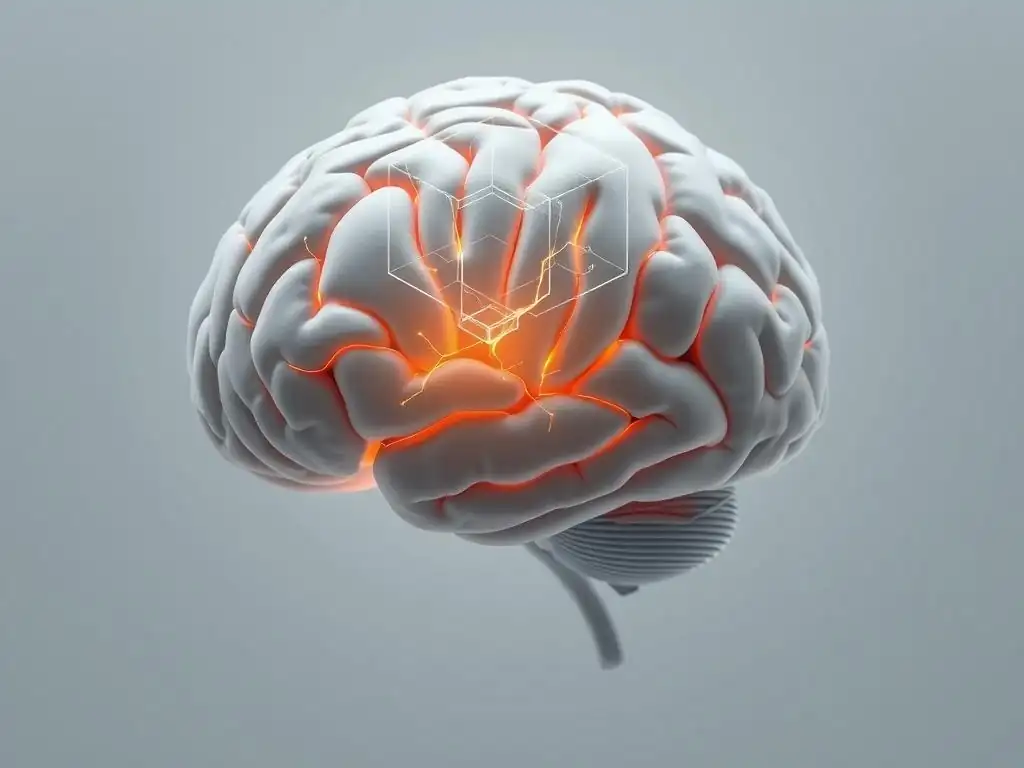
Amygdala Modulation: fMRI Evidence of Reduced Fear Response After Therapy
The amygdala is critically involved in detecting threats and initiating fear responses. In individuals with trauma, the amygdala often becomes hyperactive, reacting strongly even to non-threatening stimuli that are vaguely associated with the trauma. fMRI studies consistently show that effective trauma therapy is associated with a significant modulation of amygdala activity. For example, a meta-analysis summarizing findings from several fMRI studies on PTSD treatment reported an average reduction in amygdala reactivity to trauma cues by approximately 20-30% following successful therapy.
This reduction in amygdala activity is considered a key neural correlate of reduced fear and anxiety symptoms experienced by trauma survivors. It suggests that therapy helps the brain learn to reappraise potential threats and differentiate between past danger and present safety. The consistent fMRI evidence of amygdala modulation provides strong support for the biological impact of therapy on the brain's core fear circuitry, underscoring the potential for genuine healing in fMRI posttrauma therapy recovery.
Hippocampal Changes: fMRI Studies on Memory Processing and Trauma Integration
The hippocampus is vital for forming new memories, recalling past events, and distinguishing between past and present. Trauma can disrupt hippocampal function and, in some cases, lead to reduced volume. This can manifest as difficulty forming coherent narratives about the trauma, problems with contextualizing trauma memories (e.g., feeling like the trauma is happening now), and impaired general memory.
fMRI studies examining hippocampal activity post-trauma therapy have shown varied but promising results. Some research indicates that therapy can be associated with changes in hippocampal activation patterns during memory tasks, potentially reflecting an improved ability to process and integrate traumatic memories into a coherent life narrative. While volumetric changes might be slower or less consistently observed in the short term, functional changes in how the hippocampus interacts with other brain regions during memory recall and emotional regulation are often noted, contributing to the understanding of fMRI posttrauma therapy recovery.
Prefrontal Cortex Activation: fMRI Indicators of Improved Emotional Regulation
The prefrontal cortex (PFC), particularly the medial and ventral regions, plays a crucial role in executive functions, decision-making, and, importantly, regulating emotions by inhibiting responses from areas like the amygdala. Trauma often impairs the PFC's ability to perform this regulatory function effectively.
fMRI studies frequently demonstrate increased activation in specific areas of the PFC, such as the vmPFC and dorsolateral prefrontal cortex (dlPFC), following successful trauma therapy. For instance, studies might show greater vmPFC activity when participants are asked to reappraise negative images or memories after therapy compared to before. This increased activation is interpreted as the brain's enhanced capacity for top-down control over emotional responses, allowing individuals to better manage distress triggered by trauma reminders. This strengthened prefrontal control is a significant indicator observed through fMRI posttrauma therapy recovery.
Comparing fMRI Results Across Different Trauma Therapy Approaches
fMRI Analysis of CBT vs. Eye Movement Desensitization and Reprocessing (EMDR) in Promoting Brain Recovery
Comparing the neural effects of different therapy modalities like CBT and EMDR using fMRI is an active area of research. While both approaches are effective for PTSD, fMRI studies sometimes reveal potentially distinct or overlapping neural pathways of change. For example, some research suggests that trauma-focused CBT might lead to more pronounced changes in prefrontal cortex control over the amygdala, consistent with its emphasis on cognitive restructuring and emotional regulation skills.
On the other hand, studies exploring EMDR's effects using fMRI have sometimes pointed towards changes in the connectivity between the amygdala and other brain regions, potentially reflecting a different mechanism of processing and integrating traumatic memories. While findings can vary between studies, the use of fMRI allows researchers to investigate whether different therapies achieve similar clinical outcomes through potentially different neural routes, enriching our understanding of fMRI posttrauma therapy recovery.
Identifying Common Neural Correlates of Effective Trauma Therapies Using fMRI
Despite potential differences in the specific pathways emphasized, fMRI research across various effective trauma therapies (including CBT, EMDR, and others) often identifies common neural correlates of clinical improvement. The most consistently reported findings involve the modulation of the fear circuit, including reduced amygdala reactivity and increased activity or connectivity within the prefrontal cortex, particularly the vmPFC.
These shared fMRI findings suggest that regardless of the specific techniques used, successful trauma therapy tends to facilitate a fundamental shift in how the brain processes threat and regulates emotions. This convergence in neural outcomes provides a biological basis for the effectiveness of various evidence-based therapies and helps consolidate our understanding of what constitutes brain-based healing in fMRI posttrauma therapy recovery.
Limitations of fMRI Studies in Trauma Recovery Research
Challenges in Interpreting fMRI Data and Drawing Causal Inferences
While fMRI is a powerful tool, interpreting the data in the context of trauma recovery presents challenges. fMRI measures correlations between brain activity and behavior or psychological states; it doesn't directly prove that brain changes are caused by therapy. It's possible that other factors contribute to both clinical improvement and observed fMRI changes. Furthermore, the BOLD signal is an indirect measure of neural activity, and complex physiological processes can influence it.
Interpreting findings requires careful consideration of study design and potential confounds. For instance, simply seeing reduced amygdala activity post-therapy doesn't automatically mean the therapy caused the change; it could be influenced by symptom reduction itself, which is also occurring. Researchers use sophisticated analytical techniques and rigorous experimental designs to try and isolate the effects of therapy, but definitively establishing causality remains complex in fMRI posttrauma therapy recovery research.
Sample Size and Study Design Considerations in fMRI Trauma Research
fMRI studies are often costly and time-consuming, which can lead to relatively small sample sizes compared to behavioral studies. Small sample sizes can limit the generalizability of findings and increase the risk of both false positives and false negatives. Variability among individuals, including differences in trauma history, symptom profiles, and individual responses to therapy, can also make it challenging to detect statistically significant brain changes across a group.
Study design choices, such as whether to use a control group (e.g., waitlist control, non-trauma control), the specific tasks participants perform during scanning, and the timing of scans relative to therapy, all significantly impact the results and their interpretation. Replicating findings across different research groups and larger samples is crucial for building robust evidence about the neural mechanisms of fMRI posttrauma therapy recovery.
The Future of fMRI in Guiding Trauma Therapy and Monitoring Progress
Using fMRI as a Biomarker to Predict Therapy Response
A significant area of future research involves exploring whether fMRI patterns before therapy could serve as biomarkers to predict who is most likely to benefit from a particular treatment approach. For example, could specific patterns of connectivity between the amygdala and PFC predict a better response to CBT versus EMDR? Preliminary research in this area is ongoing, with some studies attempting to identify baseline neural markers associated with successful treatment outcomes.
If successful, this could revolutionize personalized medicine in mental health, allowing clinicians to potentially tailor treatment recommendations based on an individual's unique brain profile as revealed by fMRI. While this is still a nascent field, the potential for fMRI to help match patients to the most effective therapy, thereby optimizing fMRI posttrauma therapy recovery, is a powerful motivator for continued research.
Personalized Trauma Therapy: Tailoring Treatment Based on fMRI Brain Scans
Beyond predicting overall response, fMRI could potentially inform the tailoring of therapy itself. If fMRI scans reveal that a specific individual has particularly strong hyperconnectivity within their fear network, therapy might focus more intensely on techniques aimed at modulating that specific circuit. Conversely, if scans suggest challenges primarily in prefrontal control, interventions targeting executive functions and regulation might be prioritized.
This vision of personalized, brain-informed therapy is still some way off from routine clinical application. It requires much more research to understand the clinical significance of specific fMRI patterns and develop therapeutic interventions directly targeting these neural profiles. However, the potential for fMRI to contribute to more effective and personalized approaches to fMRI posttrauma therapy recovery represents an exciting frontier.
Implications for Understanding Brain-Based Trauma Recovery
Empowering Individuals with Knowledge of fMRI and Trauma Therapy
Understanding that trauma has a tangible impact on the brain and that therapy can be associated with observable brain changes can be incredibly empowering for individuals on their healing journey. Knowing that symptoms like heightened fear responses or difficulty regulating emotions have a basis in altered brain function, which can potentially be modulated through therapy, can reduce self-blame and instill hope.
Sharing the findings from fMRI research on fMRI posttrauma therapy recovery in an accessible way helps demystify the therapeutic process. It provides concrete, scientific evidence that healing is possible and that engaging in therapy is not just about talking through problems, but potentially facilitating real, positive changes in brain function. This knowledge can motivate individuals to seek and commit to treatment.
Resources for Finding Trauma-Informed Therapists and fMRI Research
For those interested in pursuing trauma therapy informed by the latest understanding of brain science, seeking out therapists with specific training in trauma-focused modalities like Trauma-Focused CBT, EMDR, or Somatic Experiencing is recommended. Professional organizations often provide directories of certified or trained therapists.
- Check professional psychology or social work association websites for directories.
- Look for therapists specializing in PTSD or trauma.
- Inquire about specific training in evidence-based trauma therapies.
Staying informed about fMRI research on trauma recovery often involves following publications from major universities or research institutions studying the neuroscience of mental health. While direct access to complex research papers might be challenging for the general public, summaries and articles from reputable science communicators or mental health organizations can provide updates on the latest findings regarding fMRI posttrauma therapy recovery.
Conclusion: fMRI as a Powerful Tool for Understanding and Optimizing Trauma Recovery
fMRI has emerged as a powerful tool, offering unprecedented insights into the brain's response to trauma and its capacity for healing through therapy. By allowing researchers to visualize changes in neural activity and connectivity, fMRI studies provide objective evidence that trauma therapy is associated with tangible alterations in key brain regions involved in fear processing, memory, and emotional regulation. Findings consistently point towards reduced amygdala reactivity and increased prefrontal cortex control as key neural correlates of symptom improvement, offering a biological basis for fMRI posttrauma therapy recovery.
While challenges in interpretation and study design exist, the ongoing advancements in fMRI technology and research methodologies continue to deepen our understanding of the complex neural processes underlying trauma recovery. The future holds the potential for fMRI to not only enhance our understanding but also to potentially guide personalized treatment approaches, helping to predict therapy response and tailor interventions based on individual brain profiles. Ultimately, fMRI research reinforces the profound impact of therapeutic interventions on the brain, offering hope and validation for trauma survivors on their path to healing.
Frequently Asked Questions
What is an fMRI and how is it used in the context of trauma therapy recovery?
An fMRI, or functional Magnetic Resonance Imaging, is a brain imaging technique that measures brain activity by detecting changes associated with blood flow. In trauma therapy recovery, it's used to visualize and understand how the brain changes during and after treatment. It offers objective data on neural pathways.
How does fMRI research contribute to our understanding of post-trauma therapy recovery?
fMRI research provides tangible evidence of brain restructuring following trauma therapy. It helps identify specific brain regions that are affected by trauma and how they respond to different therapeutic interventions. This leads to more targeted and effective treatments.
What specific brain changes might be observed using fMRI during successful post-trauma therapy recovery?
Successful therapy often shows increased activity in areas related to emotional regulation and cognitive processing, like the prefrontal cortex. Conversely, activity may decrease in regions associated with fear and anxiety, such as the amygdala. Overall, it reveals improved neural connectivity.
Can fMRI be used to predict the effectiveness of different types of trauma therapy for individual patients?
While still developing, fMRI shows promise in predicting treatment response. By analyzing brain activity before therapy, researchers may identify biomarkers that indicate which therapies are most likely to be beneficial for a particular individual, leading to personalized treatment plans.
Are there any limitations or ethical considerations when using fMRI in post-trauma therapy research and clinical practice?
fMRI is expensive and requires specialized equipment and expertise. Ethical considerations include data privacy and informed consent, especially with vulnerable populations. The interpretation of fMRI data also requires caution to avoid overstating the conclusions regarding individual patient outcomes.
Final Reflection: Charting the Brain's Path to Healing
The remarkable capacity of the human brain to heal after trauma is not just a theoretical concept; it's a demonstrable reality, vividly illustrated by the fMRI evidence presented in this article. Understanding the neural mechanisms behind post-trauma recovery offers not just scientific insight, but a powerful message of hope and the potential for profound transformation. This research underscores the tangible impact of effective therapeutic interventions, paving the way for more targeted and personalized treatments.
The journey towards healing from trauma is deeply personal, yet the fMRI data presented here reveals a shared, biological pathway to recovery. Witnessing the brain's reorganization and restoration through neuroimaging provides a compelling visual representation of the inner work involved in overcoming adversity. The brain is plastic, and it can change. This is the fundamental message of neuroplasticity.
This quote encapsulates the core message of our findings, emphasizing the brain's inherent ability to adapt and heal, offering hope to those struggling with the lasting effects of trauma.
Consider the implications of this research for your own life, and the lives of those around you. Are you ready to embrace the potential for healing and growth within yourself and others? This article is not just a collection of data points; it's a testament to the resilience of the human spirit and the power of therapeutic intervention.
Take the time to reflect on the journey to recovery, and remember that the brain's capacity for healing is a powerful resource available to all of us. Let this knowledge inspire you to seek support, to advocate for improved mental health services, and to champion the ongoing research that promises to further illuminate the path to healing.
Unlock Your Brain's Healing Power
Thanks for exploring the fascinating world of brain recovery with us! You've just learned about the incredible potential of the brain to heal after trauma. Now it's time to take the next step.
- Prioritize self-care: Identify one small, manageable self-care activity you can implement daily – whether it's a mindful walk, a relaxing bath, or a few minutes of deep breathing. This is your foundation for healing.
- Share your insights: Talk to a trusted friend or family member about what you learned today. Sharing your knowledge can be empowering for both of you.
- Explore further: Research local trauma-informed therapists or support groups. Discovering resources tailored to your needs is crucial on your healing journey.
What specific self-care practice will you integrate into your routine today to support your brain's remarkable healing capacity?