
Navigating the Brain: A Beginner's Guide to Mental Health Imaging Terminology

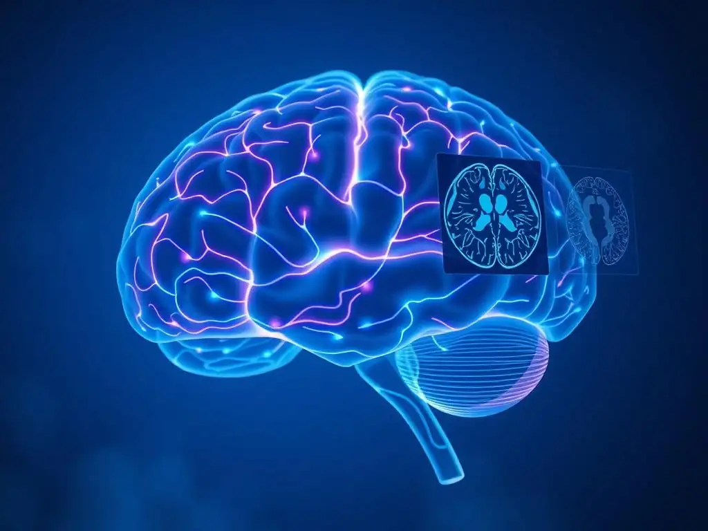
Welcome to Mind Care Guide, a sanctuary for understanding and nurturing your mental well-being.
Ever wondered what those complex brain scans actually mean when discussing mental health? It can feel overwhelming, right? This guide helps demystify it, because understanding is the first step towards empowerment. Dive into our article, " Navigating the Brain: A Beginner's Guide to Mental Health Imaging Terminology," and unlock the basics of mental health imaging terminology. Let's explore the fascinating world of neuroscience together, providing you with the knowledge to better understand your mental health journey. What secrets do those images hold?
- Introduction to Brain Imaging in Mental Health
- Mental Health Imaging Terminology Basics: A Foundation
-
Key Mental Health Imaging Modalities Explained
- MRI (Magnetic Resonance Imaging): Principles and Terminology
- CT (Computed Tomography): Basic Principles and Applications in Mental Health
- PET (Positron Emission Tomography): Exploring Brain Function at a Molecular Level
- EEG (Electroencephalography): Measuring Brain Electrical Activity
- MEG (Magnetoencephalography): A Non-Invasive Technique for Brain Activity Mapping
- Common Brain Regions and Structures Relevant to Mental Health
- Understanding Technical Terms in Mental Health Imaging
- Interpreting Brain Imaging Results: What to Look For
- Ethical Considerations in Mental Health Imaging
- Future Directions in Mental Health Imaging
- Resources for Further Learning about Brain Imaging
- Conclusion: Empowering Understanding of Brain Imaging in Mental Health
-
Frequently Asked Questions
- What is mental health imaging and why is it used?
- What are some common types of brain imaging used in mental health?
- What does 'functional imaging' specifically reveal about mental health?
- What are some limitations of using brain imaging for mental health diagnosis?
- How can understanding mental health imaging terminology benefit someone seeking mental healthcare?
- Final Reflection: Charting the Unseen Landscape of the Mind
- Unlock Your Brain's Potential: Dive Deeper into Mental Health Imaging!
Introduction to Brain Imaging in Mental Health
Brain imaging techniques have revolutionized our understanding of the complex relationship between the brain and mental health. These powerful tools allow researchers and clinicians to peer inside the living brain, observing its structure, function, and connectivity in unprecedented detail. From understanding the neural basis of emotions to identifying potential biomarkers for conditions like depression or schizophrenia, brain imaging plays a crucial role in advancing the field. For anyone seeking a comprehensive guide to mental well-being, grasping the fundamentals of mental health imaging terms is an empowering step towards understanding current research and potential future directions in treatment and diagnosis.
Why Understanding Mental Health Imaging Terminology Matters
Understanding mental health imaging terminology basics is essential for several reasons, particularly in an age where neuroscientific findings are increasingly discussed in public forums. As brain imaging research continues to uncover insights into mental health conditions, knowing the language helps individuals critically evaluate information and engage more deeply with the topic. It demystifies complex scientific concepts, making studies and findings more accessible, whether encountered in academic papers, news reports, or discussions with healthcare professionals.
Furthermore, a basic grasp of mental health brain scanning language empowers individuals to better understand their own health journey. While imaging isn't typically used for routine diagnosis in most mental health conditions, it's a vital research tool. Understanding the terms used in studies can provide context to discussions about the biological underpinnings of various challenges, fostering a more informed perspective on mental well-being and the potential for new interventions. It moves beyond simply hearing about "brain scans" to understanding what specific techniques reveal and their relevance.
The Role of Brain Imaging in Mental Health Research and Diagnosis
Brain imaging serves primarily as a research tool in mental health, providing crucial insights into the neural correlates of various conditions. Techniques like functional magnetic resonance imaging (fMRI) and positron emission tomography (PET) allow scientists to investigate which brain regions are active during specific tasks or in different emotional states, or to examine the distribution of neurotransmitter receptors.
This research helps build models of how conditions like anxiety disorders, bipolar disorder, or obsessive-compulsive disorder might affect brain function or structure. For example, studies using fMRI have consistently shown altered activity patterns in the amygdala and prefrontal cortex in individuals with major depressive disorder compared to control groups.
While brain imaging is not typically the sole basis for diagnosing most mental health conditions, which rely heavily on clinical interviews and behavioral assessments, it can occasionally play a supporting role in diagnosis or differential diagnosis. For instance, structural imaging like magnetic resonance imaging (MRI) or computed tomography (CT) might be used to rule out other potential causes of symptoms, such as brain tumors or stroke, which could present with psychiatric manifestations.
In some specific cases, imaging findings might lend support to a clinical diagnosis, though this is more common in neurological conditions that have psychiatric symptoms. The ongoing goal is to identify specific imaging markers that could eventually aid in more precise diagnosis or prediction of treatment response, though this is still largely an area of active research rather than routine clinical practice.
Mental Health Imaging Terminology Basics: A Foundation
Embarking on the journey to understand mental health imaging terminology basics requires familiarizing oneself with some foundational concepts that cut across different scanning techniques. These core ideas help explain what the various machines are measuring and how the resulting images or data are interpreted. Grasping these fundamentals is key to making sense of the more specific terms associated with each imaging modality and the findings reported in neuroimaging studies related to mental health.
Core Concepts in Brain Imaging Techniques
A fundamental concept in brain imaging is the distinction between structural and functional imaging. Structural imaging techniques, such as standard MRI or CT, provide detailed pictures of the brain's physical anatomy, showing its size, shape, and the presence of lesions or abnormalities. They offer high spatial resolution, meaning they can distinguish fine details of brain tissue. Functional imaging techniques, conversely, measure brain activity.
Examples include fMRI, PET, and EEG. These techniques capture changes related to neural activity over time, offering insights into how different brain regions work together or respond to stimuli. They often prioritize temporal resolution (how quickly changes can be detected) over spatial resolution compared to structural methods.
Another essential concept is resolution, which applies to both spatial and temporal dimensions. Spatial resolution refers to the smallest feature size that an imaging system can distinguish; higher spatial resolution means sharper, more detailed images. Temporal resolution refers to the smallest time interval over which changes in brain activity can be detected; higher temporal resolution means capturing faster brain events.
Different techniques excel in different types of resolution. For instance, fMRI has relatively good spatial resolution (down to a few millimeters) but limited temporal resolution (seconds), whereas EEG has excellent temporal resolution (milliseconds) but poor spatial resolution. Understanding these trade-offs is part of grasping the fundamentals of mental health imaging terms.
Essential Terminology for Interpreting Brain Scans
Interpreting brain scans requires understanding basic anatomical directions and image properties. Spatial orientation is crucial, with terms like axial (slice viewed from top or bottom), coronal (slice viewed from front or back), and sagittal (slice viewed from side) describing the plane of the image. Intensity refers to the brightness or darkness of different areas in the image, which corresponds to different tissue types or levels of activity depending on the imaging modality. For example, on certain types of MRI scans, fluid appears bright while bone appears dark.
Other essential mental health imaging terminology basics include concepts like signal-to-noise ratio (SNR), which indicates the strength of the desired signal (from brain tissue or activity) relative to random background noise. A higher SNR results in clearer images and more reliable data. Artifacts are distortions or unwanted features in the image that are not part of the actual brain anatomy or activity, often caused by patient movement, metal objects, or scanner imperfections. Identifying and understanding common artifacts is important for accurate interpretation and analysis of brain imaging data.
Key Mental Health Imaging Modalities Explained
Exploring the landscape of mental health imaging involves understanding the various techniques used to visualize the brain. Each modality operates on different physical principles and provides unique types of information about brain structure or function. Learning the specific mental health imaging terminology basics associated with each technique is crucial for understanding their applications and limitations in research and potential clinical contexts.
MRI (Magnetic Resonance Imaging): Principles and Terminology
Magnetic Resonance Imaging (MRI) is one of the most widely used imaging techniques in neuroscience and mental health research due to its excellent spatial resolution and ability to create detailed images of soft tissues without using ionizing radiation. The technique works by placing the patient in a strong magnetic field, which aligns the protons in the water molecules of the body. Radiofrequency pulses are then applied, knocking these protons out of alignment.
As the protons realign with the magnetic field, they emit radio signals that are detected by the scanner. The rate at which the protons realign (relaxation times) varies depending on the surrounding tissue properties, allowing the scanner to build detailed images.
Key mental health imaging terminology basics for MRI include terms related to its sequences and the properties they highlight. Different pulse sequences manipulate the radiofrequency pulses and timing to emphasize different tissue characteristics, resulting in various types of images.
The strength of the magnetic field is also significant, measured in Teslas (T); higher field strengths (e.g., 3T or 7T) generally provide higher resolution and signal-to-noise ratio, enabling more detailed studies. Gradient coils are used to create spatial variations in the magnetic field, allowing the location of the emitted signals to be precisely determined, which is essential for reconstructing the 3D image of the brain.
Understanding T1-weighted and T2-weighted Images
When discussing MRI, two fundamental types of images are T1-weighted and T2-weighted scans, named after the relaxation times they emphasize. In T1-weighted images, tissues with short T1 relaxation times appear bright, while those with long T1 times appear dark. This sequence is excellent for visualizing anatomical details. Gray matter typically appears gray, white matter appears white (due to its shorter T1), and cerebrospinal fluid (CSF) appears dark. T1-weighted images are commonly used for structural analysis, such as measuring the volume of different brain regions using techniques like Voxel-Based Morphometry (VBM).
T2-weighted images, conversely, emphasize tissues with long T2 relaxation times. In these images, fluids like CSF appear bright, while gray matter is typically a lighter gray and white matter a darker gray. T2-weighted images are particularly sensitive to detecting pathology such as inflammation, edema, or lesions, which often have increased water content and thus appear bright.
A variation called FLAIR (Fluid Attenuated Inversion Recovery) is a type of T2-weighted image where the signal from CSF is suppressed, making lesions near the ventricles or surface of the brain more conspicuous. Understanding the difference between T1 and T2 contrast is a vital part of mental health imaging terminology basics for anyone looking at MRI scans.
Functional MRI (fMRI) and Neural Activity Mapping
Functional MRI (fMRI) is a specialized type of MRI that measures brain activity by detecting changes in blood flow. It relies on the principle that when a brain region is active, it consumes more oxygen, leading to an increase in blood flow to that area. This increased blood flow delivers more oxygenated hemoglobin than is consumed. Oxygenated and deoxygenated hemoglobin have different magnetic properties. fMRI detects the ratio of oxygenated to deoxygenated hemoglobin, known as the Blood-Oxygen-Level Dependent (BOLD) signal. An increase in the BOLD signal is interpreted as an increase in neural activity in that region.
fMRI allows researchers to map brain activity during specific tasks (task-based fMRI) or while the participant is resting quietly (resting-state fMRI). Task-based fMRI might involve showing participants emotional stimuli and observing which brain areas become more active. Resting-state fMRI examines spontaneous fluctuations in the BOLD signal to understand how different brain regions are functionally connected, forming neural networks.
For example, studies using fMRI have identified altered connectivity within the default mode network (active during rest) in conditions like depression and anxiety. Understanding the BOLD signal and the concepts of task-based vs. resting-state fMRI are key components of functional mental health imaging terminology basics.
CT (Computed Tomography): Basic Principles and Applications in Mental Health
Computed Tomography (CT), also known as CAT scanning, uses X-rays to create cross-sectional images of the body. The CT scanner rotates an X-ray beam around the patient, and detectors on the opposite side measure the amount of X-rays that pass through the tissue. Denser tissues, like bone, absorb more X-rays and appear bright white on the image, while less dense tissues, like brain matter, appear in shades of gray, and fluid or air appears dark. A computer then processes these measurements to reconstruct detailed 2D slices and potentially 3D volumes of the scanned area.
In the context of mental health, CT scans are less frequently used for primary research into the neural basis of conditions compared to MRI or PET, but they have important applications, particularly in clinical settings. CT is fast, widely available, and relatively inexpensive.
Its primary use in patients presenting with psychiatric symptoms is often to rule out acute structural issues that could be causing or contributing to those symptoms. For instance, a CT scan might be performed in an emergency setting to check for stroke, hemorrhage, tumors, or significant head trauma that could manifest with cognitive or behavioral changes mimicking psychiatric disorders.
Limitations of CT Scans in Mental Health Research
Despite its advantages in speed and accessibility, CT has significant limitations as a primary tool for mental health research. A major limitation is its relatively poor contrast resolution for soft tissues compared to MRI. This means that subtle differences between gray matter, white matter, and different types of brain lesions are harder to distinguish on a CT scan. Many of the structural changes associated with mental health conditions, such as subtle volume differences in specific brain regions, are not clearly visible or quantifiable with CT.
Furthermore, CT uses ionizing radiation, which carries a small risk, especially with repeated scans. While the dose from a single head CT is generally considered low, it is a factor that makes it less suitable for research studies involving repeated scanning of healthy volunteers or patient populations, particularly children or young adults.
Unlike fMRI or PET, standard CT provides only structural information and cannot directly measure brain activity or molecular processes relevant to mental health conditions, limiting its utility for investigating the functional or neurochemical underpinnings of these disorders. This makes understanding the distinction between structural and functional techniques a key part of mental health imaging terminology basics.
PET (Positron Emission Tomography): Exploring Brain Function at a Molecular Level
Positron Emission Tomography (PET) is a powerful neuroimaging technique that provides insights into brain function and molecular processes by detecting gamma rays emitted from a radioactive tracer introduced into the body. A small amount of a radiotracer (also called a radioligand) is injected, which binds to specific molecules in the brain, such as neurotransmitter receptors, metabolic markers (like glucose), or amyloid plaques.
As the radioactive isotope in the tracer decays, it emits positrons, which quickly annihilate with electrons, producing two gamma rays that travel in opposite directions. Detectors surrounding the patient capture these gamma rays, and a computer reconstructs an image showing the distribution and concentration of the tracer in the brain.
PET is invaluable for studying the neurochemical basis of mental health conditions. For example, using radiotracers that bind to dopamine or serotonin receptors allows researchers to investigate potential abnormalities in these neurotransmitter systems, which are implicated in conditions like schizophrenia, depression, and addiction. PET scans can also measure regional cerebral glucose metabolism (using a tracer like FDG, a glucose analog), which is an indicator of brain activity. Areas with higher metabolic rates appear brighter on the scan. Understanding radiotracers and what they target is a crucial aspect of mental health imaging terminology basics related to PET.
Radioligands and Their Role in PET Imaging
Radioligands are the cornerstone of PET imaging, acting as molecular probes that selectively bind to specific targets within the brain. These targets can be neurotransmitter receptors (like dopamine D2 receptors or serotonin transporters), enzymes, or other proteins involved in neural function. The radioligand consists of a molecule that has an affinity for the target and a radioactive isotope (such as Carbon-11 or Fluorine-18) attached to it. The choice of radioligand depends entirely on the specific biological process or molecule being investigated.
The role of radioligands is to make the target visible to the PET scanner. Once injected, the radioligand travels through the bloodstream and distributes throughout the brain, binding to its specific target molecules. The amount of binding in a particular brain region reflects the density or availability of the target.
For instance, a radioligand that binds to dopamine D2 receptors can be used to measure the density of these receptors in areas like the striatum. Changes in receptor density or function, as revealed by the PET signal from the radioligand, can be associated with various psychiatric conditions. For example, studies have used PET with specific radioligands to show altered dopamine receptor availability in individuals with schizophrenia compared to healthy controls.
PET Scans and Neurotransmitter Activity
PET scans are uniquely suited among imaging modalities for directly assessing aspects of neurotransmitter systems in the living brain. By using radioligands that mimic neurotransmitters or bind to their receptors or transporters, researchers can quantify the distribution and availability of these key components of brain signaling. This provides a window into the neurochemical imbalances or alterations that are hypothesized to underlie many mental health disorders.
For example, PET studies have investigated serotonin transporter availability in individuals with depression, showing reduced availability in certain brain regions. Similarly, studies using PET with tracers that bind to dopamine receptors have explored the role of the dopaminergic system in addiction and psychotic disorders like schizophrenia, sometimes showing altered receptor binding potential in areas like the striatum.
While interpreting these findings is complex and requires considering factors like the specific tracer used and individual variability, PET provides invaluable data on the molecular level that complements the structural and functional information obtained from other imaging techniques, contributing significantly to our understanding of the biological basis of mental health challenges and the mental health imaging terminology basics surrounding neurochemistry.
EEG (Electroencephalography): Measuring Brain Electrical Activity
Electroencephalography (EEG) is a non-invasive technique that measures the electrical activity generated by neurons in the brain. It involves placing electrodes on the scalp, which detect the tiny electrical potentials produced by large populations of neurons firing together. These electrical signals are amplified and recorded over time, resulting in a trace of brain waves.
EEG has very high temporal resolution, meaning it can capture brain activity changes occurring within milliseconds, making it excellent for studying the timing of neural events. However, its spatial resolution is relatively poor because the electrodes on the scalp are far from the source of the electrical activity deep within the brain, and the skull and tissue distort the signals.
In mental health research and clinical practice, EEG is used for various purposes. Clinically, it is essential for diagnosing seizure disorders, which can sometimes present with psychiatric symptoms. In research, EEG is used to study sleep patterns (often disrupted in mental health conditions), monitor states of consciousness, and investigate how the brain responds to stimuli or performs cognitive tasks. The characteristic patterns of electrical activity, known as brain rhythms or oscillations, provide insights into different brain states and processes. Understanding these rhythms is a key part of mental health imaging terminology basics for EEG.
EEG Rhythms and Their Significance
EEG recordings are characterized by different frequency bands, or rhythms, which are associated with different brain states and cognitive functions. These rhythms are typically categorized by their frequency range, measured in Hertz (Hz). Common rhythms include:
- Delta waves (0.5-4 Hz): Slow, high-amplitude waves associated with deep sleep and certain brain disorders.
- Theta waves (4-8 Hz): Associated with drowsiness, early stages of sleep, meditation, and sometimes linked to memory processing. Increased theta activity has been observed in some studies of attention deficit hyperactivity disorder (ADHD).
- Alpha waves (8-13 Hz): Prominent when a person is awake but relaxed, particularly with eyes closed. Reduced alpha activity can be seen during mental effort.
- Beta waves (13-30 Hz): Associated with active thinking, concentration, and alertness.
- Gamma waves (>30 Hz): Linked to higher-level cognitive functions such as perception, learning, and problem-solving. Alterations in gamma band activity have been reported in conditions like schizophrenia.
Analyzing the power (amplitude) and connectivity of these different rhythms in specific brain regions (as estimated from scalp electrodes) can provide insights into the neural dynamics associated with various mental health conditions. For instance, studies might examine differences in alpha or theta power during resting states or cognitive tasks in groups of individuals with depression or anxiety.
Event-Related Potentials (ERPs) in Mental Health Studies
Event-Related Potentials (ERPs) are a specific application of EEG that involves measuring the brain's electrical response to a particular stimulus or event. When a stimulus (e.g., a sound, image, or word) is presented, it elicits a characteristic pattern of electrical activity in the brain.
By averaging the EEG signals recorded immediately after many repetitions of the same stimulus, the background random brain activity is canceled out, leaving only the consistent response triggered by the event – the ERP. ERPs appear as characteristic positive and negative deflections (peaks and troughs) in the averaged waveform, occurring at specific times after the stimulus.
Different peaks in the ERP waveform are associated with specific cognitive processes and are named according to their polarity (P for positive, N for negative) and their latency (time after the stimulus, in milliseconds) or their order (e.g., P1, N1, P2, N2, P3). For example, the P300 component, a positive peak occurring around 300 milliseconds after an unexpected or significant stimulus, is thought to reflect attention allocation and working memory updating.
Abnormalities in ERP components, such as reduced P300 amplitude or altered latency, have been observed in various mental health conditions, including schizophrenia, depression, and substance use disorders, suggesting alterations in the underlying cognitive processing. Studying ERPs provides another layer of understanding through mental health imaging terminology basics related to the timing of neural events.
MEG (Magnetoencephalography): A Non-Invasive Technique for Brain Activity Mapping
Magnetoencephalography (MEG) is a non-invasive neuroimaging technique that measures the magnetic fields produced by electrical currents generated by neuronal activity in the brain. Just as electrical currents produce magnetic fields, the coordinated firing of large groups of neurons generates weak magnetic signals that can be detected outside the head using highly sensitive sensors called SQUIDs (Superconducting QUantum Interference Devices). Like EEG, MEG measures direct neural activity and offers excellent temporal resolution, capturing brain changes on the millisecond scale.
MEG provides a complementary view to EEG. While both measure neural activity, MEG is less susceptible to distortion from the skull and scalp because magnetic fields pass through tissues more freely than electrical currents. This allows for better spatial localization of the source of the activity within the brain compared to EEG, although still not as precise as fMRI. MEG is particularly useful for studying rapid brain processes involved in perception, cognition, and motor control. Understanding that MEG measures magnetic fields, in contrast to EEG's electrical fields, is a key distinction in mental health imaging terminology basics.
Advantages of MEG over EEG
MEG offers several key advantages over EEG, primarily related to its spatial resolution and source localization capabilities. Because magnetic fields are less distorted by the conductivity differences in the scalp, skull, and meninges compared to electrical fields, MEG signals provide a more accurate representation of the location of the neural activity within the brain. This allows researchers to pinpoint the origin of brain activity with greater precision, typically within a few millimeters, especially for activity originating in the sulci (folds) of the cortex.
While EEG is very sensitive to activity in the gyri (crests) of the cortex and is easier and less expensive to use, MEG is particularly sensitive to activity in the sulci. This complementary sensitivity means that sometimes combining data from both MEG and EEG can provide a more complete picture of brain activity. The superior spatial localization of MEG makes it a valuable tool for mapping brain networks and identifying the sources of abnormal activity, which is increasingly relevant in mental health research seeking to understand the precise brain regions involved in various conditions.
MEG Applications in Studying Mental Disorders
MEG is increasingly being applied in research to study the neural basis of various mental disorders, leveraging its excellent temporal resolution and improved spatial localization compared to EEG. It is particularly useful for examining oscillatory brain activity and connectivity patterns, which are thought to be disrupted in many psychiatric conditions. For example, studies have used MEG to investigate altered gamma oscillations in individuals with schizophrenia, which may be related to impaired sensory processing and cognitive function.
MEG is also used to study the timing and processing of information in conditions like autism spectrum disorder, anxiety disorders, and depression, by examining event-related fields (ERFs), the magnetic equivalent of ERPs. These studies might look at how quickly and strongly the brain responds to social cues or emotional stimuli. While still less common in clinical practice than EEG (primarily due to cost and availability), MEG is a powerful research tool contributing to our understanding of the dynamic brain processes underlying mental health challenges and adding depth to the mental health imaging terminology basics landscape.
Common Brain Regions and Structures Relevant to Mental Health
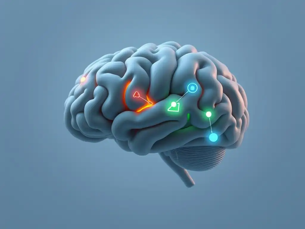
Understanding mental health imaging terminology basics also involves familiarizing oneself with the key brain regions that are frequently discussed in the context of mental health research. Different areas of the brain are specialized for different functions, and disruptions in these areas or the connections between them are often implicated in various psychiatric conditions. Imaging techniques allow us to examine the structure, activity, and connectivity of these specific regions.
The Prefrontal Cortex: Executive Functions and Emotional Regulation
The prefrontal cortex (PFC), located at the very front of the brain, is the executive control center. It is involved in a wide range of higher-level cognitive functions, collectively known as executive functions, including planning, decision-making, working memory, impulse control, and goal-directed behavior. The PFC is also critically involved in the regulation of emotions, helping to modulate responses generated by subcortical areas like the amygdala. It plays a role in evaluating situations, considering consequences, and inhibiting inappropriate emotional or behavioral reactions.
Dysfunction in the prefrontal cortex is strongly implicated in a variety of mental health conditions. For example, structural MRI studies have sometimes reported subtle volume differences in parts of the PFC in groups of individuals with major depression or schizophrenia. Functional imaging studies using fMRI often show altered activity or connectivity in the PFC during tasks requiring executive control or emotional regulation in patients with conditions like ADHD, depression, anxiety disorders, and substance use disorders. For instance, reduced activity in certain areas of the PFC during effortful tasks is a common finding in studies of depression.
The Amygdala: Processing Emotions, Especially Fear
The amygdala is a small, almond-shaped structure located deep within the temporal lobe, part of the limbic system. It is primarily known for its crucial role in processing emotions, particularly fear, and in the formation of emotional memories. The amygdala rapidly evaluates incoming sensory information for potential threat and triggers appropriate physiological and behavioral responses, such as the "fight or flight" response. It also plays a role in processing positive emotions and social cues.
Alterations in the structure or function of the amygdala are frequently observed in anxiety disorders, phobias, and post-traumatic stress disorder (PTSD). Functional MRI studies have often shown hyperactivity of the amygdala in response to fearful or negative stimuli in individuals with anxiety disorders or PTSD compared to healthy controls. Conversely, some research suggests altered amygdala connectivity with the prefrontal cortex in conditions like depression. Understanding the amygdala's role is fundamental to interpreting imaging findings related to emotional processing and fear circuitry, which is part of the broader mental health imaging terminology basics.
The hippocampus is another structure located within the temporal lobe, adjacent to the amygdala, and is also part of the limbic system. Its primary function is the formation of new long-term memories, particularly episodic memories (memories of specific events). It also plays a crucial role in spatial navigation, helping us form mental maps of our environment. The hippocampus is highly interconnected with other brain regions, including the prefrontal cortex and amygdala.
The hippocampus is particularly vulnerable to stress and trauma, and structural changes, such as reduced volume, have been consistently observed on average in groups of individuals with chronic major depression, PTSD, and certain neurodegenerative conditions. Functional imaging studies might examine hippocampal activity during memory tasks or its connectivity with other brain regions.
For instance, altered functional connectivity between the hippocampus and the prefrontal cortex has been reported in studies of depression. Understanding the hippocampus's role in memory and its sensitivity to stress is important when interpreting imaging findings in trauma-related and mood disorders, adding another layer to mental health imaging terminology basics.
The Basal Ganglia: Motor Control and Reward Systems
The basal ganglia are a group of subcortical nuclei located deep within the brain, including the striatum (composed of the caudate nucleus and putamen), globus pallidus, substantia nigra, and subthalamic nucleus. While traditionally known for their role in motor control and the planning and execution of movements, the basal ganglia are also heavily involved in reward processing, motivation, habit formation, and certain aspects of learning and decision-making. They receive extensive input from the cortex and project to the thalamus and brainstem.
Dysfunction of the basal ganglia is implicated in several mental health conditions. Alterations in the dopaminergic pathways that project to the striatum are particularly relevant to conditions like addiction (due to their role in reward) and schizophrenia. For example, PET studies using dopamine radioligands have investigated dopamine receptor availability and release in the striatum in individuals with these conditions, sometimes showing altered dopaminergic function. The basal ganglia are also implicated in obsessive-compulsive disorder (OCD), with some imaging studies suggesting altered activity or connectivity in circuits involving the striatum and prefrontal cortex.
The Thalamus: Sensory Relay Station
The thalamus is a large, central structure located deep within the brain, situated between the cerebral cortex and the midbrain. It serves as a major relay station for most sensory information (except smell) on its way to the cerebral cortex. Sensory signals from the body and sense organs are processed and filtered by the thalamus before being transmitted to the appropriate areas of the cortex for further processing. The thalamus also plays a role in regulating consciousness, sleep, and alertness.
The thalamus is interconnected with many other brain regions, including the cortex, limbic system, and basal ganglia, and plays a role in integrating information. Dysfunction or altered connectivity of the thalamus has been implicated in several mental health conditions, although its role is often viewed in the context of broader network disruptions.
For example, altered thalamic connectivity with cortical regions has been observed in some studies of schizophrenia and depression, potentially affecting sensory processing, attention, and information filtering, which can contribute to symptoms like altered perception or difficulty concentrating. Understanding the thalamus as a central relay point is helpful when considering how information flow might be disrupted in mental health conditions, contributing to the overall mental health imaging terminology basics related to brain networks.
Understanding Technical Terms in Mental Health Imaging
Beyond the names of the imaging techniques and brain regions, several technical terms describe specific analysis methods used to extract meaningful information from the raw imaging data. These methods allow researchers to quantify aspects of brain structure, connectivity, or activity and compare findings across individuals or groups. Familiarizing oneself with these terms is part of developing a solid foundation in mental health imaging terminology basics.
Voxel-Based Morphometry (VBM): Analyzing Brain Structure
Voxel-Based Morphometry (VBM) is a computational neuroimaging technique used to analyze structural MRI scans and identify differences in brain tissue composition (specifically, gray matter, white matter, and cerebrospinal fluid) across groups of subjects. Instead of manually tracing specific brain regions, VBM processes the entire brain image in a voxel-by-voxel manner. A voxel is the 3D equivalent of a pixel, representing a tiny cube of tissue in the brain scan.
The VBM process typically involves several steps: first, all individual brain scans are spatially normalized to a standard anatomical template (like the MNI or Talairach space) to align corresponding brain regions across subjects. Then, the images are segmented into gray matter, white matter, and CSF tissue types.
Finally, the segmented images are smoothed, and statistical tests are performed at each voxel to compare the local concentration or volume of gray matter or white matter between groups (e.g., patients vs. controls) or relate it to behavioral measures. VBM studies have reported average group differences in gray matter volume in various conditions, such as reduced gray matter density in the prefrontal cortex or hippocampus observed on average in groups with depression, though these findings can be variable and are not used for individual diagnosis.
Diffusion Tensor Imaging (DTI): Mapping White Matter Tracts
Diffusion Tensor Imaging (DTI) is a specialized MRI technique that measures the diffusion (movement) of water molecules in the brain. Water molecules undergo random motion called Brownian motion, but their movement is restricted by the presence of structures like cell membranes and the myelin sheaths that surround axons (the long projections of neurons that form white matter tracts). Water diffusion is not random (isotropic) in white matter but is preferentially directed along the orientation of the myelinated nerve fibers (anisotropic).
DTI measures this anisotropy and the principal direction of diffusion in each voxel. By tracking the direction of diffusion from voxel to voxel, DTI allows researchers to reconstruct the pathways of white matter tracts, which are bundles of axons that connect different brain regions. Key DTI terms include fractional anisotropy (FA), a measure of the degree of directional diffusion (higher FA indicates more organized white matter), and mean diffusivity (MD), a measure of the overall magnitude of diffusion.
Alterations in FA or MD along specific white matter tracts have been observed in various mental health conditions, suggesting disruptions in brain connectivity. For example, studies have reported altered FA in tracts connecting the prefrontal cortex and limbic regions in groups of individuals with mood disorders, suggesting potential issues with communication between these areas.
Resting-State fMRI: Exploring Brain Connectivity
Resting-state fMRI is a functional MRI technique that measures spontaneous fluctuations in the BOLD signal while participants are at rest, typically lying quietly in the scanner with their eyes closed or fixated on a cross. Unlike task-based fMRI, which examines activity during a specific behavior, resting-state fMRI explores the intrinsic organization of the brain's functional networks. Even at rest, different brain regions exhibit correlated patterns of activity, suggesting they are functionally connected and form resting-state networks (RSNs).
Analysis of resting-state fMRI data often involves identifying these RSNs, such as the Default Mode Network (DMN), the Salience Network, and the Executive Control Network, which are consistently found across individuals. Researchers then examine the functional connectivity within and between these networks, which refers to the statistical correlation between the BOLD signals of spatially distant brain regions. Altered functional connectivity within or between specific RSNs is a common finding in mental health research.
For example, studies have reported altered connectivity within the DMN and between the DMN and other networks in conditions like depression, anxiety, and schizophrenia, suggesting disrupted intrinsic brain organization. Understanding resting-state networks and functional connectivity is crucial for grasping these mental health imaging terminology basics.
Statistical Parametric Mapping (SPM): Data Analysis in Neuroimaging
Statistical Parametric Mapping (SPM) is not an imaging technique itself but a widely used software package and statistical framework for analyzing neuroimaging data, particularly fMRI, PET, and VBM data. The core idea behind SPM is to create statistical parametric maps that show where in the brain there are statistically significant differences in activity, structure, or connectivity between groups or conditions.
The process typically involves several steps: pre-processing the data (e.g., aligning images, smoothing), applying a statistical model (like the general linear model) to determine how brain activity or structure relates to the experimental design or group differences, and then performing statistical tests (like t-tests or ANOVAs) at each voxel across the brain. The result is a 3D map where each voxel is assigned a statistical value (e.g., a t-statistic or F-statistic).
These maps are then thresholded using statistical criteria (like p-values, often corrected for multiple comparisons) to identify voxels or clusters of voxels that show statistically significant effects. Understanding that SPM is an analysis tool used to generate these statistical maps is key to interpreting the findings from many neuroimaging studies and is part of essential mental health imaging terminology basics.
Interpreting Brain Imaging Results: What to Look For
Interpreting brain imaging results, especially those from research studies, requires more than just looking at colorful pictures of the brain. It involves understanding the statistical methods used, the types of abnormalities being assessed (structural or functional), and how patterns of connectivity might be disrupted. This section delves into some key aspects of interpreting findings in mental health imaging, building on the mental health imaging terminology basics already discussed.
Understanding Statistical Significance and Effect Size
When reviewing neuroimaging studies, two important concepts are statistical significance and effect size. Statistical significance, often indicated by a p-value, tells us the probability of obtaining the observed results (e.g., a difference in brain activity between two groups) if there were truly no difference in the population. A commonly used threshold for significance is p < 0.05, meaning there is less than a 5% chance the result occurred randomly.
However, because brain imaging involves analyzing thousands or millions of voxels simultaneously, a rigorous correction for multiple comparisons is essential to avoid false positives. Techniques like Bonferroni correction or False Discovery Rate (FDR) correction are used to adjust the p-value threshold accordingly.
Effect size, on the other hand, quantifies the magnitude of the difference or relationship observed, independent of sample size. A statistically significant result might have a small effect size, meaning the difference is real but perhaps not very large or clinically meaningful. Conversely, a non-significant result might mask a potentially important effect in a study with a small sample size.
In mental health imaging, effect sizes are crucial because findings are often group averages, and the variability within groups can be large. Understanding both statistical significance (the likelihood the result is real) and effect size (how large the difference is) provides a more complete picture of the study's findings and is part of navigating mental health imaging terminology basics in practice.
Identifying Structural Abnormalities
Structural imaging techniques like MRI and CT are used to identify structural abnormalities in the brain. In a clinical context, this might involve looking for gross abnormalities such as tumors, cysts, signs of stroke, or significant atrophy (shrinkage) that could be contributing to symptoms. Radiologists interpret these scans by examining the shape, size, and intensity of different brain structures. For example, a large tumor might displace surrounding brain tissue, or a stroke might appear as an area of tissue damage.
In mental health research, structural analysis often involves more subtle assessments, frequently using techniques like VBM or surface-based morphometry to quantify differences in brain volume, cortical thickness, or surface area. These studies might identify average group differences, such as slightly reduced hippocampal volume in a group of patients with PTSD compared to controls.
It's important to note that while these group differences contribute to our understanding of the neural correlates of conditions, they are typically subtle and not diagnostic for individuals. An individual's scan showing a slightly smaller hippocampus does not automatically mean they have PTSD; structural findings must be interpreted in the context of clinical symptoms and other information.
Analyzing Functional Connectivity Patterns
Functional imaging techniques, particularly fMRI and increasingly MEG, allow for the analysis of functional connectivity patterns within the brain. As discussed with resting-state fMRI, this involves examining the statistical dependencies (correlations) between the activity of different brain regions over time. Regions that show highly correlated activity are considered to be functionally connected, suggesting they are part of the same network or communicate frequently.
Analysis of functional connectivity involves identifying these networks and quantifying the strength of connections within and between them. Researchers might look for areas of hyperconnectivity (abnormally strong connections) or hypoconnectivity (abnormally weak connections) in specific networks in individuals with mental health conditions. For example, studies have reported altered functional connectivity within the default mode network and between the DMN and areas involved in cognitive control in conditions like depression and anxiety.
These findings suggest disruptions in the way different brain regions communicate and coordinate their activity, which could contribute to symptoms like rumination (excessive focus on negative thoughts) or difficulty regulating emotions. Analyzing these network-level disruptions is a key area of modern mental health imaging research and adds complexity to the mental health imaging terminology basics.
Ethical Considerations in Mental Health Imaging
As brain imaging technology advances and its use in research expands, several important ethical considerations arise, particularly in the sensitive context of mental health. These issues pertain to how data is handled, how participants are informed, and the potential societal impact of imaging findings. Addressing these ethical aspects is as crucial as understanding the mental health imaging terminology basics.
Privacy and Data Security
Brain imaging data is highly sensitive personal information. Scans can potentially reveal not only information relevant to mental health research but also incidental findings (like potential tumors) or information about an individual's unique brain anatomy. Ensuring the privacy and data security of imaging data is paramount. This involves implementing robust measures to protect data from unauthorized access, breaches, or misuse.
Researchers and institutions utilize various strategies, including anonymization or de-identification, where personal identifiers are removed from the imaging data and linked only through a secure, separate key. Data is often stored on secure servers with restricted access.
Sharing imaging data for collaborative research is common and beneficial for advancing the field, but it requires strict protocols to maintain privacy and security, often involving data use agreements that specify how the data can be accessed and analyzed. The increasing size of neuroimaging datasets and the potential for combining them with other types of personal data (like genetic information) underscore the importance of vigilant data protection practices.
Informed Consent and Patient Understanding
Obtaining truly informed consent is a fundamental ethical requirement in any research involving human participants, including mental health imaging studies. Participants must be fully informed about the nature of the study, the procedures involved (including the imaging process itself, potential risks like claustrophobia or discomfort, and the use of contrast agents or radiotracers if applicable), how their data will be used and protected, and their right to withdraw at any time without penalty.
In the context of mental health, ensuring participants fully understand the information can present unique challenges, especially if individuals are experiencing cognitive difficulties or symptoms that might affect their capacity to comprehend complex information. Researchers must take extra care to explain the study in clear, accessible language, answer all questions, and ensure the participant has the capacity to provide voluntary consent.
Participants should also understand what, if anything, will be communicated back to them or their clinician regarding any findings from the scan, particularly incidental findings. This careful process is vital for ethical research practice, complementing the understanding of mental health imaging terminology basics.
Potential for Stigmatization
One significant ethical concern in mental health imaging is the potential for stigmatization. While imaging research aims to understand the biological underpinnings of mental health challenges to reduce blame and promote empathy, findings about brain differences associated with conditions could inadvertently be misinterpreted or misused in ways that increase stigma. For example, oversimplified media reports might portray mental health conditions as purely "brain diseases" caused by "faulty wiring," which could reduce empathy for the individual's experience or lead to deterministic views that overlook the complex interplay of biological, psychological, and social factors.
There is also a concern that identifying potential imaging "biomarkers" could lead to labeling or discrimination based on brain scans, even if those markers are not definitive for diagnosis or predictive of an individual's future. Researchers and communicators have an ethical responsibility to present imaging findings accurately, emphasizing that observed brain differences are often subtle group averages, not individual destinies, and that the brain is dynamic and capable of change. Mindful communication about the limitations and nuances of mental health imaging is essential to mitigate the risk of stigmatization and foster a more compassionate understanding of mental well-being.
Future Directions in Mental Health Imaging
The field of mental health imaging is constantly evolving, driven by technological advancements and the increasing integration of different types of data. Future directions hold exciting possibilities for gaining deeper insights into the brain and potentially transforming how mental health conditions are understood, diagnosed, and treated. Staying abreast of these developments requires an evolving understanding of mental health imaging terminology basics and advanced concepts.
Advancements in Imaging Technology
Technological advancements are continually pushing the boundaries of what brain imaging can reveal. This includes the development of higher field strength MRI scanners (e.g., 7T and beyond), which provide even greater spatial resolution and signal-to-noise ratio, allowing for visualization of finer brain structures and more detailed functional mapping. New MRI sequences are being developed to measure different tissue properties or metabolic markers.
In PET imaging, new radioligands are being developed to target a wider range of neurotransmitter systems, protein aggregates (like tau and alpha-synuclein implicated in some neurodegenerative diseases that can have psychiatric symptoms), and inflammatory processes in the brain. Hybrid scanners, such as simultaneous PET-MRI, allow researchers to acquire both metabolic/molecular information (from PET) and detailed structural and functional information (from MRI) at the same time, providing a more comprehensive picture of brain health and disease.
These technological leaps promise to provide richer, more detailed data, enhancing our understanding of the brain's role in mental health and refining mental health imaging terminology basics.
Integration of Imaging Data with Other Clinical Information
A major future direction is the integration of brain imaging data with other types of information to build a more complete picture of mental health. This includes combining imaging data with genetic information (e.g., linking specific gene variants to brain structure or function), clinical assessments (symptoms, history, response to treatment), cognitive test results, and data from wearable sensors (e.g., sleep patterns, activity levels).
Analyzing these diverse datasets together, often using advanced computational approaches like machine learning and artificial intelligence (AI), holds the potential to identify complex patterns that are not visible when looking at each data type in isolation. For example, researchers are exploring whether combining fMRI connectivity data with genetic profiles can help predict an individual's risk for developing a certain condition or their likelihood of responding to a specific therapy.
This multimodal approach is expected to yield more personalized insights into mental health, requiring an understanding of how imaging data fits within this broader context and familiarity with related mental health imaging terminology basics from other fields.
Personalized Mental Health Treatment Based on Imaging Biomarkers
Perhaps one of the most ambitious future goals is the development of personalized mental health treatment based on imaging biomarkers. Currently, treatment decisions for many mental health conditions are largely based on trial and error, as it is difficult to predict which individual will respond best to a particular medication or therapy. The hope is that brain imaging might identify specific characteristics or patterns in an individual's brain that could serve as biomarkers to guide treatment selection.
For example, research is exploring whether specific patterns of functional connectivity seen on resting-state fMRI could predict response to different types of antidepressants or psychotherapy. Similarly, PET imaging of neurotransmitter systems might help determine if a medication targeting a specific system is likely to be effective for an individual. While this is still an area of active research and not yet routine clinical practice, the potential for imaging to contribute to more precise, personalized mental health care is a significant future direction, underscoring the long-term relevance of understanding mental health imaging terminology basics.
Resources for Further Learning about Brain Imaging
For those interested in delving deeper into the fascinating world of brain imaging and its applications in mental health, numerous resources are available. From online courses to academic texts and professional organizations, these avenues can help solidify your understanding of mental health imaging terminology basics and explore more advanced topics.
Online Courses and Tutorials
Many universities and educational platforms offer online courses and tutorials on neuroscience, neuroimaging techniques, and their applications in mental health. Platforms like Coursera, edX, and칸Academy often host introductory courses that explain the principles behind different imaging modalities (MRI, fMRI, PET, EEG, MEG) and basic data analysis concepts. Some courses are specifically designed for a general audience, while others are geared towards students or professionals in related fields. These resources can provide structured learning pathways and often include videos, quizzes, and supplementary materials to enhance understanding of mental health imaging terminology basics and beyond.
Specialized tutorials focusing on specific software packages used for neuroimaging data analysis (like SPM, FSL, or AFNI) are also available, often provided by the software developers themselves or research labs. While these might be more technical, they offer hands-on experience with how imaging data is processed and analyzed. Searching for "introduction to neuroimaging," "brain imaging basics," or "fMRI for beginners" on these platforms can yield valuable learning opportunities.
Books and Articles on Neuroimaging
A wealth of books and articles provide in-depth information on brain imaging. Introductory textbooks on cognitive neuroscience or neuroimaging often dedicate chapters to explaining the principles of different techniques, basic anatomy, and common analysis methods. These books can serve as comprehensive references for clarifying mental health imaging terminology basics and exploring the underlying physics and biology.
For those interested in the application of imaging to mental health, searching for review articles or book chapters specifically on "neuroimaging of depression," "fMRI in anxiety disorders," or "PET in schizophrenia," for example, can provide summaries of research findings in specific areas. Academic journals specializing in neuroimaging (like NeuroImage, Human Brain Mapping, or Biological Psychiatry: Cognitive Neuroscience and Neuroimaging) publish the latest research, though these articles can be quite technical. Websites of research labs or universities often feature accessible summaries of their work for a broader audience.
Professional Organizations and Societies
Professional organizations and societies dedicated to neuroscience, neuroimaging, and mental health research are excellent resources for staying updated and connecting with the field. Examples include the Society for Neuroscience (SfN), the Organization for Human Brain Mapping (OHBM), the Society of Biological Psychiatry, and the American College of Neuropsychopharmacology (ACNP).
These organizations host conferences, publish journals, and often have educational resources available on their websites. While membership might be required for full access, many societies offer public resources or summaries of recent findings. Following experts and research labs in the field on social media can also provide insights into the latest developments and clarify mental health imaging terminology basics through examples of current research. These resources offer pathways for continued learning and engagement with the dynamic field of mental health imaging.
Conclusion: Empowering Understanding of Brain Imaging in Mental Health
Navigating the world of brain imaging in mental health can seem daunting at first, but by building a foundation in mental health imaging terminology basics, you gain a powerful lens through which to view the fascinating relationship between the brain and our mental well-being. We've explored key techniques like MRI, CT, PET, EEG, and MEG, learned about the brain regions most relevant to mental health, and touched upon the technical terms used in analysis. Understanding these fundamentals empowers you to better interpret research findings, appreciate the complexity of the brain, and follow the exciting advancements in the field.
While brain imaging is primarily a research tool that is not yet used for routine diagnosis of most mental health conditions, its contribution to our understanding is undeniable. It has revealed subtle structural differences, altered functional connectivity patterns, and disruptions in neurotransmitter systems associated with various challenges.
As technology progresses and analysis methods become more sophisticated, imaging holds increasing promise for contributing to more personalized approaches to mental health care. By continuing to learn and engage with these mental health imaging terminology basics, you contribute to a more informed and empathetic perspective on mental health, recognizing the intricate biological factors at play alongside psychological and social influences.
Frequently Asked Questions
What is mental health imaging and why is it used?
Mental health imaging uses various imaging techniques like MRI or PET scans to visualize brain structures and activity, aiding in the diagnosis and understanding of mental health conditions. It helps researchers and clinicians identify potential abnormalities.
What are some common types of brain imaging used in mental health?
Common types include MRI (magnetic resonance imaging) which provides detailed images of the brain, and PET (positron emission tomography) scans which show brain activity based on glucose metabolism. fMRI (functional MRI) is also used to see brain activity changes.
What does 'functional imaging' specifically reveal about mental health?
Functional imaging, like fMRI, highlights areas of the brain that are active during specific tasks or emotional states. This can help understand how different brain regions contribute to mental health disorders and response to treatment.
What are some limitations of using brain imaging for mental health diagnosis?
Brain imaging is not a standalone diagnostic tool. It should be used in conjunction with clinical evaluations and patient history. Imaging results can be influenced by various factors and might not always provide definitive answers.
How can understanding mental health imaging terminology benefit someone seeking mental healthcare?
Familiarity with terms allows individuals to better understand their diagnosis, proposed treatments, and research findings. It empowers them to have informed discussions with their healthcare providers and actively participate in their care.
Final Reflection: Charting the Unseen Landscape of the Mind
Understanding the language of mental health imaging is not just about deciphering acronyms; it's about gaining access to a deeper understanding of the human brain and its intricate relationship with mental well-being. This knowledge empowers us to approach mental health challenges with greater nuance and empathy, fostering a more informed and compassionate approach to treatment and research.
The journey into the world of fMRI, EEG, and other imaging techniques has profoundly impacted my own perspective on mental health. It's revealed the incredible complexity of the brain, highlighting the limitations of simple explanations for complex conditions. This understanding underscores the importance of continued research and development in this field. The brain is the most complex organ in the known universe. Understanding its intricacies is a journey, not a destination.
- (Paraphrased, original source needed for publication). This quote perfectly encapsulates the ongoing exploration and the need for continued learning within the field of mental health imaging.
Now, it's your turn to navigate this newly charted territory. Consider how this newfound knowledge might shift your perspective on mental health, both personally and within your community. Perhaps you'll approach conversations about mental illness with greater sensitivity, or maybe you'll feel inspired to learn more about the cutting-edge research shaping our understanding of the brain. The possibilities are vast, and the journey of understanding the mind is a lifelong endeavor. Embrace the challenge, and let this guide be your first step on the path to greater understanding.
Unlock Your Brain's Potential: Dive Deeper into Mental Health Imaging!
Thanks for taking the time to explore the fascinating world of mental health imaging! We hope this article equipped you with a clearer understanding of the terminology involved. Ready to take the next step?
- Explore further: Delve into our recommended resources for learning more about specific imaging techniques mentioned in the article – some surprising insights await you!
- Share your thoughts: Discuss this article with others; what aspect resonated most with you, and what questions do you still have?
- Start your personalized research: Identify a specific mental health imaging technique that interests you most and begin researching its applications and latest advancements.
What specific area of mental health imaging are you most curious to learn more about now? Let's continue the conversation in the comments!