
The Neuroscience of Trauma and Biological Brain Changes

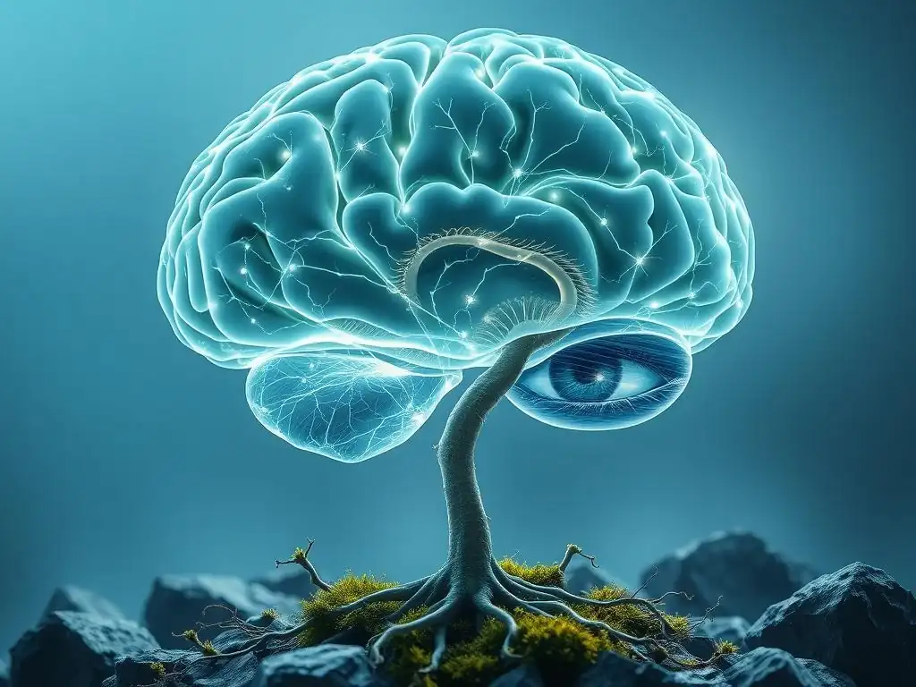
Welcome to Mind Care Guide, a sanctuary for understanding and nurturing your mental well-being.
Ever wondered how trauma can leave its mark, shaping the very structure of your brain? This article delves into the fascinating, and sometimes unsettling, neuroscience of trauma, specifically exploring the Trauma brain changes biological effects. Discover the profound impact of adverse experiences and, more importantly, begin to understand the pathways to healing. Read on to unlock the secrets of resilience and empower yourself with knowledge.
The Neuroscience of Trauma: Biological Changes and Recovery
- Understanding Trauma's Impact on the Brain
- Key Brain Regions Affected by Trauma
- Biological Mechanisms Underlying Trauma-Induced Brain Changes
- The Impact of Childhood Trauma on Brain Development
- Trauma and Neuroinflammation
- Genetic and Environmental Factors
- Diagnosing Trauma-Related Brain Changes
- Therapeutic Interventions Targeting Brain Changes
- Promoting Brain Health and Resilience
- Future Directions in Trauma Research
-
Frequently Asked Questions
- What are some of the biological brain changes that can occur after experiencing trauma?
- How does trauma specifically impact the amygdala's function?
- What role does the hippocampus play in the context of trauma and memory?
- Can trauma brain changes biological effects be reversed or mitigated?
- How can understanding the neuroscience of trauma help individuals struggling with its effects?
- Final Reflection: The Enduring Impact of Trauma on the Brain
- Unlock Your Brain's Resilience: Steps to Healing
Understanding Trauma's Impact on the Brain
Defining Trauma: Beyond Psychological Effects
Trauma is often understood through its psychological manifestations, such as flashbacks, anxiety, and emotional numbness. However, at its core, trauma is an experience that overwhelms an individual's capacity to cope, often involving a threat to life or safety. This overwhelming experience doesn't just reside in the mind; it profoundly impacts the biological architecture and function of the brain.
The definition extends beyond a single event to encompass chronic exposure to adversity, like neglect or abuse, which can have cumulative and equally damaging effects on neural pathways. Understanding trauma requires acknowledging this intricate interplay between psychological distress and underlying trauma brain changes biological effects.
The Diagnostic and Statistical Manual of Mental Disorders (DSM-5) defines a traumatic event as exposure to actual or threatened death, serious injury, or sexual violence. This exposure can be direct, witnessed, learned about happening to a close loved one, or experienced through repeated or extreme indirect exposure (e.g., first responders).
While the DSM focuses on the event type, the biological impact is determined by the individual's response and the subsequent neurobiological alterations. These alterations are not merely transient stress responses but can lead to long-lasting structural and functional changes, fundamentally altering how the brain processes information, regulates emotions, and stores memories.
Trauma and Brain Function: A Neurobiological Overview
Trauma disrupts the brain's intricate network of communication, primarily impacting regions responsible for threat processing, memory formation, and emotional regulation. The brain's primary goal during a traumatic event is survival, which triggers a cascade of biological responses mediated by the sympathetic nervous system and the hypothalamic-pituitary-adrenal (HPA) axis. This acute stress response is adaptive in the short term, preparing the body for "fight or flight." However, chronic or severe trauma can lead to a sustained state of hyperarousal or dissociation, fundamentally altering the baseline activity and connectivity within key neural circuits.
The long-term consequences of trauma on brain function include altered activity in the amygdala (involved in fear), hippocampus (involved in memory), and prefrontal cortex (involved in executive functions and emotional control). Studies using neuroimaging techniques have shown consistent patterns of altered activity and even volume changes in these areas in individuals with trauma-related disorders like PTSD.
These trauma brain changes biological effects can manifest as heightened reactivity to perceived threats, difficulty forming coherent memories of the trauma, impaired emotional regulation, and challenges with decision-making and impulse control. The brain essentially becomes wired to anticipate danger, even when none is present.
Key Brain Regions Affected by Trauma
The Amygdala: Emotional Dysregulation and Threat Detection
The amygdala, a pair of almond-shaped nuclei deep within the temporal lobes, is a central player in processing emotions, particularly fear. It acts as the brain's alarm system, quickly assessing potential threats in the environment. In the face of a perceived danger, the amygdala triggers the stress response cascade. After trauma, the amygdala often becomes hyperactive, leading to an exaggerated fear response and heightened vigilance, even in safe situations. This persistent state of alarm contributes significantly to symptoms like hyperarousal, exaggerated startle response, and irritability commonly seen in trauma survivors.
Research indicates that individuals with PTSD often exhibit increased amygdala activity when exposed to trauma-related stimuli or even general stress. For example, a 2019 meta-analysis combining data from multiple fMRI studies found that veterans with PTSD showed significantly greater amygdala activation compared to healthy controls when viewing fearful faces. This heightened reactivity means that the brain is constantly on high alert, interpreting ambiguous cues as threatening, which underlies the persistent feeling of being in danger, even years after the traumatic event has passed.
Amygdala's Role in Fear Conditioning and Trauma Memories
The amygdala is crucial for fear conditioning, a type of learning where a neutral stimulus becomes associated with a fearful outcome. During trauma, the amygdala links sensory information (sights, sounds, smells) present during the event with the intense fear experienced. This creates powerful, often fragmented, trauma memories that are highly emotionally charged and easily triggered by similar cues in the environment. Unlike normal memories stored in the hippocampus, these amygdala-based memories are often processed as immediate threats, leading to flashbacks and intense emotional reactions as if the trauma is happening again.
The persistence of these fear associations is a hallmark of trauma disorders. For instance, a survivor of a car accident might develop intense fear and anxiety whenever they hear screeching tires, even in a completely safe context. This is because the amygdala has strongly associated the sound of screeching tires with the life-threatening event.
Studies show that the connectivity between the amygdala and other brain regions, such as the sensory cortices, is altered after trauma, reinforcing these conditioned fear responses. This biological wiring explains why triggers can evoke such visceral and overwhelming reactions, highlighting the profound trauma brain changes biological effects on how fear is learned and remembered.
The Hippocampus: Memory Encoding and Contextual Processing
The hippocampus, located next to the amygdala in the temporal lobe, is essential for forming new long-term memories, particularly episodic memories (memories of specific events) and spatial navigation. It helps to contextualize experiences – integrating sensory details, time, place, and emotional information into a cohesive narrative. In the context of trauma, the hippocampus plays a critical role in processing the event and storing it as a past memory, distinguishing it from the present.
However, trauma can significantly impair hippocampal function. High levels of stress hormones, particularly cortisol, released during prolonged stress can damage hippocampal neurons and even reduce the volume of the hippocampus. This damage interferes with the hippocampus's ability to properly process and store the traumatic event as a coherent memory with a clear beginning, middle, and end. Instead, fragments of the trauma – sensory details, intense emotions, physical sensations – may be stored without proper context, leading to intrusive flashbacks and difficulty distinguishing between past and present.
Hippocampal Changes and Memory Deficits After Trauma
Numerous studies using MRI have documented reduced hippocampal volume in individuals with chronic PTSD compared to those without the disorder. While the exact percentage varies across studies, reductions ranging from 5% to 10% have been reported in some populations. This structural change is associated with functional impairments, including difficulty forming new memories, problems recalling specific details of the trauma, and challenges with spatial memory. The impaired contextualization by the hippocampus contributes to the feeling that trauma memories are happening now rather than belonging to the past.
Furthermore, research suggests that the connectivity between the hippocampus and other brain regions, such as the prefrontal cortex, is often disrupted after trauma. This impaired communication hinders the brain's ability to regulate the stress response and integrate emotional information with cognitive processing. The resulting memory deficits and contextual confusion are not simply psychological quirks but are direct consequences of the biological impact of trauma on the hippocampus, illustrating significant trauma brain changes biological effects on memory function.
The Prefrontal Cortex: Executive Function and Emotional Regulation
The prefrontal cortex (PFC), the most forward part of the frontal lobe, is the brain's executive control center. It is responsible for higher-level cognitive functions such as planning, decision-making, working memory, impulse control, and regulating emotions. The PFC receives input from the amygdala and hippocampus and helps to interpret the significance of threats, inhibit inappropriate responses, and modulate emotional reactions based on the current context. It plays a crucial role in bringing the stress response back to baseline after a threat has passed.
Trauma can impair PFC function, particularly the medial PFC, which is involved in evaluating threats and regulating amygdala activity. Reduced activity or volume in certain areas of the PFC is frequently observed in trauma survivors. This impairment weakens the brain's ability to dampen the amygdala's fear response and regulate intense emotions. Consequently, individuals may struggle with emotional lability, impulsivity, difficulty concentrating, impaired decision-making, and challenges in planning for the future – all functions mediated by a healthy PFC.
PFC's Role in Inhibitory Control and Cognitive Appraisal
A key function of the PFC is inhibitory control – the ability to suppress unwanted thoughts, actions, or emotions. After trauma, the impaired connection between the PFC and the amygdala reduces the PFC's capacity to inhibit the amygdala's fear signals. This contributes to the difficulty trauma survivors have in controlling their fear and anxiety responses. For example, someone with PTSD might have trouble suppressing intrusive thoughts or flashbacks, or they might react with intense fear in situations that rationally pose no threat because their PFC is less effective at overriding the amygdala's alarm.
The PFC is also involved in cognitive appraisal – how we interpret and assign meaning to events. Trauma can alter this process, leading individuals to appraise neutral or ambiguous situations as threatening. This negative bias in appraisal is linked to reduced activity in specific PFC subregions. Restoring healthy PFC function is a key goal in trauma treatment, as it is essential for regaining control over emotions, improving decision-making, and developing more adaptive responses to stress. These significant trauma brain changes biological effects on the PFC highlight its central role in cognitive and emotional recovery.
Biological Mechanisms Underlying Trauma-Induced Brain Changes
Neurotransmitter Systems: Serotonin, Dopamine, and Norepinephrine
Neurotransmitters are chemical messengers that transmit signals between neurons, playing a critical role in mood, cognition, and behavior. Trauma can disrupt the balance of several key neurotransmitter systems. Norepinephrine, involved in the body's stress response and alertness, is often found in elevated levels or shows dysregulated activity in trauma survivors, contributing to hyperarousal and exaggerated startle responses. Serotonin, which influences mood, sleep, and anxiety, may show reduced activity or altered receptor sensitivity, potentially contributing to symptoms of depression, anxiety, and emotional dysregulation.
Dopamine, associated with reward, motivation, and pleasure, can also be affected. While the picture is complex, alterations in dopaminergic pathways may contribute to anhedonia (inability to experience pleasure) and difficulties with motivation often seen in trauma survivors. The intricate balance between these neurotransmitters is crucial for maintaining stable mood and adaptive responses to stress. Trauma-induced imbalances are not just symptomatic but are fundamental trauma brain changes biological effects that underpin many of the psychological and behavioral consequences.
Neurotransmitter Imbalances and Their Impact on Mood and Behavior
The dysregulation of neurotransmitter systems directly impacts mood, behavior, and cognitive function. For instance, chronically elevated norepinephrine can lead to persistent feelings of anxiety, irritability, and difficulty sleeping. Altered serotonin levels are strongly linked to symptoms of depression, including low mood, loss of interest, and fatigue. Changes in dopamine signaling can affect motivation, concentration, and the ability to experience joy.
These imbalances create a biological environment in the brain that perpetuates distress and makes it harder to cope with everyday challenges. For example, impaired serotonin function can reduce resilience to stress, making individuals more vulnerable to further anxiety or depressive episodes. Understanding these specific trauma brain changes biological effects at the neurotransmitter level is vital for developing targeted pharmacological interventions aimed at restoring balance and alleviating symptoms.
HPA Axis Dysregulation: The Stress Response System
The hypothalamic-pituitary-adrenal (HPA) axis is the body's central stress response system. When faced with a stressor, the hypothalamus releases corticotropin-releasing hormone (CRH), which signals the pituitary gland to release adrenocorticotropic hormone (ACTH). ACTH then signals the adrenal glands to release cortisol, the primary stress hormone. Cortisol mobilizes energy resources and prepares the body to respond to the threat. Normally, once the threat passes, the HPA axis activity decreases, and cortisol levels return to baseline through a negative feedback loop involving the hippocampus and prefrontal cortex.
However, chronic or severe trauma can disrupt this delicate balance, leading to HPA axis dysregulation. The pattern of dysregulation can vary; some individuals exhibit a hyperactive HPA axis with elevated cortisol levels, while others, particularly those with chronic or early-life trauma, may show blunted cortisol responses or altered diurnal rhythms. This persistent dysregulation means the body struggles to return to a state of calm, maintaining a heightened state of physiological alert or, conversely, becoming chronically fatigued and disengaged due to a blunted stress response.
Chronic Stress, Cortisol, and Brain Function
Chronic activation of the HPA axis and prolonged exposure to high levels of cortisol have detrimental effects on brain structure and function. As mentioned earlier, high cortisol can damage neurons in the hippocampus, impairing memory and contextual processing. It can also affect the prefrontal cortex, hindering executive functions and emotional regulation. Furthermore, chronic cortisol exposure can sensitize the amygdala, making it even more reactive to future stressors.
Studies have linked HPA axis dysregulation to increased risk for various mental health conditions, including PTSD, depression, and anxiety disorders. For example, research shows that individuals with PTSD often have altered cortisol profiles – sometimes lower baseline levels but exaggerated responses to stress, or chronically elevated levels depending on the trauma type and individual. These persistent hormonal imbalances are critical trauma brain changes biological effects that perpetuate vulnerability to stress and make recovery more challenging without targeted interventions.
Neuroplasticity: Adaptive and Maladaptive Brain Changes
Neuroplasticity refers to the brain's remarkable ability to reorganize itself throughout life by forming new neural connections. This capacity allows the brain to adapt to new experiences, learn new skills, and recover from injury. In the context of trauma, neuroplasticity is a double-edged sword. It is the mechanism by which the brain undergoes the biological changes in response to trauma, some of which are maladaptive and contribute to symptoms. However, it is also the mechanism by which the brain can heal and rewire itself in response to therapeutic interventions and positive experiences, enabling recovery.
Maladaptive neuroplasticity after trauma can strengthen pathways associated with fear and threat detection (e.g., between the amygdala and sensory areas) while weakening pathways involved in emotional regulation and cognitive control (e.g., between the PFC and amygdala). This creates a brain state that is hypervigilant and reactive. Adaptive neuroplasticity, conversely, involves strengthening the regulatory pathways, forming new connections that support emotional processing, and helping the brain to integrate traumatic memories into a coherent narrative. Understanding these processes is key to developing effective treatments.
Brain Rewiring After Trauma: Implications for Recovery
The concept of neuroplasticity offers hope for recovery from trauma. Therapeutic interventions like psychotherapy, medication, and even lifestyle changes work, in part, by promoting adaptive neuroplasticity. For example, trauma-focused therapies like Prolonged Exposure or Eye Movement Desensitization and Reprocessing (EMDR) are thought to help the brain reprocess traumatic memories and strengthen connections between the hippocampus and PFC, allowing the trauma to be stored as a past event rather than an ongoing threat.
Research using fMRI has shown that effective trauma treatment can lead to measurable changes in brain activity, such as decreased amygdala reactivity and increased PFC regulation. This "rewiring" of the brain is the biological basis of healing. It demonstrates that the trauma brain changes biological effects are not necessarily permanent. By engaging in therapeutic processes and fostering a supportive environment, individuals can harness the power of neuroplasticity to build resilience and create new, healthier neural pathways.
The Impact of Childhood Trauma on Brain Development

Adverse Childhood Experiences (ACEs) and Long-Term Effects
Adverse Childhood Experiences (ACEs) are potentially traumatic events that occur in childhood (0-17 years), such as experiencing violence, abuse, neglect, or witnessing violence in the home, as well as aspects of the child's environment that can undermine their sense of safety, stability, and bonding, like growing up in a household with substance misuse, mental health problems, or instability due to parental separation or incarceration. Exposure to ACEs, especially chronic exposure or multiple types of ACEs, has profound and lasting effects on brain development and function.
Studies have shown a strong dose-response relationship between the number of ACEs experienced and the risk of developing a wide range of physical and mental health problems later in life, including depression, anxiety, substance use disorders, cardiovascular disease, and chronic pain. The developing brain is particularly vulnerable to the effects of stress hormones and trauma, as critical neural circuits are being formed and myelinated. Early trauma can disrupt the typical developmental trajectories of key brain regions, leading to long-term alterations in stress response systems, emotional regulation, and cognitive abilities.
Sensitive Periods and Vulnerability to Trauma
Brain development occurs in stages, with specific periods being particularly sensitive to environmental influences. Early childhood, especially the first few years, is a critical sensitive period for the development of the HPA axis, amygdala, and hippocampus. Trauma during these periods can have a more pervasive and enduring impact than trauma experienced later in life because it occurs while these systems are still being wired. For example, chronic neglect during infancy can impair the development of healthy attachment circuits and stress response systems.
The prefrontal cortex undergoes significant development during adolescence. Trauma experienced during this period can disrupt the maturation of executive functions and emotional regulation abilities, increasing vulnerability to impulsive behavior and mental health issues. Understanding these sensitive periods helps explain why the timing of trauma exposure is a critical factor in determining the nature and severity of long-term trauma brain changes biological effects. Early intervention and support during these vulnerable stages are therefore crucial for mitigating developmental disruption.
Differences Between Childhood and Adulthood Trauma
While trauma at any age can have significant biological effects on the brain, childhood trauma often results in more pervasive and long-lasting changes due to the brain's ongoing development. In adults, trauma typically impacts a relatively mature brain, often leading to functional alterations and potentially some structural changes (like hippocampal volume reduction). In children, trauma can disrupt the process of development itself, altering how circuits are built and connected from the ground up.
For example, studies show that childhood trauma is more strongly associated with structural changes in the amygdala and hippocampus and more widespread alterations in brain connectivity compared to trauma first experienced in adulthood. The developing brain may also become sensitized to stress, leading to exaggerated physiological responses to stressors throughout life. These differences underscore the unique vulnerability of the developing brain and highlight why childhood trauma can lay the groundwork for a lifetime of challenges related to trauma brain changes biological effects.
Trauma and Neuroinflammation
Inflammatory Cytokines and Brain Function
Beyond the immediate stress response and structural changes, trauma can also trigger and perpetuate neuroinflammation – inflammation within the brain and spinal cord. The immune system and the nervous system are closely linked, and psychological stress, particularly chronic stress and trauma, can activate inflammatory pathways. Trauma can lead to the release of pro-inflammatory cytokines, such as Interleukin-6 (IL-6), Tumor Necrosis Factor-alpha (TNF-alpha), and C-reactive protein (CRP), which can cross the blood-brain barrier or be produced by immune cells within the brain (microglia).
These inflammatory molecules are not just indicators of stress; they can directly impact neuronal function, synaptic plasticity, and neurotransmitter metabolism. Elevated levels of cytokines in the brain can impair communication between neurons, disrupt the balance of neurotransmitters like serotonin and dopamine, and even contribute to neuronal damage. This neuroinflammatory state can contribute to the cognitive deficits, mood disturbances, and fatigue often experienced by trauma survivors.
Chronic Inflammation and Mental Health
Persistent low-grade inflammation, often a consequence of chronic stress and trauma, is increasingly recognized as a contributor to various mental health conditions, including depression, anxiety, and PTSD. Research has found elevated levels of inflammatory markers in the blood and cerebrospinal fluid of individuals with these disorders. This chronic inflammatory state can disrupt the delicate balance required for optimal brain function, affecting neural circuits involved in mood regulation, motivation, and stress processing.
For instance, inflammation can increase the metabolism of tryptophan down the kynurenine pathway, leading to the production of neurotoxic metabolites that can impair neuronal function and contribute to depressive symptoms. Addressing this neuroinflammatory component is becoming an important area of research for developing new therapeutic strategies for trauma-related disorders. The link between trauma and chronic inflammation adds another layer to understanding the complex trauma brain changes biological effects and their widespread impact on physical and mental health.
Genetic and Environmental Factors
Gene-Environment Interactions in Trauma Response
An individual's response to trauma is not solely determined by the event itself but is also shaped by their genetic predisposition and the interplay between their genes and environment. Certain genetic variations can influence an individual's vulnerability or resilience to developing trauma-related disorders. For example, variations in genes related to the HPA axis, neurotransmitter systems (like serotonin transporters), or brain-derived neurotrophic factor (BDNF, important for neuronal growth and survival) have been studied in relation to PTSD risk.
However, genes rarely act in isolation. Their expression and impact are often modified by environmental experiences. This concept of gene-environment interaction suggests that a genetic vulnerability may only manifest as increased risk for a disorder if an individual is exposed to a specific environmental trigger, such as trauma. Conversely, protective genetic factors or supportive environments can buffer the effects of trauma. Understanding these interactions helps explain why not everyone exposed to trauma develops a disorder and highlights the complex biological underpinnings of individual differences in trauma response.
Epigenetic Changes and Trauma
Epigenetics refers to changes in gene expression that do not involve alterations to the underlying DNA sequence but are influenced by environmental factors. These epigenetic modifications, such as DNA methylation or histone modification, can turn genes "on" or "off," affecting protein production and ultimately altering cell function. Trauma, particularly during critical developmental periods, can induce lasting epigenetic changes that influence the function of genes involved in stress response, emotional regulation, and neurodevelopment.
Studies have identified specific epigenetic modifications in genes related to the HPA axis (e.g., the glucocorticoid receptor gene, NR3C1) and neurotransmitter systems in individuals exposed to trauma, particularly early-life trauma. These epigenetic "marks" can be relatively stable and may even be transmitted across generations (though this is a complex and active area of research). These findings demonstrate that trauma can leave a biological imprint on our genome, influencing how our genes are expressed throughout life and contributing to the enduring trauma brain changes biological effects.
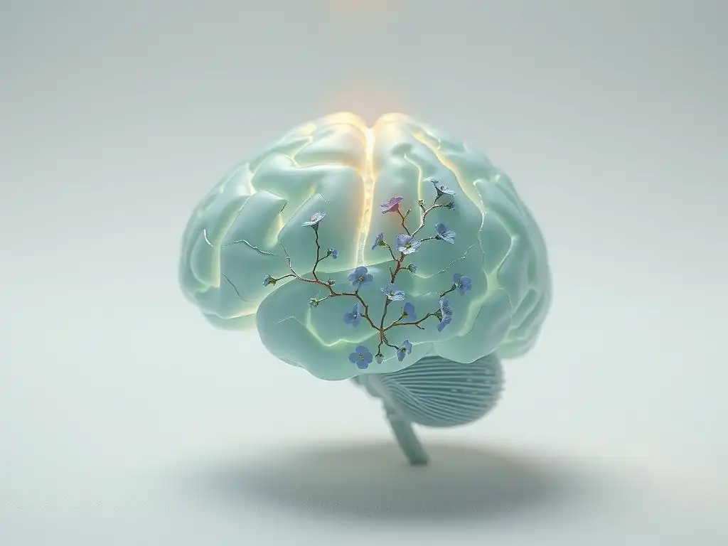
Neuroimaging Techniques: MRI, fMRI, and DTI
Neuroimaging techniques provide powerful tools for visualizing the brain and studying its structure and function in vivo. Structural Magnetic Resonance Imaging (MRI) can detect differences in brain volume and shape, allowing researchers to observe reductions in the hippocampus or changes in the prefrontal cortex associated with trauma. Functional MRI (fMRI) measures brain activity by detecting changes in blood flow, revealing altered patterns of activation in the amygdala, PFC, and other regions when individuals are at rest or performing specific tasks related to emotional processing or memory.
Diffusion Tensor Imaging (DTI) is a type of MRI that measures the movement of water molecules to infer the structure and integrity of white matter tracts – the bundles of nerve fibers that connect different brain regions. DTI studies in trauma survivors have shown alterations in the connectivity between the amygdala, hippocampus, and PFC, providing insights into how communication pathways are disrupted after trauma. While these techniques are primarily used in research settings, they provide crucial evidence for the existence and nature of trauma brain changes biological effects.
Biomarkers of Trauma: Research and Potential Applications
Researchers are actively searching for biological markers (biomarkers) that could potentially aid in diagnosing trauma-related disorders, predicting treatment response, or even identifying individuals at higher risk. These biomarkers could include:
- Neurotransmitter levels: Measuring levels or activity of serotonin, norepinephrine, or dopamine metabolites in blood, urine, or cerebrospinal fluid.
- HPA axis indicators: Measuring cortisol levels in blood, saliva, or hair (for long-term exposure), or assessing the HPA axis response to challenge tests.
- Inflammatory markers: Measuring levels of pro-inflammatory cytokines (IL-6, TNF-alpha) or CRP in the blood.
- Genetic/Epigenetic markers: Identifying specific genetic variations or epigenetic modifications associated with trauma vulnerability or resilience.
- Neuroimaging patterns: Specific patterns of structural or functional changes seen on MRI or fMRI.
While promising, most of these biomarkers are still in the research phase and are not yet used for routine clinical diagnosis. The complexity of trauma responses and the variability across individuals make finding single, definitive biomarkers challenging. However, the ongoing research into these biological indicators holds potential for more personalized approaches to trauma care in the future, further illuminating the biological impact of trauma on the brain.
Therapeutic Interventions Targeting Brain Changes
Pharmacological Approaches: Medications and Their Mechanisms
Medications are often used in the treatment of trauma-related disorders to alleviate specific symptoms, working primarily by modulating neurotransmitter systems or the HPA axis. Selective Serotonin Reuptake Inhibitors (SSRIs) are commonly prescribed and are thought to work by increasing serotonin availability in the brain, which can help improve mood, reduce anxiety, and dampen amygdala reactivity. Other antidepressants, like SNRIs, may also be used.
Certain medications may target norepinephrine system activity, which can help reduce hyperarousal and improve sleep. Prazosin, an alpha-1 adrenergic receptor antagonist, for example, has shown some efficacy in reducing trauma-related nightmares. While medications can provide significant symptom relief for some individuals, they typically do not address the core memory processing or contextualization issues directly impacted by trauma. They primarily manage the downstream biological effects, such as neurotransmitter imbalances.
Medications Used in Trauma Treatment: Benefits and Limitations
Common medications used for trauma-related symptoms include SSRIs (e.g., sertraline, paroxetine, fluoxetine), SNRIs (e.g., venlafaxine, duloxetine), and sometimes alpha-blockers like prazosin. Benzodiazepines may be used short-term for severe anxiety but are generally avoided long-term due to dependence risk and potential interference with memory processing needed for psychotherapy. Antipsychotics or mood stabilizers might be used for co-occurring symptoms like psychosis or severe mood swings.
The benefits of these medications include reducing core symptoms like anxiety, depression, and hyperarousal, which can make it easier for individuals to engage in psychotherapy. However, limitations include variable effectiveness across individuals, potential side effects, and the fact that they often manage symptoms without addressing the underlying trauma processing deficits. They target the biological consequences of trauma, such as neurotransmitter dysregulation, but typically do not directly reverse the structural or fundamental functional trauma brain changes biological effects in the same way that some psychotherapies are believed to.
Neurofeedback and Brain Training
Neurofeedback is a type of biofeedback that measures brain activity (often using electroencephalography or EEG) and provides real-time information to the individual, allowing them to learn to regulate their own brainwave patterns. The goal is to train the brain to operate in healthier patterns associated with states of calm and focus, rather than hyperarousal or dissociation. For example, neurofeedback protocols for trauma might aim to decrease activity in specific brain regions (like the amygdala) or improve connectivity between regions (like the PFC and amygdala).
While more research is needed to establish its efficacy definitively for trauma, preliminary studies and clinical reports suggest that neurofeedback may help reduce symptoms like hyperarousal, anxiety, and difficulty concentrating by teaching the brain to self-regulate. It is hypothesized to work by promoting adaptive neuroplasticity, directly influencing brain activity patterns that were altered by trauma. This approach represents a direct attempt to influence the biological brain changes resulting from trauma.
Psychotherapy's Role in Neural Rewiring
Psychotherapy, particularly trauma-focused therapies, is considered the first-line treatment for trauma-related disorders. Therapies like Trauma-Focused Cognitive Behavioral Therapy (TF-CBT), Prolonged Exposure (PE), and Eye Movement Desensitization and Reprocessing (EMDR) are not just "talk therapy"; they are believed to facilitate significant changes in brain structure and function. These therapies help individuals process the traumatic memory, challenge maladaptive beliefs, and develop new coping strategies.
The mechanisms by which psychotherapy impacts the brain involve promoting adaptive neuroplasticity. For instance, PE helps individuals gradually approach trauma-related memories and situations, which can lead to habituation of the fear response and strengthen the PFC's ability to regulate the amygdala. EMDR is hypothesized to facilitate the reprocessing of traumatic memories, integrating them into the brain's standard memory networks (mediated by the hippocampus and PFC) rather than remaining fragmented and emotionally charged in the amygdala. These therapies aim to reverse or mitigate the maladaptive trauma brain changes biological effects.
Effective Therapy Approaches: Mechanisms of Change
Effective trauma therapies work by engaging the brain's natural healing processes and promoting the formation of new, healthier neural pathways. Key mechanisms of change include:
- Memory Consolidation/Reconsolidation: Helping the brain integrate traumatic memories into autobiographical memory, contextualizing them as past events. This likely involves strengthening hippocampal-PFC connections.
- Fear Extinction: Reducing the conditioned fear response to trauma-related cues. This involves learning that triggers are no longer associated with danger, a process mediated by the PFC inhibiting amygdala activity.
- Cognitive Restructuring: Identifying and challenging distorted thoughts and beliefs about the trauma, oneself, and the world. This engages the PFC and helps to create more balanced appraisals.
- Emotional Regulation: Learning skills to manage intense emotions, which strengthens the regulatory capacity of the PFC.
Studies using fMRI before and after trauma-focused psychotherapy have shown changes consistent with these mechanisms, such as decreased amygdala reactivity, increased PFC activity during emotional tasks, and changes in connectivity between key brain regions. This provides compelling evidence that psychotherapy leads to tangible trauma brain changes biological effects, facilitating recovery at a neurobiological level.
Promoting Brain Health and Resilience
Nutrition and Brain Health
Proper nutrition plays a vital role in supporting brain health and resilience, which can be particularly important for individuals healing from trauma. The brain requires a steady supply of essential nutrients, including omega-3 fatty acids, vitamins (especially B vitamins and vitamin D), minerals (like zinc and magnesium), and antioxidants, to function optimally. Omega-3 fatty acids, found in fatty fish, are crucial components of brain cell membranes and have anti-inflammatory properties, potentially helping to counteract neuroinflammation associated with trauma.
A diet rich in fruits, vegetables, whole grains, and lean proteins provides the necessary building blocks and protective compounds for brain repair and maintenance. Conversely, diets high in processed foods, sugar, and unhealthy fats can contribute to inflammation and oxidative stress, potentially exacerbating the biological impact of trauma. While nutrition alone cannot "cure" trauma, supporting overall brain health through diet can enhance neuroplasticity and resilience, aiding the brain's capacity for recovery from trauma brain changes biological effects.
Exercise and Physical Activity
Regular physical exercise is a powerful tool for promoting brain health and can be particularly beneficial for trauma survivors. Exercise increases blood flow to the brain, promotes the production of neurotrophic factors like BDNF (which supports the growth and survival of neurons), and can stimulate neurogenesis (the birth of new neurons), particularly in the hippocampus. It also helps regulate stress hormones and can improve mood by increasing the release of endorphins and positively impacting neurotransmitter levels.
Aerobic exercise has been shown to reduce anxiety and depressive symptoms, common comorbidities of trauma. Strength training can improve body image and self-efficacy. Engaging in regular physical activity can help individuals feel more in control of their bodies and can provide a healthy outlet for managing stress and hyperarousal. The neurobiological effects of exercise, such as increased BDNF and improved HPA axis regulation, directly counteract some of the negative trauma brain changes biological effects, promoting resilience and recovery.
Mindfulness and Meditation
Mindfulness and meditation practices involve focusing attention on the present moment without judgment. These practices have been shown to have measurable effects on brain structure and function, particularly in regions affected by trauma. Regular meditation has been linked to increased gray matter density in the prefrontal cortex and hippocampus and decreased amygdala volume and reactivity.
By training the ability to observe thoughts and emotions without being overwhelmed by them, mindfulness can help individuals develop greater emotional regulation skills and reduce the intensity of the stress response. It can also improve connectivity between the PFC and amygdala, enhancing the brain's ability to manage fear and anxiety. Incorporating mindfulness into daily life can be a powerful way to promote adaptive neuroplasticity and support the brain's healing process after trauma, addressing the trauma brain changes biological effects through conscious regulation.
Future Directions in Trauma Research
Personalized Medicine Approaches
Future research in the neuroscience of trauma is increasingly focusing on personalized medicine approaches. Given the significant variability in how individuals respond to trauma and treatment, understanding the specific biological profile of each person – including their genetic background, epigenetic marks, hormonal status, and unique pattern of brain changes – could lead to more tailored interventions. This involves identifying biomarkers that can predict who is most likely to develop PTSD after trauma, who will respond best to a particular therapy (e.g., medication vs. psychotherapy), or who is at risk for relapse.
For example, genetic testing might identify individuals with variations in stress-related genes who could benefit from early, targeted preventative strategies or specific pharmacological interventions. Neuroimaging patterns could potentially guide treatment decisions, suggesting that someone with pronounced amygdala hyperreactivity might benefit more from therapies specifically designed to dampen fear responses. Personalized medicine holds the promise of moving beyond a one-size-fits-all approach to trauma care, optimizing treatment based on an individual's unique trauma brain changes biological effects.
Novel Therapeutic Targets and Interventions
The growing understanding of the biological mechanisms underlying trauma's impact is paving the way for the development of novel therapeutic targets and interventions. Research is exploring compounds that could modulate neuroinflammation, promote neurogenesis in the hippocampus, or enhance synaptic plasticity in key brain regions. For instance, studies are investigating the potential of compounds like ketamine or psychedelics, which affect glutamate neurotransmission and have been shown to rapidly increase synaptic connections, to treat severe, treatment-resistant trauma symptoms.
Other research is exploring interventions that target specific receptors involved in the stress response or emotional memory consolidation. Non-invasive brain stimulation techniques, such as Transcranial Magnetic Stimulation (TMS), are also being investigated for their potential to modulate activity in affected brain regions like the PFC. These novel approaches aim to intervene more directly at the biological level to reverse or mitigate the enduring trauma brain changes biological effects.
Early Intervention and Prevention Strategies
A critical future direction is the development and implementation of effective early intervention and prevention strategies, particularly for individuals exposed to trauma during critical developmental periods. Understanding the biological vulnerability created by childhood trauma highlights the importance of addressing adversity early to prevent long-lasting changes in brain architecture and function. This includes:
- Trauma-informed care: Integrating knowledge about the prevalence and impact of trauma into all systems that interact with children and families (healthcare, education, social services).
- Parenting support programs: Helping caregivers create stable, nurturing environments that buffer the effects of stress and promote healthy brain development.
- Targeted interventions for at-risk youth: Providing therapeutic support to children exposed to ACEs to mitigate the biological and psychological impacts early on.
- Post-trauma early interventions: Implementing brief interventions immediately following a traumatic event to reduce the likelihood of developing chronic trauma-related disorders by facilitating adaptive processing and preventing the consolidation of maladaptive biological changes.
By focusing on early intervention and prevention, researchers and clinicians aim to minimize the initial trauma brain changes biological effects and build resilience from the outset, shifting the focus from treating chronic illness to promoting lifelong mental well-being.
Frequently Asked Questions
What are some of the biological brain changes that can occur after experiencing trauma?
Trauma can lead to alterations in brain structures like the amygdala (fear response), hippocampus (memory), and prefrontal cortex (executive function). These changes impact emotional regulation and cognitive processes.
How does trauma specifically impact the amygdala's function?
The amygdala, responsible for processing emotions like fear, can become hyperactive after trauma, leading to increased anxiety and reactivity to perceived threats, even in safe environments.
What role does the hippocampus play in the context of trauma and memory?
Trauma can impair the hippocampus's ability to form and retrieve consolidated memories, potentially leading to fragmented or incomplete recollections of the traumatic event.
Can trauma brain changes biological effects be reversed or mitigated?
Yes, through therapies like EMDR, CBT, and neurofeedback, along with lifestyle changes, individuals can often experience significant improvements in brain function and overall well-being after trauma.
How can understanding the neuroscience of trauma help individuals struggling with its effects?
Understanding the biological basis of trauma can help individuals reduce self-blame, access appropriate treatments, and develop coping mechanisms tailored to their specific neurological changes.
Final Reflection: The Enduring Impact of Trauma on the Brain
The neuroscience of trauma reveals not just psychological suffering, but profound and lasting biological changes in the brain—a reality with far-reaching implications for how we understand, treat, and prevent trauma's devastating effects. This understanding is no longer a niche area of research; it's crucial for shaping effective interventions and fostering a more compassionate and informed society. The implications extend beyond individual suffering, impacting public health, criminal justice, and social policy.
The ongoing influence of trauma research is undeniable. We are only beginning to grasp the intricate ways in which adverse experiences reshape brain structure and function, influencing everything from emotional regulation to cognitive abilities. The body keeps the score,
as Bessel van der Kolk eloquently puts it, highlighting the enduring physical manifestation of trauma. This understanding necessitates a shift away from simplistic narratives of resilience and towards a more nuanced appreciation of the biological underpinnings of trauma's impact. It challenges us to move beyond blame and towards empathy and effective support systems.
Consider the implications of this knowledge for your own life. How might understanding the biological impact of trauma inform your interactions with others, your approach to self-care, or your advocacy for social change? Take a moment to reflect on your own experiences and the experiences of those around you. By acknowledging the profound biological effects of trauma, we can cultivate a more compassionate and informed approach to healing, prevention, and building a more resilient society for all.
Unlock Your Brain's Resilience: Steps to Healing
Thanks for taking the time to explore the fascinating world of trauma and the brain! We hope you gained valuable insights into the biological processes involved in healing. Now, it's time to put that knowledge into action. Ready to start your journey towards greater well-being?
- Prioritize self-compassion: Take 5 minutes today to practice a mindfulness exercise or engage in a self-soothing activity. This is your first step towards building resilience.
- Explore additional resources: Seek out a trusted therapist or support group specializing in trauma-informed care. Connecting with others who understand can make a profound difference.
- Share your story (optional): If you feel comfortable, consider sharing your takeaways from this article with a friend or family member. Open communication is a powerful tool for healing and building support systems.
What small step will you take today to nurture your brain's incredible capacity for healing?