
How Brain Imaging Helps Identify Risk Factors for Mental Health Conditions

Welcome to Mind Care Guide, your sanctuary for understanding and navigating the complexities of mental well-being.
Ever wondered what secrets lie hidden within the intricate pathways of your brain, and how these secrets might influence your mental health? In this article, we delve into the fascinating world of brain imaging mental health risks, exploring how cutting-edge technology is helping us identify potential risk factors for a range of conditions. Discover the potential benefits – and the ethical considerations – involved in this rapidly evolving field. Are we closer than ever to predicting and preventing mental health challenges? Read on to find out...
How Brain Imaging Helps Identify Risk Factors for Mental Health Conditions
- Introduction: Unveiling Mental Health Risks with Brain Imaging
- Understanding Brain Imaging Techniques for Mental Health Assessment
- Identifying Early Risk Factors: Brain Imaging Insights
- Specific Mental Health Conditions and Brain Imaging Risk Markers
- Advanced Brain Imaging Analysis and Predictive Modeling
- Ethical Considerations and Limitations of Brain Imaging in Risk Assessment
- Future Directions: Advancing Brain Imaging for Mental Health Risk Prediction
- Conclusion: Brain Imaging as a Powerful Tool for Proactive Mental Healthcare
-
Frequently Asked Questions
- How can brain imaging techniques help in identifying mental health risks?
- What are some common brain imaging methods used in mental health research?
- Are there any potential risks associated with brain imaging for mental health?
- Can brain imaging predict the likelihood of developing a specific mental health condition?
- How is the information gathered from brain imaging used to improve mental health care?
- Final Reflection: Unveiling the Mind's Landscape: A Path to Prevention
- Unlock Your Brain's Potential: Take Charge of Your Mental Wellbeing
Introduction: Unveiling Mental Health Risks with Brain Imaging
Brain imaging techniques are revolutionizing our understanding of the complex landscape of mental health. By providing non-invasive windows into the living brain, these technologies allow researchers and clinicians to observe structural, functional, and chemical characteristics that may predispose individuals to various psychological conditions. The quest to identify brain imaging mental health risks is a critical frontier, offering the potential for earlier detection and more targeted interventions. This article explores how different brain imaging modalities contribute to uncovering these vulnerabilities, from subtle structural differences to dynamic functional patterns, paving the way for a more proactive approach to mental well-being.
The Role of Neuroscience in Understanding Mental Health Vulnerabilities
Neuroscience plays a pivotal role in dissecting the biological underpinnings of mental health conditions. It seeks to understand how disruptions in brain circuits, neurotransmitter systems, and neural connectivity contribute to the onset and progression of disorders like depression, anxiety, schizophrenia, and bipolar disorder. By focusing on brain structure and function, neuroscience provides a framework for identifying specific neural correlates associated with increased vulnerability. This foundational knowledge is essential for interpreting the findings from various brain imaging techniques aimed at pinpointing brain imaging mental health risks.
Research in neuroscience, often leveraging post-mortem studies and animal models alongside human brain imaging, has illuminated the intricate interplay between genetic predisposition, environmental factors, and brain development in shaping mental health trajectories. For instance, studies have shown variations in brain regions like the hippocampus and prefrontal cortex that may correlate with a higher likelihood of developing stress-related disorders. Understanding these complex biological pathways is crucial for developing effective strategies to mitigate brain imaging mental health risks before symptoms become severe.
Personalized Mental Healthcare and Early Intervention
The insights gained from identifying brain imaging mental health risks hold immense promise for personalizing mental healthcare. Instead of a one-size-fits-all approach, understanding an individual's unique neural profile could allow for tailored prevention strategies and interventions. Imagine a scenario where brain imaging results, combined with genetic and lifestyle data, could predict an elevated risk for a specific condition years before its potential onset.
Early intervention is paramount in mental health, as addressing issues in their nascent stages can significantly improve outcomes and reduce the long-term burden of illness. By using brain imaging to identify individuals at higher risk, resources and support could be directed proactively.
For example, individuals showing early signs of altered brain connectivity associated with schizophrenia risk might benefit from targeted cognitive behavioral therapy or social skills training programs designed to build resilience and potentially delay or even prevent the full manifestation of the disorder. This shift towards predictive and preventative care is a major goal in the field of brain imaging for mental health risks.
Understanding Brain Imaging Techniques for Mental Health Assessment
Various brain imaging techniques offer distinct ways to visualize and measure brain activity and structure. Each modality provides unique insights, contributing to a comprehensive picture of brain health and potential vulnerabilities. Understanding the strengths and limitations of each is key to interpreting findings related to brain imaging mental health risks.
An Overview of Neuroimaging Modalities: MRI, fMRI, PET, and EEG
Several prominent neuroimaging techniques are used in mental health research.
- Magnetic Resonance Imaging (MRI) provides detailed structural images of the brain, revealing anatomy, tissue types, and potential abnormalities like volume changes or lesions.
- Functional Magnetic Resonance Imaging (fMRI) measures brain activity by detecting changes in blood flow. It highlights which brain regions are active during specific tasks or at rest, allowing researchers to study functional connectivity and brain networks.
- Positron Emission Tomography (PET) uses radioactive tracers to measure metabolic activity, blood flow, or the distribution of specific molecules like neurotransmitter receptors. It offers insights into the brain's chemical environment.
- Electroencephalography (EEG) measures electrical activity in the brain via electrodes placed on the scalp. It has excellent temporal resolution, capturing rapid changes in brain states, and is often used to study sleep patterns, seizure activity, and cognitive processing.
Each of these techniques contributes uniquely to the study of brain imaging mental health risks. MRI can identify structural correlates of risk, fMRI reveals functional dysregulation, PET provides insights into neurochemical imbalances, and EEG captures aberrant electrical patterns associated with various conditions. Researchers often combine these modalities to gain a more complete understanding of brain function and structure in relation to mental health vulnerability.
How Each Technique Reveals Brain Structure and Function Relevant to Mental Health
MRI's ability to provide high-resolution anatomical images is crucial for identifying structural markers associated with mental health risks. For instance, studies might use MRI to measure the volume of specific brain regions, such as the hippocampus or amygdala, or assess the integrity of white matter tracts connecting different brain areas. Differences in these structural features compared to healthy control groups can indicate potential vulnerabilities. For example, reduced hippocampal volume has been observed in individuals with chronic stress and depression, suggesting a structural risk factor.
fMRI is invaluable for studying brain function and connectivity, which are often disrupted in mental health conditions. By observing brain activity during cognitive tasks (like emotional processing or decision-making) or during periods of rest (resting-state fMRI), researchers can map functional networks and identify areas of hypo- or hyper-activity. Altered connectivity between the amygdala and prefrontal cortex, for example, is a well-documented functional marker associated with anxiety disorders and a potential brain imaging mental health risk factor.
PET allows for the study of specific neurochemical systems implicated in mental health, such as dopamine or serotonin pathways, by imaging the distribution of tracer molecules that bind to receptors or transporters. EEG, with its high temporal resolution, is effective for capturing rapid neural oscillations and event-related potentials (ERPs) that may be abnormal in conditions like schizophrenia or ADHD, providing insights into the timing and efficiency of neural processing.
Identifying Early Risk Factors: Brain Imaging Insights
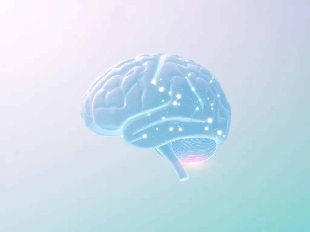
Brain imaging provides a powerful lens through which to observe subtle deviations in brain structure and function that may predate the onset of overt mental health symptoms. This capacity to detect early indicators is fundamental to identifying brain imaging mental health risks.
Detecting Subtle Brain Differences Associated with Mental Health Predisposition
Research utilizing brain imaging has consistently shown that individuals with a family history of certain mental health conditions, or those exhibiting subclinical symptoms, often display subtle structural or functional differences in their brains compared to those without such predispositions. These differences are not necessarily indicative of current illness but rather suggest an increased vulnerability.
For instance, a study using structural MRI might find that adolescents with a high genetic risk for schizophrenia show slightly reduced gray matter volume in specific frontal or temporal lobe regions compared to their peers without the same genetic risk, years before any psychotic symptoms emerge.
These subtle variations, sometimes only detectable through advanced image analysis techniques, represent potential early risk markers. They highlight specific brain circuits or regions that may be inherently less resilient or more susceptible to the impact of stress or other environmental triggers. Identifying these subtle brain imaging mental health risks allows researchers to focus on the underlying biological pathways that confer vulnerability, offering targets for preventative interventions aimed at strengthening these circuits or mitigating their susceptibility.
One prominent area of research in brain imaging mental health risks for anxiety disorders focuses on the connectivity between the amygdala, a region heavily involved in processing fear and emotion, and the prefrontal cortex (PFC), which is critical for regulating emotions and executive function. In healthy individuals, the PFC typically exerts inhibitory control over the amygdala, helping to modulate fearful responses. However, studies using fMRI have consistently shown altered connectivity in individuals with anxiety disorders.
Specifically, research suggests that individuals prone to anxiety may exhibit hyperactivity in the amygdala and/or reduced functional connectivity between the PFC and the amygdala. For example, during tasks involving viewing fearful faces, individuals with high trait anxiety might show exaggerated amygdala activation and weaker coupling with ventromedial PFC regions compared to low-anxiety individuals. These patterns of altered amygdala-PFC connectivity are considered significant brain imaging mental health risks for developing anxiety conditions.
Examining Hippocampal Volume Changes Linked to Increased Risk of Depression
The hippocampus, a brain structure vital for learning, memory, and stress regulation, has been extensively studied in the context of depression. Chronic stress, a known risk factor for depression, can impair neurogenesis (the birth of new neurons) in the hippocampus and lead to a reduction in its volume. Structural MRI studies have frequently reported smaller hippocampal volumes in individuals diagnosed with major depressive disorder.
Importantly, prospective studies examining individuals at risk for depression (e.g., those with a history of childhood trauma or a first-degree relative with depression) have sometimes observed smaller hippocampal volumes even before the onset of a depressive episode. While not a definitive predictor, reduced hippocampal volume is considered a structural brain imaging mental health risk factor, suggesting a vulnerability in the brain's stress response and emotional regulation systems that may contribute to the development of depressive symptoms later in life.
Specific Mental Health Conditions and Brain Imaging Risk Markers
Brain imaging research has identified specific neural signatures associated with the risk for various mental health conditions. These markers provide crucial insights into the distinct neurobiological pathways underlying different disorders.
Brain Imaging Biomarkers for Depression Risk
Identifying neural circuits vulnerable to depressive episodes is a key focus of brain imaging research into depression risk. Studies using fMRI have highlighted dysfunction within the brain's reward circuitry, particularly involving regions like the ventral striatum and prefrontal cortex. Individuals prone to depression may show reduced activation in these areas in response to positive stimuli or rewards, a pattern known as anhedonia, which is a core symptom of depression.
Furthermore, alterations in the default mode network (DMN), a network of brain regions active when the mind is at rest or self-reflecting, are also considered brain imaging mental health risks for depression. Overactivity or aberrant connectivity within the DMN, particularly involving the medial prefrontal cortex and posterior cingulate cortex, can be associated with excessive rumination and negative self-referential thinking, both of which are hallmarks of depression. These functional biomarkers, observed before the full clinical syndrome develops, can signal an increased predisposition.
Brain Imaging Biomarkers for Anxiety Disorder Risk
Examining amygdala hyperactivity and prefrontal cortex dysfunction is central to understanding brain imaging mental health risks for anxiety disorders. As mentioned earlier, an overactive amygdala is a common finding in individuals with heightened anxiety or anxiety disorders. This heightened reactivity can be observed during tasks involving emotional stimuli or even during resting states.
Coupled with amygdala hyperactivity, dysfunction in the prefrontal cortex, particularly areas involved in cognitive control and emotion regulation (like the ventromedial and dorsolateral PFC), is also frequently implicated. Reduced inhibitory control from the PFC over the amygdala is thought to underpin the exaggerated fear and worry responses characteristic of anxiety. These functional connectivity patterns, detectable via fMRI, serve as important brain imaging mental health risks, indicating a biological vulnerability to experiencing excessive fear and anxiety.
Brain Imaging Biomarkers for Schizophrenia Risk
Investigating brain volume abnormalities and connectivity issues is a major avenue for identifying brain imaging mental health risks for schizophrenia. Schizophrenia is associated with a range of structural differences, including reduced gray matter volume in various brain regions, such as the temporal lobes (including the hippocampus and amygdala), frontal lobes, and thalamus. While these changes are often more pronounced after the onset of illness, subtle volume differences can sometimes be detected in individuals at high genetic risk or during the prodromal phase (the period before the onset of full symptoms).
Connectivity issues, particularly disruptions in the connections between different brain regions, are also considered key brain imaging mental health risks for schizophrenia. fMRI studies have shown altered functional connectivity within and between various neural networks, including the salience network, default mode network, and executive control network. These disruptions are thought to contribute to the cognitive disorganization, hallucinations, and delusions characteristic of the disorder. White matter abnormalities, assessed using diffusion tensor imaging (DTI), which measures the integrity of fiber tracts, also represent structural risk markers.
Brain Imaging Biomarkers for Bipolar Disorder Risk
Observing changes in brain activity during mood swings provides insights into brain imaging mental health risks for bipolar disorder, a condition characterized by alternating periods of mania/hypomania and depression. Neuroimaging studies have identified abnormalities in brain regions involved in emotional processing and regulation, including the amygdala, prefrontal cortex (particularly the ventrolateral and orbitofrontal regions), and anterior cingulate cortex.
During manic states, individuals with bipolar disorder often show increased activity in limbic regions like the amygdala, while during depressive states, activity in prefrontal regions involved in emotional regulation may be reduced. Longitudinal studies tracking individuals at high genetic risk for bipolar disorder have sometimes observed subtle differences in the structure or function of these regions even during euthymic periods (stable mood), suggesting potential trait markers. For instance, altered connectivity within fronto-limbic circuits has been proposed as a functional brain imaging mental health risk for developing bipolar disorder.
Advanced Brain Imaging Analysis and Predictive Modeling
The sheer volume and complexity of brain imaging data necessitate sophisticated analytical approaches to identify meaningful patterns and predict future outcomes. Advanced computational methods are transforming how we interpret brain imaging mental health risks.
Machine Learning and AI in Predicting Mental Health Outcomes
Machine learning and artificial intelligence (AI) are increasingly being applied to brain imaging data to build predictive models for mental health outcomes. These algorithms can analyze complex patterns across multiple brain regions and features (e.g., volume, connectivity, activity levels) that might be subtle or too intertwined for traditional statistical methods to detect. By training models on large datasets of brain scans from individuals who later developed a mental health condition compared to those who did not, researchers aim to identify combinations of neural features that predict risk with a certain degree of accuracy.
For example, a machine learning model might be trained on fMRI data from adolescents at high risk for psychosis. The model could analyze patterns of functional connectivity across hundreds of brain regions and learn to identify specific patterns that are highly associated with transitioning to a psychotic episode within a few years.
Early studies using such approaches have reported promising results, with some models achieving prediction accuracies ranging from approximately 60% to 80% for certain conditions, though these models are still largely experimental and require further validation in larger, independent datasets. This application of AI to brain imaging data is a major step forward in leveraging brain imaging mental health risks for prediction.
Using Brain Imaging Data to Personalize Mental Health Interventions
Beyond prediction, brain imaging data analyzed with advanced methods can potentially inform personalized intervention strategies. If a brain scan reveals specific patterns of dysfunction associated with a particular risk factor, interventions could be tailored to target those specific neural circuits. For instance, an individual showing reduced connectivity between the prefrontal cortex and amygdala, identified as a brain imaging mental health risk for anxiety, might benefit more from therapies specifically designed to strengthen emotion regulation pathways, such as certain forms of cognitive behavioral therapy or neurofeedback training, compared to someone with a different neural profile.
Furthermore, brain imaging could potentially be used to predict an individual's likely response to different types of treatment. For example, research is exploring whether specific brain activity patterns before starting antidepressant medication can predict whether a patient will respond well to that particular drug class. While still an active area of research, the integration of brain imaging data into personalized treatment planning holds significant promise for improving the effectiveness and efficiency of mental healthcare, moving towards interventions guided by an individual's unique brain characteristics and identified brain imaging mental health risks.
Ethical Considerations and Limitations of Brain Imaging in Risk Assessment

While brain imaging offers powerful insights into mental health risks, its use raises important ethical considerations and faces inherent limitations that must be carefully navigated.
Privacy and Data Security Concerns in Neuroimaging Research
Brain imaging data is incredibly personal and potentially revealing about an individual's cognitive and emotional state. As datasets grow larger and are shared across research institutions, ensuring the privacy and security of this sensitive information becomes paramount. There is a significant risk of data breaches or unauthorized access, which could potentially lead to discrimination or stigmatization based on an individual's identified brain imaging mental health risks.
Developing robust data anonymization protocols, secure storage systems, and clear ethical guidelines for data sharing are critical. Participants in neuroimaging studies must be fully informed about how their data will be used, stored, and protected, and their consent must be truly informed. The potential for re-identification, even from anonymized data, remains a concern that researchers and institutions must actively address to maintain public trust in the use of brain imaging for identifying mental health risks.
The Importance of Responsible Interpretation of Brain Imaging Results
Interpreting brain imaging findings, especially in the context of risk prediction, requires extreme caution and responsibility. A brain scan showing a pattern associated with an increased risk for a condition does not mean that the individual will definitely develop that condition. Brain imaging markers are probabilistic, indicating a likelihood or vulnerability, not a deterministic fate. Attributing a diagnosis or making definitive pronouncements based solely on a brain scan is scientifically inaccurate and ethically problematic.
It is crucial to communicate brain imaging findings with nuance, emphasizing that they represent just one piece of a complex puzzle that includes genetic, environmental, psychological, and social factors. Oversimplification or misinterpretation of brain imaging mental health risks can lead to undue anxiety or false reassurance. Clinicians and researchers must receive adequate training in interpreting neuroimaging data within the broader clinical context and avoid deterministic language when discussing risk.
Potential for Stigmatization and the Need for Comprehensive Assessment
The concept of identifying brain imaging mental health risks, while intended to be empowering for early intervention, also carries the potential for increased stigmatization. Labeling individuals as "at risk" based on their brain patterns could lead to prejudice in areas like employment, insurance, or social interactions. This is particularly concerning given the probabilistic nature of the risk markers and the fact that many individuals identified as high-risk may never develop the condition.
Therefore, brain imaging should never be used in isolation for risk assessment or diagnosis. It must always be part of a comprehensive evaluation that includes clinical interviews, psychological assessments, consideration of personal history, genetics, and environmental factors. The focus should remain on supporting individuals and providing resources, rather than labeling them based on potential future risks. The ethical imperative is to use insights from brain imaging mental health risks to empower and support, not to stigmatize or predict destiny.
Future Directions: Advancing Brain Imaging for Mental Health Risk Prediction
The field of brain imaging for mental health risk prediction is rapidly evolving, driven by technological advancements and innovative analytical approaches. The future holds exciting possibilities for enhancing our ability to identify, understand, and mitigate mental health vulnerabilities.
Exploring Novel Brain Imaging Techniques and Analysis Methods
Future advancements will likely involve the development of novel brain imaging techniques offering even higher resolution, better sensitivity, or the ability to measure new aspects of brain function or chemistry. For instance, ultra-high field MRI scanners (7T and above) provide unprecedented anatomical detail, while advanced PET tracers could allow for imaging of a wider range of neurotransmitter systems or inflammatory markers implicated in mental health.
Beyond hardware, significant progress is expected in analysis methods. Diffusion MRI techniques like neurite orientation dispersion and density imaging (NODDI) offer more detailed insights into tissue microstructure. Graph theory is increasingly used to analyze complex brain networks derived from fMRI, revealing subtle disruptions in network topology associated with risk. Furthermore, the integration of multimodal data – combining insights from MRI, fMRI, PET, and EEG – promises a more comprehensive picture of brain health and risk factors than any single modality can provide. These advancements will refine our ability to detect subtle brain imaging mental health risks.
Integrating Brain Imaging with Genetic and Environmental Data
A key future direction is the integration of brain imaging data with other powerful sources of information, particularly genetic and environmental data. Mental health conditions are complex, resulting from the interplay of genetic predispositions, environmental exposures (like stress, trauma, or toxins), and individual experiences. Brain imaging provides a crucial intermediate layer, showing how these factors converge to impact brain structure and function.
By combining large-scale genetic datasets (e.g., from genome-wide association studies) with brain imaging data and detailed information about environmental exposures and life experiences, researchers can build more sophisticated models of risk. For example, studies could investigate how specific genetic variants influence brain connectivity patterns and how these patterns, in turn, interact with early life stress to increase the risk for depression or anxiety. This integrated approach is essential for moving beyond simply identifying brain imaging mental health risks to understanding the complex causal pathways involved.
The Potential for Early Intervention and Prevention Strategies Guided by Brain Imaging
Ultimately, the goal of identifying brain imaging mental health risks is to facilitate early intervention and prevention. As our ability to predict risk improves and we gain a deeper understanding of the underlying neural mechanisms, we can develop and target preventative strategies more effectively. For individuals identified as high-risk based on brain imaging and other data, interventions could be initiated proactively.
These interventions might include targeted psychological therapies (e.g., resilience training, stress management), lifestyle modifications (e.g., exercise, sleep hygiene), or even novel approaches like neurofeedback or targeted neuromodulation techniques aimed at strengthening vulnerable brain circuits. For instance, if brain imaging suggests a weakened prefrontal control circuit as a brain imaging mental health risk, interventions could focus on therapies known to enhance prefrontal function. The future envisions a system where brain imaging insights inform personalized, preventative mental healthcare, helping individuals build resilience and navigate potential vulnerabilities before they manifest as debilitating conditions.
Conclusion: Brain Imaging as a Powerful Tool for Proactive Mental Healthcare
Brain imaging stands as a powerful and evolving tool in the quest to understand and address mental health vulnerabilities. By offering unprecedented views into the living brain, techniques like MRI, fMRI, PET, and EEG allow us to identify structural and functional patterns associated with increased risk for various conditions, from depression and anxiety to schizophrenia and bipolar disorder. The ongoing effort to pinpoint brain imaging mental health risks is transforming our approach to mental well-being, shifting the focus from reactive treatment to proactive prevention.
Empowering Individuals with Knowledge About Their Mental Health Risks
The insights gained from brain imaging mental health risks have the potential to empower individuals by providing them with knowledge about their specific vulnerabilities. Understanding potential predispositions can enable people to take proactive steps, such as adopting healthier lifestyle habits, seeking early support, or engaging in preventative therapies. This knowledge, when communicated responsibly and empathetically, can reduce uncertainty and foster a sense of agency in managing one's mental health journey.
It is crucial, however, that this information is presented within a supportive framework that avoids deterministic interpretations or stigmatization. The goal is to equip individuals with information that helps them make informed choices about their well-being, not to label them based on probabilistic risk markers. Providing resources and support alongside the communication of brain imaging findings is essential for truly empowering individuals.
The Future of Brain Imaging in Promoting Mental Well-being Through Early Detection
The future of brain imaging in mental health lies in its potential to facilitate early detection and truly preventative care. As techniques become more refined, analysis methods more sophisticated (especially with the aid of AI), and our understanding of the complex interplay between brain, genes, and environment deepens, brain imaging mental health risks will become clearer and more actionable.
The integration of brain imaging with other data sources promises a holistic view of individual vulnerability, enabling the development of highly personalized prevention strategies. This move towards precision mental health, guided by insights from brain imaging, holds the promise of reducing the incidence and severity of mental health conditions, ultimately promoting greater mental well-being across populations. The journey to fully harness the power of brain imaging for this purpose is ongoing, but the potential to transform mental healthcare through early risk identification is immense.
Frequently Asked Questions
How can brain imaging techniques help in identifying mental health risks?
Brain imaging like MRI and fMRI can reveal structural and functional abnormalities in the brain associated with mental disorders. These scans can highlight early indicators of risk, such as altered connectivity or volume changes in specific regions. This helps in understanding the biological basis of these conditions.
What are some common brain imaging methods used in mental health research?
Common methods include Magnetic Resonance Imaging (MRI) for structural details, functional MRI (fMRI) for brain activity, and Positron Emission Tomography (PET) for metabolic processes. Each provides unique insights into brain function and structure, aiding in the identification of risk factors.
Are there any potential risks associated with brain imaging for mental health?
While generally considered safe, some risks exist. MRI involves strong magnetic fields, posing risks for individuals with certain metallic implants. PET scans involve low-level radiation exposure. It's crucial to discuss these potential risks and benefits with a healthcare professional.
Can brain imaging predict the likelihood of developing a specific mental health condition?
Brain imaging can identify potential risk factors and biomarkers, but it's not a definitive predictor. It's used in conjunction with clinical assessments, genetic information, and environmental factors to assess the overall risk. It provides valuable insights, but not a guaranteed prediction.
How is the information gathered from brain imaging used to improve mental health care?
Brain imaging findings can lead to more targeted and personalized treatments. They help researchers develop new therapies and improve the understanding of how existing treatments affect the brain. This ultimately leads to improved diagnosis and management of mental health conditions.
Final Reflection: Unveiling the Mind's Landscape: A Path to Prevention
The ability to visualize the brain's intricate workings through advanced imaging techniques is revolutionizing our understanding of mental health, offering a powerful new lens through which to identify and potentially prevent debilitating conditions. This isn't just about diagnosis; it's about proactively identifying individuals at risk, paving the way for early intervention and personalized treatment strategies that could dramatically alter the trajectory of countless lives.
The impact of this research extends far beyond the clinical setting, influencing our very understanding of the mind-body connection and challenging long-held stigmas surrounding mental illness. By visualizing the biological underpinnings of these conditions, we move away from simplistic explanations and towards a more nuanced appreciation of the complex interplay of genetics, environment, and individual experience. The greatest advances in medicine are often made not by treating disease, but by preventing it.
This quote underscores the transformative potential of brain imaging in mental health, shifting the focus from reactive treatment to proactive prevention.
Now, consider this: what if we could identify individuals at high risk for depression or anxiety before symptoms even manifest? What if personalized interventions, tailored to specific brain patterns, could significantly reduce the burden of mental illness on individuals and society? This is the future brain imaging promises. You have the power to share this knowledge, to advocate for increased research funding, and to encourage open conversations about mental health. Let's work together to build a future where early detection and personalized care become the norm, not the exception, in mental health.
Unlock Your Brain's Potential: Take Charge of Your Mental Wellbeing
Thanks for taking the time to explore the fascinating world of brain imaging and mental health with us! You've now gained valuable insights into how early identification can make a real difference. Ready to put this knowledge into action?
- Schedule a consultation with your doctor to discuss your mental health and explore potential risk factors. This is the most important step you can take to proactively manage your wellbeing.
- Share this article with a friend or family member who might benefit from understanding the link between brain imaging and mental health. Knowledge is power!
- Explore MindCareGuide.org for more resources on mental health and brain wellness. Discover additional articles and tools to support your journey towards a healthier mind.
What proactive steps will you take today to prioritize your mental wellbeing?