
Beyond Symptoms: Brain Scan Proof of Therapy's Deep Impact

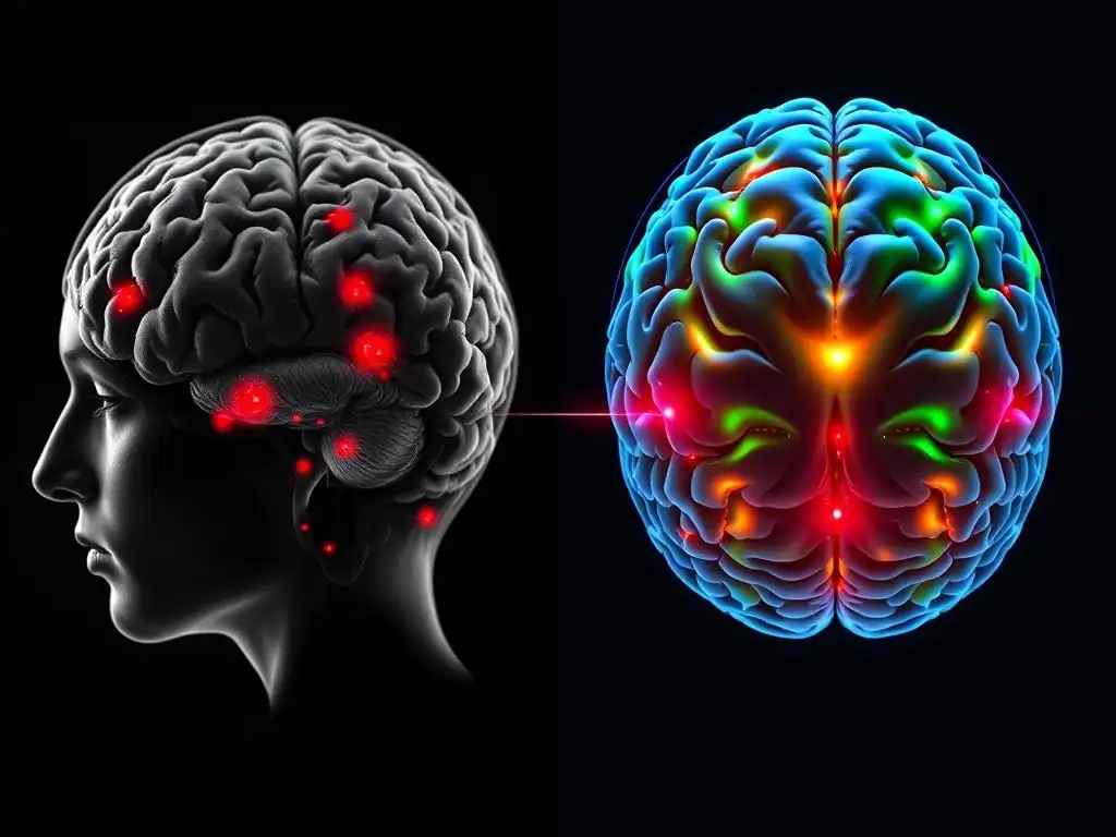
Welcome to Mind Care Guide, a space dedicated to nurturing your mental well-being.
Have you ever wondered if therapy actually changes your brain? In this compelling exploration, "Beyond Symptoms: Brain Scan Proof of Therapy's Deep Impact," we delve into the fascinating world of neuroscience to uncover therapy’s impact on brain scans. Get ready to see compelling evidence that goes beyond just feeling better, revealing the tangible and lasting changes that therapy can bring. Let's explore this exciting intersection of neuroscience and mental health together...
- Beyond Symptoms: Brain Scan Validation of Therapy's Impact - A Neuroscientific Guide
- Neuroscience of Mental Health: Understanding the Brain's Response to Therapeutic Interventions
- Long-Term Therapy Outcomes: Brain Scan Evidence
- Clinical Significance of Brain Scan Findings
- Addressing Misconceptions about Brain Scans and Therapy
- The Future of Neuroimaging in Mental Health Care
- Conclusion: The Profound Impact of Therapy
-
Frequently Asked Questions
- How does therapy actually change the brain, according to the Mind Care Guide article?
- What types of brain scans are typically used to demonstrate the impact of therapy?
- What specific mental health conditions have shown brain scan improvements after therapy?
- Is brain scan evidence necessary to prove the effectiveness of therapy?
- Can any type of therapy lead to observable changes in brain scans?
- Final Reflection: The Rewiring of Resilience: Seeing Therapy's Impact on the Brain
- Unlock Your Brain's Potential: Take Charge of Your Wellbeing
Beyond Symptoms: Brain Scan Validation of Therapy's Impact - A Neuroscientific Guide
The Subjectivity of Symptom-Based Assessment in Mental Health
Assessing mental health challenges often relies heavily on subjective reports from individuals. Symptoms like persistent sadness, excessive worry, intrusive thoughts, or difficulty concentrating are crucial indicators, forming the basis of diagnosis and treatment planning. However, the nature of these symptoms can be highly variable; they fluctuate in intensity, can be challenging for individuals to articulate precisely, and are subject to personal interpretation and cultural context. While essential, this reliance on subjective experience can sometimes make it difficult to objectively measure the subtle or gradual shifts that occur during therapeutic interventions.
Furthermore, symptom-based assessments primarily capture the manifestations of mental health conditions rather than the underlying biological processes. A person might report reduced anxiety, but it can be challenging to determine how their brain is processing fear differently or if the neural pathways associated with distress have truly been modulated. This inherent subjectivity highlights the need for complementary tools that can offer a more objective perspective on mental well-being and provide concrete evidence of therapy's impact on brain scans and function.
Brain Scans: Objective Measurement of Therapy's Effectiveness
Neuroimaging techniques offer a powerful lens into the brain's structure and function, providing a more objective measure of the changes that occur in response to therapeutic interventions. Unlike symptom checklists, which rely on self-report, brain scans capture physiological activity and structural characteristics. By comparing scans taken before and after a course of therapy, researchers and clinicians can visualize and quantify alterations in specific brain regions, neural circuits, and neurochemical levels.
This objective data provides compelling evidence of therapy's impact on brain scans, demonstrating that talking therapies are not merely psychological constructs but interventions that induce tangible, physical changes in the brain. This neurobiological validation strengthens the understanding of mental health conditions as disorders with biological underpinnings and reinforces the efficacy of therapy as a potent treatment modality capable of reshaping the very organ responsible for our thoughts, feelings, and behaviors.
Neuroscience of Mental Health: Understanding the Brain's Response to Therapeutic Interventions
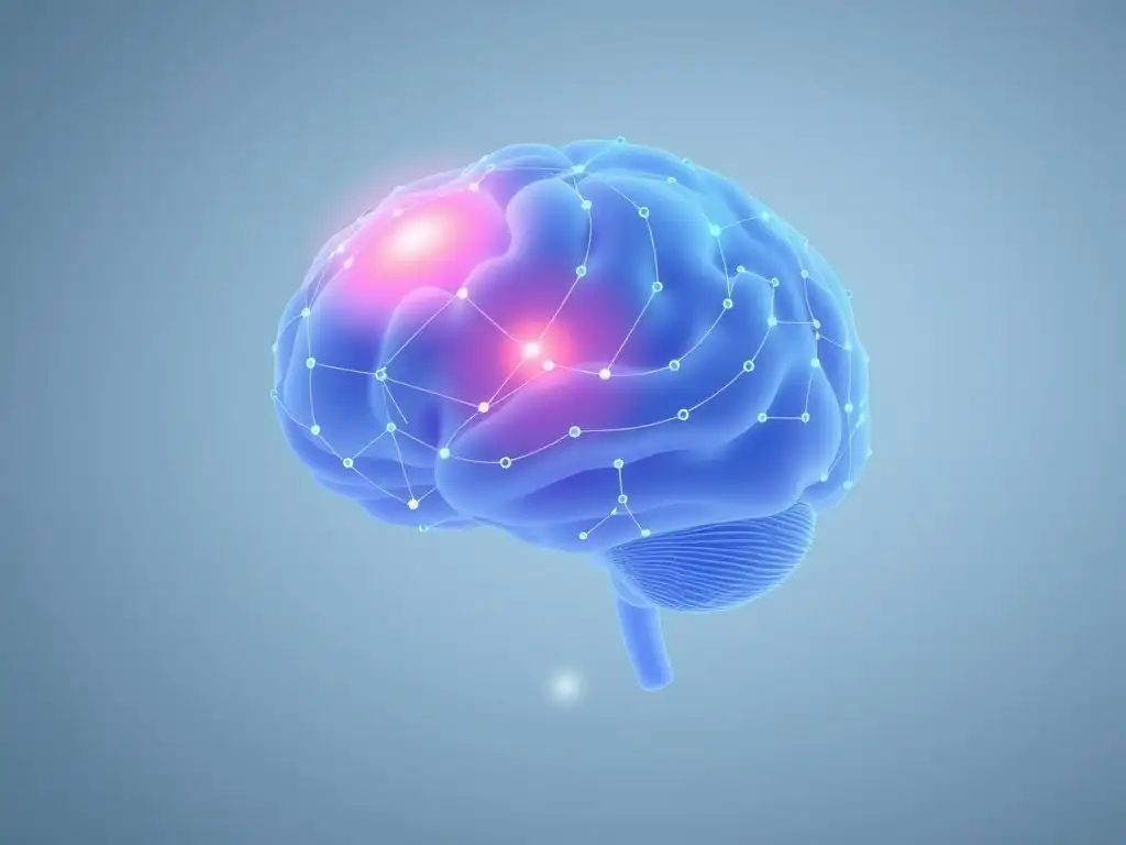
Brain Plasticity: Reshaping Neural Pathways Through Therapy
A cornerstone of understanding therapy's impact on brain scans is the concept of neuroplasticity. The human brain is not a static organ but possesses a remarkable ability to reorganize itself throughout life – forming new neural connections, strengthening existing ones, and even generating new neurons in certain areas. This plasticity allows the brain to adapt to new experiences, learning, and environmental changes. Mental health conditions often involve maladaptive patterns of neural activity or connectivity, such as an overactive amygdala in anxiety or altered reward pathways in depression.
Therapeutic interventions leverage this inherent plasticity. By engaging in specific therapeutic techniques, individuals are essentially providing the brain with new experiences and learning opportunities. For example, cognitive restructuring in CBT helps retrain thought patterns, while exposure therapy helps the brain learn that previously feared stimuli are safe. These processes involve repeated activation of new or alternative neural pathways, gradually weakening the old, maladaptive connections and strengthening healthier ones. This physical rewiring is the basis for the observable changes seen when examining therapy’s impact on brain scans.
Neuroimaging Techniques: Visualizing Therapy's Impact
Understanding therapy’s impact on brain scans relies heavily on advanced neuroimaging techniques. These technologies allow researchers to peer inside the living brain and observe its activity and structure in detail, providing visual evidence of the neural changes induced by therapeutic processes. Each technique offers a unique perspective, capturing different facets of brain function and structure.
fMRI (Functional Magnetic Resonance Imaging): Mapping Brain Activity During Therapeutic Processes
Functional Magnetic Resonance Imaging (fMRI) measures brain activity by detecting changes in blood flow. When a brain region is more active, it requires more oxygenated blood, and fMRI can pick up the subtle magnetic differences between oxygenated and deoxygenated blood.
This allows researchers to see which brain areas are more or less active during specific tasks, or even at rest (resting-state fMRI, which measures connectivity). Studies using fMRI have shown, for instance, decreased activity in the amygdala (a fear-processing region) and increased activity in the prefrontal cortex (involved in regulation) in individuals with anxiety disorders following successful cognitive behavioral therapy (CBT), providing direct fMRI evidence of therapy's impact on brain scans.
PET Scans (Positron Emission Tomography): Examining Neurochemical Changes in Response to Therapy
Positron Emission Tomography (PET) scans use small amounts of radioactive tracers that bind to specific molecules in the brain, such as neurotransmitter receptors or metabolic markers. This allows researchers to measure levels of certain neurochemicals or metabolic activity in different brain regions. PET scans can reveal changes in neurotransmitter systems (like serotonin or dopamine) or glucose metabolism that occur as a result of therapy. For example, some PET studies have indicated changes in serotonin transporter binding in individuals with depression following psychotherapy, adding a neurochemical dimension to the understanding of therapy’s impact on brain scans.
EEG (Electroencephalography): Assessing Brainwave Patterns and Therapy's Effects
Electroencephalography (EEG) measures the electrical activity of the brain using electrodes placed on the scalp. It is particularly good at capturing rapid changes in brain activity and analyzing brainwave patterns (like alpha, beta, theta, delta waves) associated with different states of consciousness or cognitive processes.
While not providing high spatial resolution like fMRI or PET, EEG offers excellent temporal resolution. Research using EEG has shown changes in brainwave coherence or power in specific frequencies in individuals undergoing therapy for conditions like ADHD or anxiety, suggesting alterations in neural communication patterns as part of therapy's impact on brain scans at the level of electrical signaling.
Long-Term Therapy Outcomes: Brain Scan Evidence
Therapy's Impact on Brain Scans: Specific Mental Health Conditions
Research across various mental health conditions has increasingly utilized brain scans to provide objective evidence of the neural changes associated with successful therapeutic outcomes. These studies move beyond symptom reduction to demonstrate how therapy physically alters the brain's structure and function, solidifying the understanding of therapy’s impact on brain scans for specific disorders.
Depression: Brain Scan Validation of Antidepressant and Cognitive Therapies
Major depressive disorder is associated with functional and structural abnormalities in brain regions involved in mood regulation, reward processing, and cognitive control, such as the prefrontal cortex, hippocampus, and amygdala. Studies using fMRI have shown that effective psychotherapies for depression, such as Cognitive Behavioral Therapy (CBT) or Interpersonal Therapy (IPT), can lead to increased activity in areas of the prefrontal cortex associated with emotional regulation and decreased activity in the amygdala.
Some research has even suggested potential increases in hippocampal volume or function after long-term therapy, areas often reduced or impaired in chronic depression. For instance, one line of research has found that effective CBT for depression can lead to normalization of connectivity between the subgenual cingulate cortex and other brain regions, a pattern often disrupted in depressed individuals, providing clear brain scan evidence of therapy's impact.
Anxiety Disorders: Neural Correlates of CBT Success
Anxiety disorders are characterized by hyperactivation of fear circuits, particularly involving the amygdala, and often reduced regulatory control from the prefrontal cortex. Cognitive Behavioral Therapy (CBT), especially exposure therapy, is a highly effective treatment. Brain scan studies using fMRI consistently show reduced amygdala reactivity to fearful stimuli after successful CBT for various anxiety disorders, including specific phobias, social anxiety disorder, and panic disorder.
Simultaneously, activity and connectivity in prefrontal regions involved in evaluating and regulating emotional responses often increase. This pattern of changes in key fear and regulatory circuits provides robust neural correlates, demonstrating therapy's impact on brain scans by rebalancing activity in these critical areas.
PTSD (Post-Traumatic Stress Disorder): Amygdala and Hippocampus Changes Following Trauma-Focused Therapy
Post-Traumatic Stress Disorder (PTSD) involves persistent changes in brain function related to trauma processing. Individuals with PTSD often exhibit hyperactive amygdala responses to trauma-related cues and can show reduced hippocampal volume or altered function, impacting memory and context processing. Trauma-focused therapies, such as Prolonged Exposure (PE) or Eye Movement Desensitization and Reprocessing (EMDR), aim to process traumatic memories safely.
Brain scan research, particularly fMRI, has shown that these therapies can lead to a significant reduction in amygdala hyperactivity when exposed to trauma reminders. Some studies also suggest improvements in hippocampal function or connectivity, indicating that therapy helps normalize key brain areas disrupted by trauma, offering compelling brain scan evidence of therapy's impact on trauma-related neural circuits.
OCD (Obsessive-Compulsive Disorder): Neural Insights from Exposure and Response Prevention (ERP) Therapy
Obsessive-Compulsive Disorder (OCD) is associated with dysfunction in cortico-striato-thalamo-cortical circuits, which are involved in habit formation, decision-making, and error detection. Exposure and Response Prevention (ERP) therapy, a form of CBT, is the most effective psychological treatment for OCD. Brain imaging studies using PET and fMRI have demonstrated that successful ERP therapy is associated with changes in activity and connectivity within these specific circuits.
For example, research has shown decreased activity in areas like the caudate nucleus and orbitofrontal cortex, which are often hyperactive in individuals with untreated OCD. These measurable changes within the implicated neural pathways provide strong neural support, illustrating therapy's impact on brain scans by normalizing activity in dysfunctional circuits.
Case Studies: Illustrating Therapy's Impact on Brain Structure and Function
Real-world examples, often documented in case studies or larger clinical trials, provide powerful illustrations of how therapeutic interventions translate into observable changes in the brain. While individual results vary, these cases highlight the potential for significant neural reshaping.
Adolescent with Social Anxiety: Increased Prefrontal Cortex Connectivity After CBT
Consider a hypothetical case, based on research findings, of a 16-year-old adolescent diagnosed with severe social anxiety disorder. Before therapy, an fMRI scan might show reduced functional connectivity between the ventrolateral prefrontal cortex (a region involved in regulating negative emotions) and the amygdala when anticipating social interaction. After completing a 12-week course of manualized CBT specifically targeting social anxiety, the adolescent reports significant symptom reduction and increased participation in social activities.
A follow-up fMRI scan reveals a measurable increase in the functional connectivity between these two brain regions. This increased "communication" suggests that the prefrontal cortex has become more effective at regulating the amygdala's fear response, providing clear brain scan evidence of therapy's impact on key anxiety circuits in a young person.
Adult with Panic Disorder: Reduced Amygdala Reactivity Following Exposure Therapy
Imagine an adult in their late 30s struggling with frequent panic attacks and a diagnosis of panic disorder with agoraphobia. An initial fMRI scan, perhaps conducted while the individual views images related to their panic triggers (e.g., crowded spaces, physical sensations like shortness of breath), shows heightened reactivity in the amygdala compared to neutral images. After undergoing a course of exposure therapy, gradually confronting feared situations and sensations without avoidance, the individual experiences a dramatic reduction in panic attack frequency and severity.
A repeat fMRI scan, using the same trigger stimuli, now reveals a significantly blunted amygdala response. The brain is no longer reacting with the same intensity of fear to these previously triggering cues, offering compelling brain scan proof of therapy's impact on the brain's core fear response system.
Clinical Significance of Brain Scan Findings
Guiding Treatment Decisions: Personalized Therapy Based on Neuroimaging
While still largely in the research realm, the ability to visualize therapy's impact on brain scans holds significant potential for guiding future clinical decisions. Understanding the specific neural pathways or regions involved in an individual's condition, and how these might respond to different therapeutic approaches, could pave the way for more personalized treatment plans. For example, if a brain scan indicates a particular pattern of prefrontal cortex dysfunction in a patient with depression, it might suggest that therapies focusing on cognitive control and emotional regulation could be particularly effective for that individual.
Furthermore, neuroimaging could potentially help differentiate between subtypes of mental health conditions that present with similar symptoms but have different underlying neural profiles. This could lead to selecting interventions that are neurobiologically better matched to the individual's specific brain patterns, theoretically increasing the likelihood of success and optimizing therapy’s impact on brain scans for that person. Although routine clinical use is not yet widespread due to cost and complexity, the promise of precision mental health care informed by neuroimaging is a significant area of research.
Monitoring Progress: Objective Measures of Therapeutic Improvement
One of the challenges in therapy is objectively measuring progress beyond subjective symptom reduction. Brain scans offer a potential solution by providing biological markers of change. Seeing quantifiable alterations in brain activity, connectivity, or structure can serve as an objective indicator that the therapy is indeed having the desired effect at a neural level, complementing patient reports. For instance, if fMRI shows a gradual normalization of amygdala activity in an anxious patient over several months of therapy, it provides tangible evidence of therapeutic progress that aligns with, or even precedes, reported symptom improvement.
Monitoring therapy’s impact on brain scans could potentially allow clinicians to make data-driven adjustments to treatment plans. If scans show minimal or no change in targeted brain regions despite consistent therapy attendance, it might signal the need to modify the therapeutic approach, try a different modality, or consider adjunctive treatments. This neurobiological feedback loop could enhance the effectiveness and efficiency of mental health care by providing objective checkpoints on the path to recovery.
Enhancing Patient Understanding and Motivation: Visualizing Therapy's Benefits
For individuals undergoing therapy, understanding how it works can be incredibly empowering. Showing patients simplified visualizations of brain scan findings – perhaps illustrating the difference in brain activity before and after therapy in someone with a similar condition – can make the abstract concept of 'rewiring' the brain feel more concrete and achievable. This visual evidence of therapy's impact on brain scans can demystify the therapeutic process and help patients see that their efforts in therapy are leading to real, physical changes in their brain.
Seeing tangible proof that therapy is inducing positive changes can significantly boost a patient's motivation and commitment to the process, especially during challenging periods or when progress feels slow. It reinforces the idea that they are actively participating in healing their brain, fostering a sense of agency. This objective validation from brain scans can transform the patient's perspective, moving from simply managing symptoms to understanding that they are fundamentally altering the neural landscape underlying their mental health, thus maximizing therapy’s impact on brain scans and overall well-being.
Addressing Misconceptions about Brain Scans and Therapy
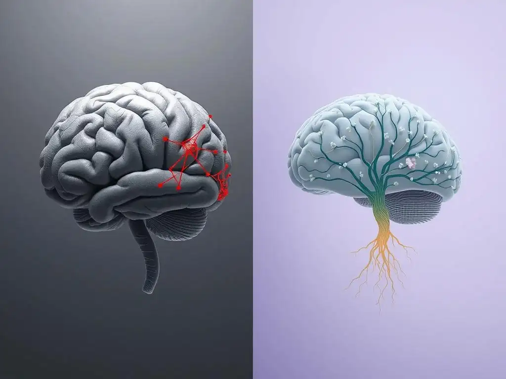
Myth: Brain Scans Guarantee Therapy Success
A common misconception is that obtaining a brain scan can predict with certainty whether a specific therapy will be successful for an individual. This is not accurate.
While brain scans can reveal insights into neural function associated with a condition and have shown patterns of change after successful therapy, they are not currently reliable predictors of individual treatment outcomes. The effectiveness of therapy is influenced by a complex interplay of factors, including the therapeutic alliance, the individual's motivation and adherence to treatment, their unique life circumstances, co-occurring conditions, and the skill of the therapist, none of which are directly captured by a single scan.
Therefore, relying solely on a brain scan to guarantee therapy success is an oversimplification. Brain imaging provides valuable data about the brain's state and how groups of people tend to respond to therapy at a neural level, contributing to our understanding of therapy’s impact on brain scans. However, it cannot account for the myriad non-biological variables that are critical determinants of an individual's journey through therapy and their ultimate outcome. Clinical assessment, patient history, and the therapeutic relationship remain paramount in predicting and facilitating successful treatment.
Reality: Brain Scans are a Tool, Not a Determinant
In reality, brain scans are powerful scientific and potentially clinical tools that provide valuable data about the brain, but they do not determine a person's destiny or the outcome of their therapy. They offer a snapshot or a dynamic view of brain activity or structure at a given time, contributing to a more comprehensive understanding of mental health conditions and how interventions affect the brain. As a tool, neuroimaging can complement clinical assessment, research, and potentially future treatment planning, but it does not replace the need for skilled therapeutic practice, patient engagement, and a holistic view of the individual.
The insights gained from studying therapy’s impact on brain scans are primarily used in research settings to deepen our understanding of mental health and validate the biological underpinnings of therapy. While there is growing interest in using neuroimaging to inform clinical practice (e.g., identifying potential biomarkers), this is still an active area of research and not yet standard clinical protocol. Understanding brain scans as valuable investigative tools, rather than deterministic crystal balls, is crucial for setting realistic expectations about their role in mental health care.
Myth: Therapy is Solely Behavioral, Not Biological
Another prevalent myth is that psychological therapies, because they involve talking and behavioral exercises, are purely "mind over matter" and do not induce physical, biological changes in the brain. This view incorrectly separates the mind and brain. Modern neuroscience clearly demonstrates that all psychological processes – thoughts, feelings, behaviors, and experiences – are the direct result of brain activity. Therefore, interventions that change thoughts, feelings, and behaviors inherently change the brain.
Therapy is a powerful form of guided experience and learning. Just as learning a new language or skill changes brain structure and function, engaging in therapeutic processes like challenging distorted thoughts or practicing new coping mechanisms creates new neural pathways and modifies existing ones. The evidence from neuroimaging studies showing measurable changes in brain activity, connectivity, and even structure after therapy definitively refutes the idea that therapy is solely behavioral; it is fundamentally biological, demonstrating therapy’s impact on brain scans as physical evidence of this change.
Reality: Therapy Induces Measurable Changes in Brain Structure and Function
The undeniable reality, supported by decades of neuroimaging research, is that effective psychological therapies induce measurable and often lasting changes in brain structure and function. Studies using fMRI, PET, and EEG have consistently shown alterations in activity levels in key brain regions (like the amygdala, prefrontal cortex, hippocampus, and cingulate cortex), changes in the strength and pattern of connections between different brain areas, and even subtle structural changes like grey matter density in specific locations following successful therapy for various conditions.
These findings provide compelling neurobiological validation for the efficacy of psychotherapy. They show that therapy doesn't just help people feel better; it helps their brains function differently. The ability to demonstrate therapy's impact on brain scans offers powerful evidence that psychological interventions are robust, biologically active treatments that can help restore healthier brain function, leading to symptom reduction and improved well-being from the ground up.
The Future of Neuroimaging in Mental Health Care
Advancements in Neuroimaging
The field of neuroimaging is rapidly advancing, promising even greater insights into the brain and therapy's impact on brain scans in the future. Technologies are becoming more sophisticated, offering higher resolution and greater specificity. For example, ultra-high field MRI scanners can provide finer detail of brain structure and function.
Developments in portable or magnetoencephalography (MEG) could potentially make brain imaging more accessible outside traditional clinical research centers. Furthermore, advancements in computational analysis, including machine learning and artificial intelligence, are enabling researchers to identify complex patterns in neuroimaging data that were previously undetectable, potentially leading to more accurate identification of biomarkers and a deeper understanding of how different therapies affect the brain at a network level.
These technological leaps mean we may soon be able to visualize therapy's impact on brain scans with unprecedented clarity and detail. This could include tracking changes in specific neural circuits with greater precision, understanding the timing of these changes, and potentially identifying individual differences in how brains respond to therapy. The ongoing innovation in neuroimaging techniques is continuously expanding the possibilities for researching and potentially integrating neurobiological data into mental health care, offering a more nuanced understanding of the brain's response to therapeutic interventions.
Integrating Neuroimaging Data into Clinical Practice
The ultimate goal for many researchers is to translate findings about therapy's impact on brain scans from the laboratory into routine clinical practice. While significant challenges remain, such as cost, accessibility, standardization, and interpreting complex data for individual patients, the potential benefits are substantial. Imagine a future where neuroimaging helps inform initial treatment selection, guiding clinicians towards therapies most likely to be effective for a patient's specific neural profile, or where follow-up scans provide objective feedback on treatment progress, allowing for timely adjustments.
Integrating neuroimaging data could lead to a paradigm shift in mental health care, moving towards a more biologically informed and personalized approach. It could help destigmatize mental illness by clearly demonstrating its basis in brain function and provide patients with objective evidence of their progress. However, this integration requires not only technological advancements but also robust research demonstrating clinical utility, training for clinicians, and the development of ethical guidelines to ensure responsible use of sensitive neurobiological information when assessing therapy's impact on brain scans.
Ethical Considerations and Responsible Use of Neuroimaging
As neuroimaging becomes more powerful and potentially integrated into clinical settings, it is crucial to address significant ethical considerations. The data obtained from brain scans is highly personal and sensitive, raising concerns about privacy and data security. There is also the risk of misinterpretation or oversimplification of scan results, potentially leading to diagnostic labels or prognostic statements that are not fully supported by the science, especially when discussing therapy's impact on brain scans in individuals. It is vital to communicate the limitations of current neuroimaging technology clearly to both clinicians and patients.
Furthermore, potential disparities in access to expensive neuroimaging technologies could exacerbate inequalities in mental health care. Ensuring equitable access and avoiding a scenario where advanced, neurobiologically informed care is only available to a privileged few is paramount. Responsible use of neuroimaging requires ongoing dialogue among researchers, clinicians, ethicists, policymakers, and the public to establish guidelines that prioritize patient well-being, data protection, informed consent, and the accurate and ethical communication of findings related to therapy’s impact on brain scans.
Conclusion: The Profound Impact of Therapy
Therapy's Capacity to Reshape the Brain
The evidence from neuroimaging research provides a powerful testament to therapy's profound capacity to reshape the brain. Moving beyond symptom management, effective psychological interventions demonstrably alter neural activity, connectivity, and sometimes even structure. Studies using fMRI, PET, and EEG across various mental health conditions like depression, anxiety, PTSD, and OCD reveal concrete changes in the very circuits and regions underlying these disorders. This neurobiological evidence solidifies the understanding that therapy is not merely a psychological conversation but a process that instigates real, physical changes in the brain, offering compelling brain scan proof of therapy's impact.
By leveraging the brain's inherent plasticity, therapy helps individuals build new, healthier neural pathways and weaken maladaptive ones. This rewiring can lead to more regulated emotional responses, altered thought patterns, and improved cognitive function, contributing significantly to recovery and long-term well-being. The ability to visualize these changes through brain scans provides objective validation for the hard work undertaken in therapy and underscores the brain's incredible ability to heal and adapt.
Evidence-Based Mental Health Care.
The growing body of research demonstrating therapy's impact on brain scans reinforces its status as a powerful, evidence-based intervention for mental health challenges. The neurobiological data complements decades of clinical outcome studies showing symptom reduction and improved functioning. By understanding how therapy works at the level of the brain, we gain a deeper appreciation for its mechanisms and validate its effectiveness through objective biological measures.
This convergence of psychological and neurobiological evidence strengthens the case for prioritizing access to quality psychotherapy. It underscores that investing in therapeutic interventions is investing in the physical health of the brain. As neuroimaging technology continues to advance and become more integrated into understanding and potentially guiding treatment, the picture of therapy's impact on brain scans will only become clearer, further solidifying its essential role in comprehensive mental health care and promoting greater understanding and acceptance of its profound benefits.
Frequently Asked Questions
How does therapy actually change the brain, according to the Mind Care Guide article?
The article highlights that therapy can lead to measurable changes in brain activity and structure. Brain scans often show increased activity in areas related to emotional regulation and decreased activity in regions associated with stress responses.
What types of brain scans are typically used to demonstrate the impact of therapy?
Common brain scans include fMRI (functional Magnetic Resonance Imaging), which shows brain activity, and PET (Positron Emission Tomography), which reveals metabolic changes. These scans help visualize how therapy affects neural pathways and brain function.
What specific mental health conditions have shown brain scan improvements after therapy?
The Mind Care Guide article suggests conditions like depression, anxiety disorders, and PTSD have demonstrated noticeable changes. Brain scans reveal improved connectivity and function in areas affected by these mental health challenges after therapy.
Is brain scan evidence necessary to prove the effectiveness of therapy?
No, brain scan evidence is not strictly necessary, but it provides objective data to support subjective reports. While subjective experiences are vital, brain scans offer tangible proof of therapy's impact on the brain.
Can any type of therapy lead to observable changes in brain scans?
The article implies that different types of therapy, such as cognitive behavioral therapy (CBT) and psychodynamic therapy, can result in changes. However, the specific brain regions affected may vary depending on the therapeutic approach.
Final Reflection: The Rewiring of Resilience: Seeing Therapy's Impact on the Brain
The tangible evidence of therapy's effectiveness, as revealed through brain scans, is no longer a futuristic concept; it's a present-day reality reshaping our understanding of mental health. This groundbreaking research demonstrates that the changes wrought by therapy aren't just feelings or behavioral shifts—they are demonstrably physical alterations in the brain's structure and function, offering a powerful testament to the transformative power of therapeutic intervention.
The implications of this neurological evidence are profound, extending far beyond the individual experiencing therapy. It provides a concrete, visible counterpoint to the stigma often surrounding mental health struggles. By showcasing the actual, measurable changes in the brain, we can begin to dismantle misconceptions and foster a more empathetic and informed understanding of mental well-being. The brain is not fixed; it is plastic, and it can be changed through experience.
This quote underscores the hope and potential inherent in the findings presented in this article, offering a powerful message of healing and growth.
Consider the impact of this knowledge on your own life. Perhaps you've experienced the benefits of therapy firsthand, or maybe you know someone who has. This article isn't just about scientific data; it's about the human experience of healing and resilience.
Take a moment to reflect on the power of change, the possibility of transformation, and the profound impact that dedicated therapeutic intervention can have on the brain and, ultimately, on the life of an individual. Let this knowledge empower you to seek support, offer understanding, and advocate for a future where mental health is prioritized and celebrated as a fundamental aspect of overall well-being.
Unlock Your Brain's Potential: Take Charge of Your Wellbeing
Thank you for taking the time to explore the profound impact therapy can have on your brain. We hope you gained valuable insights into the science behind healing and recovery. Now it's time to take action!
- Schedule a consultation with a therapist: This is the most impactful step you can take towards improving your mental and emotional well-being. Don't wait; your brain is ready to heal.
- Share this article with someone you care about: Mental health is crucial for everyone. Helping others understand the benefits of therapy is a powerful act of support.
- Explore Mind Care Guide's resource section: Discover additional articles and information that can further enhance your understanding of mental health and well-being.
What small step will you take today to invest in your mental health?