
The Science of Resilience: Brain Imaging of Therapy's Impact


Welcome to Mind Care Guide, a space dedicated to nurturing your mental well-being.
Ever wondered what actually happens in your brain when you're building resilience? In a world demanding so much, understanding how therapy can strengthen your inner fortitude is crucial. Delve into our article exploring the fascinating world of brain imaging therapy resilience benefits and discover how science is revealing the profound impact of mental health interventions. Let's unlock the secrets to a more resilient you. Are you ready to explore the science behind a stronger mind?
The Science of Resilience: Brain Imaging of Therapy's Impact
- Introduction: Unveiling Resilience Through Brain Imaging in Therapy
- Defining Resilience: A Neuroscientific Perspective
- Brain Imaging Techniques: A Window into Therapy's Effects
- Brain Imaging Studies: Demonstrating Therapy-Induced Resilience
- Specific Brain Regions and Resilience: Evidence from Imaging
- Brain Imaging and Long-Term Therapy Outcomes: A Correlation Analysis
- Limitations of Brain Imaging in Therapy Research
- Future Directions: Advancing Brain Imaging in Mental Health
- Conclusion: The Power of Brain Imaging to Inform and Improve Therapy
-
Frequently Asked Questions
- What is brain imaging therapy and how does it relate to resilience?
- How can brain imaging techniques help us understand resilience?
- What are some specific brain imaging techniques used to study resilience?
- What are the potential benefits of understanding resilience through brain imaging?
- How does "Mind Care Guide" support individuals seeking information on brain imaging therapy resilience benefits?
- Final Reflection: Unfolding the Resilient Brain
- Unlock Your Brain's Resilience: Take the Next Step
Introduction: Unveiling Resilience Through Brain Imaging in Therapy
Exploring the Neuroscience Behind Mental Resilience
Mental resilience, the ability to adapt and thrive in the face of adversity, is not merely a psychological trait but is deeply rooted in the structure and function of the brain. Advances in neuroscience, particularly in brain imaging techniques, are beginning to reveal the intricate neural pathways and regions that underpin this vital capacity. Research indicates that resilience is associated with specific patterns of brain activity and connectivity, involving areas responsible for emotional regulation, executive function, and memory processing. Understanding these neural correlates provides a scientific foundation for comprehending how individuals navigate stress and trauma effectively.
This exploration delves into the biological underpinnings of resilience, moving beyond purely behavioral explanations to examine the brain's dynamic role. Studies using advanced imaging technologies allow scientists to observe the brain in action, identifying how different regions interact when someone is faced with challenging situations. This neuroscientific perspective highlights that resilience is not a fixed state but a capacity that can be influenced and strengthened, offering hope and direction for therapeutic interventions aimed at enhancing mental well-being.
Mind Care Guide: Understanding Brain Changes from Long-Term Therapy
For those seeking to build resilience and improve their mental health, long-term therapy offers a powerful path forward. What was once understood primarily through self-report and behavioral observation can now, in part, be visualized through brain imaging. This allows us to see how therapeutic processes can lead to tangible changes in the brain over time, supporting the development of resilience. The Mind Care Guide aims to illuminate these complex connections, making the science accessible to everyone.
Studies tracking individuals undergoing sustained therapy, such as cognitive behavioral therapy (CBT) or psychodynamic therapy, show that these interventions are associated with measurable alterations in brain structure and function. These changes often correlate with improvements in symptoms and an increased ability to cope with stressors. By examining these Brain imaging therapy resilience benefits, we gain deeper insight into how therapy works at a neural level, validating its effectiveness and guiding the development of more targeted and efficient treatment strategies.
Defining Resilience: A Neuroscientific Perspective
What is Resilience? Beyond Coping Mechanisms
Resilience is more than just coping; it's an active process of adapting positively to difficult experiences. From a neuroscientific standpoint, it involves the flexible and efficient operation of specific brain networks that allow an individual to regulate emotions, make sound decisions under pressure, and maintain a positive outlook despite setbacks. This goes beyond simply enduring hardship; it's about growth and recovery, leveraging internal and external resources effectively.
While coping mechanisms might involve short-term strategies to manage immediate stress, resilience reflects a deeper, more integrated capacity built over time. It involves the intricate interplay of genetic predispositions, environmental factors, and learned behaviors, all of which shape the brain's architecture and function. Brain imaging studies provide a unique window into this complexity, revealing the neural signatures that differentiate highly resilient individuals from those more vulnerable to stress-related disorders.
The Role of Neuroplasticity in Fostering Resilience
A cornerstone of the brain's capacity for resilience is neuroplasticity – its remarkable ability to reorganize itself by forming new neural connections throughout life. This dynamic process allows the brain to adapt to new experiences, learn new skills, and recover from injury. In the context of resilience, neuroplasticity enables the brain to strengthen pathways associated with positive adaptation and weaken those linked to maladaptive responses.
Therapeutic interventions designed to build resilience often leverage this principle. By practicing new ways of thinking, feeling, and behaving in therapy, individuals can effectively rewire their brains. This rewiring involves changes at the synaptic level, altering the strength and efficiency of communication between neurons. Over time, these micro-level changes can lead to macro-level alterations in brain structure and function, enhancing the neural networks that support resilience. Understanding neuroplasticity is key to appreciating the Brain imaging therapy resilience benefits observed in research.
Brain Imaging Techniques: A Window into Therapy's Effects
Functional Magnetic Resonance Imaging (fMRI) and Resilience Research
Functional Magnetic Resonance Imaging (fMRI) is a widely used non-invasive technique that measures brain activity by detecting changes in blood flow. When a brain region is more active, it requires more oxygenated blood, which fMRI can detect. In resilience research, fMRI allows scientists to observe which brain areas are engaged during tasks related to emotional processing, decision-making, and stress response in resilient individuals compared to others.
Studies using fMRI have shown, for example, that resilient individuals often exhibit greater activity in the prefrontal cortex (PFC) – an area involved in executive function and regulation – when faced with negative stimuli. Furthermore, fMRI can track changes in brain activity patterns in individuals undergoing therapy, demonstrating how interventions can alter the neural networks associated with resilience. Research might show, for instance, a decrease in amygdala reactivity (an area associated with fear and threat detection) and an increase in connectivity between the amygdala and PFC after a course of effective therapy.
Electroencephalography (EEG) and Neural Correlates of Therapeutic Change
Electroencephalography (EEG) measures electrical activity in the brain using electrodes placed on the scalp. While it offers lower spatial resolution than fMRI, EEG provides excellent temporal resolution, meaning it can capture rapid changes in brain activity occurring in milliseconds. This makes it particularly useful for studying the dynamic processes involved in emotional regulation and cognitive processing that are central to resilience.
EEG studies can identify specific brainwave patterns associated with different emotional states or cognitive functions. Research investigating the effects of therapy using EEG might look for changes in alpha or theta wave activity, which are sometimes linked to states of relaxation or focused attention, respectively. For example, studies on mindfulness-based therapies using EEG have shown changes in gamma band activity, potentially indicating increased integration of brain functions associated with improved emotional regulation and resilience.
Other Brain Imaging Methods: PET Scans and SPECT in Mental Health
Positron Emission Tomography (PET) and Single-Photon Emission Computed Tomography (SPECT) are nuclear imaging techniques that use radioactive tracers to visualize metabolic activity or blood flow in the brain. PET, for instance, can measure glucose metabolism, an indicator of neuronal activity, or track neurotransmitter systems, such as serotonin or dopamine, which are implicated in mood regulation and stress response.
While less common in routine therapy outcome studies due to their invasiveness and cost compared to fMRI or EEG, PET and SPECT have contributed significantly to understanding the neurobiological basis of mental health conditions and the potential mechanisms by which therapies exert their effects. For example, PET studies have shown altered serotonin transporter binding in individuals with depression, and some research explores how therapy might influence these binding patterns, contributing to the Brain imaging therapy resilience benefits observed at a functional level.
Brain Imaging Studies: Demonstrating Therapy-Induced Resilience
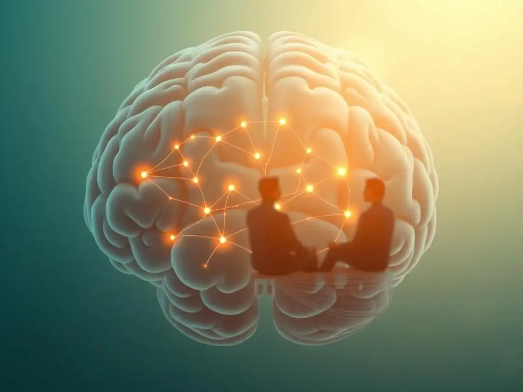
Studies on Cognitive Behavioral Therapy (CBT) and Brain Activity
Cognitive Behavioral Therapy (CBT) is a widely practiced therapy that focuses on identifying and changing negative thought patterns and behaviors. Numerous brain imaging studies have investigated its impact on neural activity. Research involving fMRI has consistently shown that successful CBT for conditions like anxiety disorders or depression is associated with altered activity in brain regions involved in emotional processing and regulation, such as the amygdala, ventromedial prefrontal cortex (vmPFC), and dorsal anterior cingulate cortex (dACC).
For instance, a study on individuals with social anxiety disorder receiving CBT showed a reduction in amygdala hyperreactivity to social threat cues after therapy, alongside increased activity in prefrontal regions involved in regulating emotional responses. These findings suggest that CBT helps individuals build resilience by strengthening top-down control over emotional reactions, a key aspect of the Brain imaging therapy resilience benefits. Another study on depression found that CBT was associated with normalization of activity in circuits related to reward processing and emotional regulation.
Research on Psychodynamic Therapy and Neural Connectivity
Psychodynamic therapy, which explores unconscious patterns and past experiences influencing present behavior, has also been subject to brain imaging research, though perhaps less extensively than CBT. Studies in this area often focus on changes in neural connectivity, particularly in networks related to self-referential processing and emotional memory.
One line of research has used fMRI to investigate the effects of psychodynamic therapy on the default mode network (DMN), a set of brain regions active when the mind is at rest or engaged in introspection. Dysregulation of the DMN is often observed in depression and anxiety. Studies have shown that successful psychodynamic therapy can be associated with changes in DMN connectivity, potentially reflecting altered ways of processing self-related information and past experiences. While specific figures can vary, findings often point towards more integrated and less rigid patterns of connectivity, contributing to greater psychological flexibility and resilience.
The Impact of Mindfulness-Based Therapies on Brain Structure and Function
Mindfulness-based therapies, such as Mindfulness-Based Stress Reduction (MBSR) or Mindfulness-Based Cognitive Therapy (MBCT), train individuals to pay attention to the present moment non-judgmentally. Brain imaging research has provided compelling evidence for their impact on brain structure and function, particularly in areas related to attention, emotional regulation, and self-awareness.
Multiple studies using structural MRI have reported increases in gray matter density in brain regions like the hippocampus (involved in memory and learning), the temporo-parietal junction (involved in perspective-taking), and areas of the prefrontal cortex after mindfulness training. Functionally, fMRI studies show that mindfulness practice can decrease amygdala reactivity to emotional stimuli and increase connectivity between the amygdala and the prefrontal cortex. These structural and functional changes are thought to underpin the enhanced emotional regulation and stress resilience observed in individuals who practice mindfulness, showcasing significant Brain imaging therapy resilience benefits.
Specific Brain Regions and Resilience: Evidence from Imaging
The Amygdala and Emotional Regulation: Brain imaging resilience benefits
The amygdala, a small almond-shaped structure deep within the temporal lobe, is a key player in processing emotions, particularly fear and threat detection. In individuals with low resilience or stress-related disorders, the amygdala often shows heightened reactivity to perceived threats. This can lead to exaggerated fear responses, anxiety, and difficulty recovering from stressful events.
Brain imaging studies consistently demonstrate that effective therapy, across various modalities, can lead to a reduction in amygdala activity in response to negative stimuli. For example, research might show a decrease in amygdala activation by approximately 20-30% after successful treatment for PTSD or anxiety.
Furthermore, therapy often strengthens the functional connection between the amygdala and the prefrontal cortex, allowing for better top-down regulation of emotional responses. This ability of therapy to modulate amygdala activity is a direct example of Brain imaging therapy resilience benefits, showing how treatment helps the brain become less reactive and more controlled in the face of emotional challenges.
The Prefrontal Cortex and Executive Function: Strengthening with Therapy
The prefrontal cortex (PFC), located at the front of the brain, is the seat of executive functions, including planning, decision-making, working memory, and regulating impulses and emotions. A well-functioning PFC is crucial for resilience, enabling individuals to assess situations rationally, inhibit maladaptive reactions, and pursue goals despite obstacles.
Brain imaging research frequently shows that therapy is associated with increased activity and improved connectivity within the PFC, as well as between the PFC and other brain regions involved in emotion and memory. For instance, studies might report increased gray matter volume or enhanced functional connectivity in specific PFC subregions after therapy for depression or anxiety.
These changes reflect a strengthening of the brain's executive control systems, allowing for more flexible and adaptive responses to stress. The enhancement of PFC function is a core component of the Brain imaging therapy resilience benefits, providing the neural basis for improved problem-solving and emotional control.
The Hippocampus and Memory Consolidation: Linking Trauma and Resilience
The hippocampus, located near the amygdala, is vital for forming and retrieving memories, particularly episodic memories (memories of specific events). It plays a critical role in contextualizing experiences and integrating them into our personal narrative. Chronic stress and trauma are known to negatively impact the hippocampus, sometimes leading to reduced volume and impaired function, which can contribute to difficulties with memory, learning, and emotional regulation.
Resilience involves the ability to process and integrate traumatic or stressful memories in a way that reduces their debilitating impact. Brain imaging studies have explored how therapy can influence hippocampal structure and function. Research on trauma-focused therapies, for example, has shown that successful treatment can be associated with increases in hippocampal volume or changes in hippocampal connectivity. These changes suggest that therapy helps individuals process traumatic memories more adaptively, reducing avoidance and promoting integration, thus linking hippocampal changes to Brain imaging therapy resilience benefits by facilitating healthier memory processing.
Brain Imaging and Long-Term Therapy Outcomes: A Correlation Analysis
Neural Signatures of Successful Therapy: Identifying Predictive Biomarkers
One exciting area of research using brain imaging is the search for neural signatures or biomarkers that predict responsiveness to therapy or indicate successful long-term outcomes. By examining baseline brain activity or structure before therapy begins, researchers hope to identify patterns that might suggest which individuals are more likely to benefit from a specific type of treatment.
Studies have explored various potential biomarkers, such as the connectivity patterns within the resting-state brain networks or the reactivity of certain brain regions to emotional stimuli. For example, some preliminary research suggests that certain patterns of connectivity between the amygdala and PFC might predict a better response to CBT for anxiety. While this field is still developing, the potential to use brain imaging to personalize therapy and predict Brain imaging therapy resilience benefits is significant, offering hope for more efficient and targeted interventions in the future.
Brain Changes Associated with Reduced Symptoms and Increased Well-being
Beyond predicting outcomes, brain imaging provides concrete evidence of the neural changes that occur alongside symptom reduction and increased well-being following long-term therapy. As individuals experience fewer symptoms of depression, anxiety, or PTSD, and report greater life satisfaction and coping ability, their brain scans often reflect these improvements.
These changes typically involve the normalization of activity in brain circuits that were previously dysregulated. This could mean reduced hyperactivity in threat-detection circuits, increased activity in reward and motivation pathways, or improved connectivity between regions involved in emotional regulation and cognitive control. These observed changes in brain function and structure provide a biological basis for the subjective experience of recovery and enhanced resilience, offering visible Brain imaging therapy resilience benefits.
Brain Scans Validate Long-Term Therapy Outcomes
For both therapists and clients, brain imaging offers a powerful form of validation for the effectiveness of long-term therapy. While subjective reports of feeling better and behavioral changes are the primary measures of success, seeing tangible changes in brain scans can reinforce the understanding that therapy leads to real, physical changes in the brain.
This validation is important for several reasons. It helps to destigmatize mental health conditions by showing they have a neurobiological basis, just like other medical conditions. It provides objective support for the efficacy of therapeutic interventions. And it can empower individuals by demonstrating that their efforts in therapy are actively reshaping their brains in ways that support recovery and resilience. The ability of brain scans to highlight Brain imaging therapy resilience benefits underscores the profound impact of psychological treatment on the brain.
Limitations of Brain Imaging in Therapy Research

Ethical Considerations and Data Interpretation Challenges
While promising, the use of brain imaging in therapy research is not without limitations. Ethical considerations are paramount, particularly regarding the privacy and potential misuse of brain data. Ensuring informed consent and protecting sensitive neural information are critical concerns that researchers must address rigorously.
Furthermore, interpreting brain imaging data is complex. Correlation does not equal causation; observing changes in brain activity alongside therapeutic progress doesn't definitively prove that the brain changes caused the improvement, although it strongly suggests a relationship. Brain activity is highly variable, influenced by many factors beyond therapy, and distinguishing therapy-specific changes from other life events or natural recovery processes can be challenging. The field is continuously refining methodologies to address these interpretation challenges and better understand the true Brain imaging therapy resilience benefits.
Cost and Accessibility of Brain Imaging Technologies
Brain imaging technologies like fMRI and PET scans are expensive to acquire and operate. This high cost limits their accessibility for routine clinical use and can pose significant barriers to conducting large-scale research studies, especially in diverse populations or resource-limited settings.
The expense means that brain imaging studies are often conducted with relatively small sample sizes, which can limit the generalizability of findings. While EEG is more affordable and accessible, it provides different types of information compared to fMRI or PET. The cost factor is a major hurdle in widely leveraging brain imaging to study Brain imaging therapy resilience benefits across the population and integrate these insights into standard clinical practice.
The Complexity of Brain-Behavior Relationships
The relationship between brain activity/structure and complex human behaviors like resilience is incredibly intricate. Resilience is not governed by a single brain region but emerges from the dynamic interaction of multiple brain networks working in concert. Understanding how these networks interact and change in response to therapy is a significant scientific challenge.
Current brain imaging techniques provide valuable snapshots or simplified models of this complexity. They cannot capture the full richness of subjective experience or the myriad external factors that influence behavior and resilience. Future research will need to employ more sophisticated analytical techniques and integrate multiple levels of analysis to fully unravel the complex brain-behavior links underpinning the Brain imaging therapy resilience benefits.
Future Directions: Advancing Brain Imaging in Mental Health
Developing Personalized Therapy Approaches Based on Brain Imaging Data
A major future direction for brain imaging in mental health is its potential to inform personalized therapy. If specific neural patterns can predict responsiveness to different types of therapy, clinicians could potentially use this information to match individuals with the most effective treatment from the outset.
For example, if research consistently shows that individuals with a certain pattern of prefrontal connectivity benefit most from CBT, while those with a different pattern benefit more from psychodynamic therapy, brain imaging could help guide treatment selection. This move towards precision mental health care, informed by neural data, holds great promise for maximizing Brain imaging therapy resilience benefits by tailoring interventions to the individual brain.
Integrating Brain Imaging with Other Measures of Psychological Functioning
To gain a more comprehensive understanding of resilience and the impact of therapy, brain imaging data needs to be integrated with other measures of psychological functioning. This includes self-report questionnaires on symptoms and well-being, behavioral assessments, genetic information, and physiological measures (like heart rate variability or cortisol levels).
Combining these diverse data streams through advanced analytical methods can provide a richer picture of the complex interplay between brain, body, and behavior. This integrated approach can help researchers validate brain imaging findings, identify mediating factors, and develop more robust models of how resilience is built and how therapy facilitates this process, enhancing our understanding of Brain imaging therapy resilience benefits in a broader context.
Exploring the Potential of Neurofeedback for Enhancing Resilience
Neurofeedback is a technique that uses real-time brain imaging (often EEG or fMRI) to show individuals their own brain activity patterns. The goal is to teach them to consciously alter their brain activity in desired ways. For example, someone might be trained to increase activity in a prefrontal region associated with emotional control or decrease activity in a hyperactive amygdala.
This technology holds potential as a therapeutic tool for directly targeting and training the neural circuits involved in resilience. While still largely experimental for enhancing general resilience, neurofeedback based on insights from brain imaging research could offer a novel way for individuals to gain more direct control over their brain states, potentially complementing traditional therapy and amplifying Brain imaging therapy resilience benefits.
Conclusion: The Power of Brain Imaging to Inform and Improve Therapy
Brain Imaging Therapy Resilience Benefits: A Summary
Brain imaging techniques like fMRI, EEG, and PET scans are revolutionizing our understanding of mental resilience and how therapy fosters this vital capacity. These technologies provide objective evidence that therapy induces measurable changes in brain structure and function, particularly in regions critical for emotional regulation (amygdala), executive function (prefrontal cortex), and memory processing (hippocampus). The Brain imaging therapy resilience benefits are seen as alterations in neural activity and connectivity that correlate with reduced symptoms, improved coping, and enhanced well-being.
Studies across various therapeutic modalities, including CBT, psychodynamic therapy, and mindfulness-based approaches, demonstrate that therapy actively reshapes the brain's landscape, strengthening the neural networks that underpin resilience. While challenges remain regarding cost, interpretation, and ethical considerations, the insights gained from brain imaging validate the profound impact of therapy and open new avenues for personalized and more effective mental health interventions.
Mind Care Guide: Empowering You Through Neuroscience Insights
At Mind Care Guide, we believe that understanding the science behind mental health can be incredibly empowering. The insights from brain imaging research not only validate the effectiveness of therapy but also provide a deeper appreciation for the brain's remarkable capacity for change and adaptation. By exploring topics like the Brain imaging therapy resilience benefits, we aim to equip you with knowledge that can inform your mental health journey.
Knowing that your efforts in therapy are supported by tangible changes in your brain can be motivating and reassuring. It underscores that resilience is not an innate, fixed trait but a skill that can be learned and strengthened, with visible effects on a biological level. We encourage you to explore these fascinating connections between mind and brain as you navigate your path towards greater well-being.
Neuroscience of Mental Health: Further Reading and Resources
The field of the neuroscience of mental health is rapidly evolving, continuously uncovering new insights into the brain's role in various psychological states and conditions. The research on Brain imaging therapy resilience benefits is just one piece of this larger puzzle. For those interested in delving deeper, numerous academic journals, books, and reputable online resources offer further information.
Exploring the neuroscience of mental health can provide a richer context for understanding challenges like anxiety, depression, and trauma, as well as the pathways to recovery and resilience. Staying informed about these scientific advancements helps to appreciate the biological underpinnings of mental well-being and the evidence-based approaches that support it.
Frequently Asked Questions
What is brain imaging therapy and how does it relate to resilience?
Brain imaging therapy isn't a therapy itself, but rather the use of brain imaging techniques (like fMRI) to understand how therapies affect the brain. Studying these changes helps us understand and potentially enhance resilience benefits.
How can brain imaging techniques help us understand resilience?
Brain imaging allows researchers to observe brain activity and structural changes associated with resilience. This reveals which brain regions are involved in coping with stress and how therapy can strengthen those areas.
What are some specific brain imaging techniques used to study resilience?
Common techniques include functional Magnetic Resonance Imaging (fMRI), which measures brain activity, and Diffusion Tensor Imaging (DTI), which assesses the integrity of white matter tracts. These help visualize how the brain responds to therapy.
What are the potential benefits of understanding resilience through brain imaging?
Understanding the neural mechanisms of resilience can lead to more effective therapeutic interventions. This could mean personalized treatment plans and strategies to enhance coping skills based on individual brain responses.
How does "Mind Care Guide" support individuals seeking information on brain imaging therapy resilience benefits?
"Mind Care Guide" provides comprehensive information on mental well-being, including the science behind resilience and how therapy impacts the brain. We offer resources and support to navigate various mental health challenges.
Final Reflection: Unfolding the Resilient Brain
The science of resilience is no longer a theoretical pursuit; it's a tangible, measurable reality revealed through brain imaging. Understanding how therapy reshapes the brain's architecture to foster resilience offers a powerful antidote to the pervasive anxieties of our modern world. This knowledge empowers us to actively cultivate mental strength and navigate life's inevitable challenges with greater ease.
The impact of this research extends far beyond the clinical setting; it's fundamentally altering our understanding of mental well-being. It challenges the outdated stigma surrounding mental health struggles, highlighting the brain's remarkable plasticity and capacity for healing. This shift in perspective is crucial for fostering a more compassionate and supportive society. The brain is not fixed; it is constantly changing and adapting in response to our experiences.
This quote encapsulates the core message of this article, emphasizing the potential for positive transformation through targeted interventions.
Consider the implications of this research on your own life. What steps can you take today to nurture your brain's resilience? Perhaps it's seeking professional support, practicing mindfulness, engaging in activities that bring you joy, or simply cultivating stronger social connections. The journey towards resilience is a personal one, but the science presented here provides a roadmap, illuminating the path towards a stronger, more adaptable, and ultimately, happier you.
Unlock Your Brain's Resilience: Take the Next Step
Thanks for exploring the fascinating science of resilience with us! You've now gained valuable insights into how therapy impacts brain function and strengthens your ability to bounce back from life's challenges. Ready to put this knowledge into action?
- Start your resilience journey: Identify one small, achievable step you can take today to build your resilience—it could be as simple as practicing mindful breathing for five minutes or connecting with a supportive friend.
- Share your insights: Discuss this article with someone you know who might benefit from learning about resilience and the power of therapeutic interventions. Let's spread awareness together!
- Explore further: Delve deeper into the resources mentioned in the article to find additional tools and techniques to strengthen your mental wellbeing. Consider researching mindfulness exercises or local therapy options.
What small step will you take today to cultivate your brain's natural resilience?