
Is Your Brain Different After Therapy? A Look at fMRI Studies

Welcome to Mind Care Guide, a safe space dedicated to understanding and nurturing your mental well-being.
Ever wondered what really happens inside your mind during therapy? Our latest exploration delves into the fascinating world of neuroscience, specifically focusing on fMRI therapy brain changes. Join us as we unravel the secrets revealed by fMRI studies and discover how therapy can reshape the very fabric of your brain. Ready to unlock the science behind healing? Let's begin.
Is Your Brain Different After Therapy? A Look at fMRI Studies
- Introduction: Exploring Brain Changes Through fMRI After Therapy
- fMRI Studies: A Window into the Therapeutic Brain
- Specific Mental Health Conditions and fMRI-Observed Brain Changes Post-Therapy
- Types of Therapy and Corresponding fMRI Brain Changes
- fMRI Therapy Brain Changes: Interpreting the Results
- Beyond fMRI: Complementary Neuroimaging Techniques
- Future Directions in fMRI Therapy Brain Changes Research
- Conclusion: The Promise of fMRI in Understanding and Enhancing Therapy
-
Frequently Asked Questions
- What is fMRI and how is it used in mental health research?
- Can therapy actually change the physical structure or function of the brain, as shown by fMRI studies?
- What types of mental health conditions have shown brain changes after therapy, according to fMRI studies?
- What specific brain regions are most commonly affected by therapy, as revealed by fMRI?
- How can understanding fMRI therapy brain changes help improve mental health treatments in the future?
- Final Reflection: The Evolving Landscape of the Therapeutic Brain
- Unlock Your Brain's Potential: Next Steps to Well-being
Introduction: Exploring Brain Changes Through fMRI After Therapy
The Growing Interest in Neuroscience and Therapy Outcomes
The intersection of neuroscience and mental health treatment is a rapidly expanding field, captivating both researchers and individuals seeking effective care. For years, the benefits of psychotherapy have been recognized through observable changes in behavior, mood, and cognitive function. However, a deeper question lingers: how exactly does talking and processing experiences translate into lasting change within the intricate network of the brain?
This growing interest stems from a desire to understand the biological underpinnings of psychological transformation and to potentially enhance therapeutic approaches. The exploration of fMRI therapy brain changes is central to this endeavor, providing a window into the neural mechanisms at play during and after successful treatment.
This fascination is fueled by technological advancements, particularly in neuroimaging techniques like functional Magnetic Resonance Imaging (fMRI). As our ability to non-invasively observe brain activity improves, so does our potential to correlate psychological interventions with specific neural shifts. Understanding these brain changes after therapy, as revealed by fMRI, can help validate the efficacy of different therapeutic modalities and offer insights into why some individuals respond more effectively than others. It moves the conversation beyond subjective reports to objective, biological evidence of healing and adaptation.
Understanding fMRI Technology and Its Role in Mental Health Research
Functional Magnetic Resonance Imaging (fMRI) is a neuroimaging technique that measures brain activity by detecting changes associated with blood flow. When a specific area of the brain is more active, it consumes more oxygen, and blood flow increases to that region. fMRI tracks these changes in blood flow, creating maps of brain activity that show which areas are engaged during particular tasks or in response to certain stimuli.
This provides researchers with a dynamic view of the brain at work, unlike structural MRI which only captures the brain's anatomy. Its non-invasive nature makes it a valuable tool for studying the living brain.
In the context of mental health research, fMRI is employed to investigate the neural correlates of various conditions, from depression and anxiety to PTSD and OCD. By scanning individuals while they perform specific tasks (like emotional processing or memory recall) or while they are at rest, researchers can identify patterns of activity or connectivity that differ between healthy individuals and those with mental health challenges.
Crucially, fMRI is increasingly used before and after therapy to examine whether and how therapeutic interventions lead to observable changes in these identified brain patterns, shedding light on the mechanisms behind fMRI therapy brain changes.
The Core Question: Does Therapy Actually Change the Brain?
The fundamental question driving much of this research is whether psychotherapy, a non-pharmacological intervention focused on thoughts, feelings, and behaviors, can induce measurable and lasting changes in the brain. While the concept of neuroplasticity – the brain's ability to reorganize itself by forming new neural connections throughout life – provides the theoretical basis for this possibility, fMRI studies aim to provide empirical evidence. They seek to demonstrate that engaging in therapeutic processes is not merely a psychological shift but is accompanied by tangible alterations in brain function and structure.
Evidence from numerous fMRI studies suggests that the answer is indeed yes. Therapy appears to influence the activity levels, connectivity patterns, and even the volume of specific brain regions known to be involved in emotion regulation, cognitive processing, and stress response. These observed fMRI therapy brain changes provide compelling support for the idea that psychological interventions have a biological impact, reinforcing the value and effectiveness of therapy as a treatment for mental health conditions. They offer a biological anchor to the subjective experience of healing and recovery.
fMRI Studies: A Window into the Therapeutic Brain
How fMRI Works: Measuring Brain Activity During Therapy
fMRI operates on the principle of Blood-Oxygen-Level Dependent (BOLD) contrast. When neurons become active, they require more energy, leading to an increase in blood flow to that region. This surge in oxygenated blood changes the magnetic properties of the blood, which the fMRI scanner detects. By tracking these magnetic signal variations over time, the fMRI machine generates detailed images showing which brain areas are more or less active during specific periods or tasks. This allows researchers to observe dynamic neural processes in real-time or near real-time.
In therapy research, participants might undergo fMRI scans while engaging in tasks designed to probe areas relevant to their condition, such as viewing emotionally charged images, recalling traumatic memories, or performing cognitive tasks related to rumination or worry. Some studies even attempt to scan participants during simulated therapy sessions, though this is technically challenging. By comparing brain activity patterns before therapy to those after a course of treatment, researchers can identify significant fMRI therapy brain changes, such as altered reactivity to stimuli or shifts in resting-state connectivity, which are presumed to be related to the therapeutic outcomes.
Key Areas of the Brain Studied in Therapy Research: [Amygdala, Prefrontal Cortex, Anterior Cingulate Cortex]
Several brain regions are consistently highlighted in fMRI studies investigating mental health conditions and the impact of therapy. The amygdala, often referred to as the brain's "fear center," plays a crucial role in processing emotions, particularly fear and threat detection. In many anxiety disorders and PTSD, the amygdala shows heightened activity.
The prefrontal cortex (PFC), especially the medial and dorsolateral areas, is involved in executive functions like decision-making, planning, working memory, and perhaps most importantly in this context, regulating emotions and inhibiting responses from areas like the amygdala. Dysfunction or reduced activity in certain PFC areas is linked to depression and anxiety.
The anterior cingulate cortex (ACC) is another key area, involved in conflict monitoring, error detection, and emotional regulation. It plays a role in attention and the integration of cognitive and emotional information. Research into fMRI therapy brain changes frequently examines how therapy influences the activity and connectivity within and between these regions. For instance, successful therapy might be associated with decreased amygdala reactivity, increased PFC activity and its regulatory influence over the amygdala, and altered activity in the ACC reflecting improved emotional processing and cognitive control.
Ethical Considerations and Limitations of fMRI Studies in Therapy Research
While fMRI offers exciting possibilities for understanding fMRI therapy brain changes, its use in research involves significant ethical considerations. Participants must be fully informed about the procedure, including the enclosed space, noise, and potential discomfort, and provide informed consent. Ensuring participant privacy and the secure handling of sensitive neuroimaging data are paramount. Furthermore, researchers must be careful not to overstate the implications of findings, avoiding neuro-essentialism or reducing complex psychological experiences solely to brain activity patterns. Interpreting fMRI data requires careful consideration of individual variability and context.
fMRI also has inherent limitations in the context of therapy research. The technology captures neural activity indirectly through blood flow, which is a relatively slow process compared to the speed of neural communication. This limits its temporal resolution.
The spatial resolution is also finite, meaning it captures activity in clusters of neurons rather than individual ones. The scanner environment itself can be stressful or unnatural, potentially influencing brain activity. Moreover, establishing clear cause-and-effect between specific therapeutic interventions and observed fMRI therapy brain changes remains challenging due to the complexity of therapy, individual differences, and the correlational nature of many fMRI studies.
Specific Mental Health Conditions and fMRI-Observed Brain Changes Post-Therapy
Depression: fMRI Evidence of Neural Pathway Restructuring After Therapy
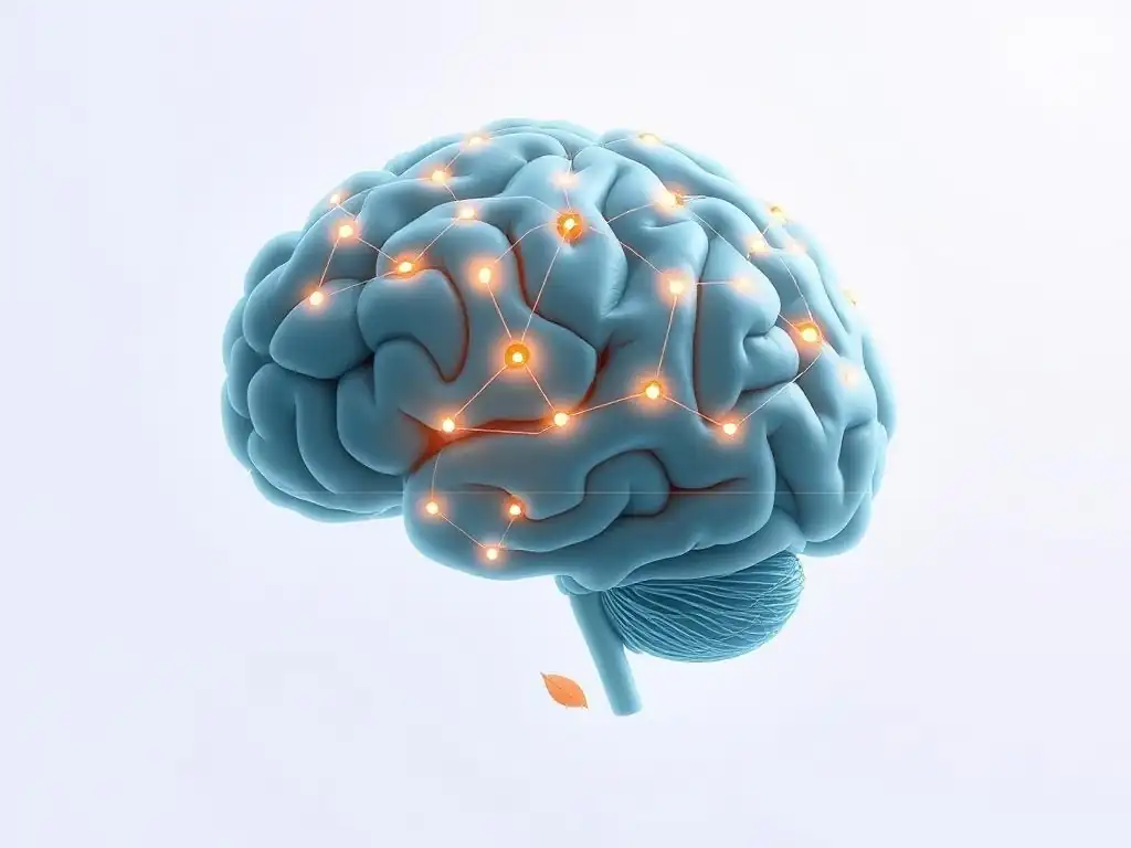
Depression is associated with altered activity and connectivity in several brain regions, including the prefrontal cortex, amygdala, hippocampus, and anterior cingulate cortex. fMRI studies before and after psychotherapy for depression, such as Cognitive Behavioral Therapy (CBT) or Interpersonal Therapy (IPT), have provided compelling evidence of neural pathway restructuring. These studies often show shifts in how these regions interact and respond to emotional stimuli. For example, areas involved in processing negative self-referential thoughts may show decreased activity, while areas associated with positive emotional processing or cognitive control may show increased engagement.
This restructuring suggests that therapy doesn't just change how someone feels about their depression; it may literally change the underlying brain circuits contributing to the condition. Observing these fMRI therapy brain changes provides biological validation for the efficacy of psychotherapy in treating depression. It reinforces the idea that psychological interventions can facilitate significant neural adaptation, helping individuals break free from maladaptive thought patterns and emotional responses rooted in altered brain function.
Changes in Amygdala Activity in Depressed Patients Following Cognitive Behavioral Therapy (CBT)
A common finding in fMRI studies of depressed patients is elevated activity in the amygdala, particularly in response to negative emotional stimuli. Following a course of Cognitive Behavioral Therapy (CBT), studies have frequently observed a significant reduction in this heightened amygdala reactivity. For instance, research has shown that after successful CBT, depressed individuals exhibit amygdala responses to negative faces that are more similar to those of healthy control groups, compared to their pre-treatment scans. This suggests that CBT helps to dampen the brain's exaggerated response to perceived threats or negative information.
This decrease in amygdala activity is often correlated with improvements in depressive symptoms. The fMRI therapy brain changes observed here indicate that CBT may work, in part, by recalibrating the brain's emotional alarm system. By teaching new ways to identify and challenge negative thoughts, CBT techniques appear to influence how the amygdala processes emotional input, leading to a less intense and persistent negative affective state.
Prefrontal Cortex Activation and Improved Emotional Regulation After Therapy
Depression is also linked to reduced activity in certain areas of the prefrontal cortex (PFC), particularly those involved in cognitive control and emotional regulation. These areas are crucial for managing emotional responses and shifting attention away from negative stimuli. fMRI studies have demonstrated that successful psychotherapy for depression is often associated with increased activation in these specific PFC regions. For example, research has shown enhanced activity in the dorsolateral PFC during tasks requiring emotional regulation after individuals completed a course of therapy.
This increase in PFC activation is thought to reflect improved top-down control over emotional centers like the amygdala. The fMRI therapy brain changes observed in the PFC suggest that therapy strengthens the brain's capacity for emotional regulation. As individuals learn new coping strategies and cognitive skills in therapy, their prefrontal cortex becomes more engaged and effective in managing challenging emotions and redirecting attention, contributing to symptom reduction and improved mood.
Anxiety Disorders: fMRI Insights into Fear Processing and Therapy Effectiveness
Anxiety disorders, such as Generalized Anxiety Disorder (GAD), Social Anxiety Disorder (SAD), and phobias, are characterized by excessive fear and worry. fMRI studies in these populations often reveal hyperactive amygdala responses to threat cues and altered connectivity within the fear circuitry. Therapy, particularly exposure-based therapies and CBT, has been highly effective in treating anxiety. fMRI is being used to understand the neural mechanisms underlying these therapeutic effects, focusing on fMRI therapy brain changes related to fear processing and regulation.
Research consistently points to therapy's ability to modulate the brain's response to anxiety-provoking stimuli. By systematically confronting feared situations or thoughts in a safe environment, therapy helps to extinguish fear responses and build new, non-fearful associations. The fMRI therapy brain changes observed reflect this process, showing shifts in activity within the limbic system and the prefrontal cortex, indicating a biological basis for overcoming excessive fear and anxiety.
Reduced Amygdala Reactivity to Threat Stimuli Post-Therapy
Similar to depression, heightened amygdala activity is a hallmark of anxiety disorders when individuals are presented with threat-related stimuli (e.g., pictures of snakes for someone with ophidiophobia, or negative social cues for someone with social anxiety). Following successful therapy, particularly exposure therapy or CBT, fMRI studies have consistently shown a significant reduction in this exaggerated amygdala response. For example, research on individuals with social anxiety disorder after receiving CBT demonstrated decreased amygdala activation when viewing critical faces, compared to their pre-treatment scans.
This decreased amygdala reactivity is a key fMRI therapy brain change indicating successful treatment. It suggests that therapy helps to desensitize the brain's threat detection system. By repeatedly confronting feared situations without experiencing actual harm, the amygdala's alarm signal becomes less intense and persistent, reducing the physiological and emotional components of anxiety.
Increased Connectivity Between Prefrontal Cortex and Amygdala in Anxious Patients
Beyond changes in activity within single regions, fMRI studies also examine functional connectivity – how different brain areas communicate with each other. In anxiety disorders, there is often reduced inhibitory connectivity from the prefrontal cortex (PFC) to the amygdala, meaning the PFC is less effective at regulating the amygdala's activity. Successful therapy has been associated with increased functional connectivity between these two crucial regions.
This increased connectivity suggests that therapy strengthens the communication pathway that allows the cognitive, regulatory parts of the brain (PFC) to exert more control over the emotional, reactive parts (amygdala). The fMRI therapy brain changes in connectivity indicate that therapy facilitates the development of more effective neural circuits for managing fear and worry, enabling individuals to better regulate their emotional responses in the face of potential threats.
PTSD: Neural Correlates of Trauma Processing and Recovery Observed with fMRI
Post-Traumatic Stress Disorder (PTSD) is characterized by persistent re-experiencing of trauma, avoidance, negative alterations in mood and cognition, and hyperarousal. fMRI studies in PTSD patients often reveal a complex pattern of altered brain activity, including hyperactive amygdala responses to trauma-related cues, hypoactive medial prefrontal cortex (mPFC), and reduced hippocampal volume. Trauma-focused therapies, such as Prolonged Exposure (PE) or Eye Movement Desensitization and Reprocessing (EMDR), are effective treatments. fMRI is used to map the neural changes associated with processing traumatic memories and achieving recovery.
Research using fMRI has provided insights into how trauma processing in therapy leads to neural normalization. By safely confronting and processing traumatic memories, individuals can integrate these experiences in a less threatening way. The fMRI therapy brain changes observed often include shifts in the activity and interaction of regions involved in memory, emotion regulation, and threat response, reflecting the brain's capacity to heal and adapt even after severe trauma.
fMRI Evidence of Hippocampal Volume Changes After Trauma-Focused Therapy
The hippocampus, a brain structure critical for memory formation and retrieval, is often found to be smaller in individuals with PTSD compared to healthy controls. While the exact relationship (whether it's a cause or consequence of trauma) is debated, fMRI studies combined with structural MRI have explored whether therapy impacts hippocampal volume. Some research suggests that successful trauma-focused therapy may be associated with an increase in hippocampal volume or a normalization of its activity patterns, particularly during memory-related tasks.
These potential fMRI therapy brain changes in the hippocampus are significant because the hippocampus plays a role in contextualizing memories – helping to distinguish between past traumatic events and present safety. Changes here could reflect an improved ability to process traumatic memories as belonging to the past, rather than feeling constantly threatened in the present, contributing to a reduction in re-experiencing symptoms.
Altered Brain Network Connectivity in PTSD Patients Post-Therapy
Beyond individual regions, PTSD is associated with altered connectivity within large-scale brain networks, such as the default mode network (DMN), salience network, and central executive network. For example, there might be increased connectivity within the salience network (involved in detecting important stimuli) and reduced connectivity between the DMN (active during self-referential thought) and executive control regions. fMRI studies have shown that successful trauma-focused therapy can lead to a normalization of these network connectivity patterns.
For instance, studies have observed increased connectivity between the ventromedial prefrontal cortex (part of the DMN involved in emotion regulation) and the amygdala after therapy, similar to findings in anxiety. There may also be shifts in connectivity within the DMN, potentially reflecting changes in how individuals process self-referential and trauma-related thoughts. These fMRI therapy brain changes in connectivity suggest that therapy helps to reconfigure the brain's large-scale networks, facilitating better integration of emotional and cognitive processing and reducing the intrusive nature of traumatic memories.
Types of Therapy and Corresponding fMRI Brain Changes
Cognitive Behavioral Therapy (CBT): fMRI Studies on Cognitive Restructuring
Cognitive Behavioral Therapy (CBT) is one of the most widely studied psychotherapies using fMRI. Its core principle is that psychological problems are based, in part, on faulty or unhelpful ways of thinking (cognitions) and learned patterns of unhelpful behavior. CBT aims to identify and change these patterns. fMRI studies have sought to understand the neural mechanisms underlying CBT's effectiveness by observing fMRI therapy brain changes associated with its techniques, particularly cognitive restructuring.
These studies often focus on brain regions involved in cognitive control, emotional regulation, and the processing of negative information. By examining brain activity as patients engage in tasks designed to mimic cognitive restructuring (e.g., evaluating negative thoughts), researchers can see how CBT influences the neural circuits involved in these processes. The observed fMRI therapy brain changes provide a biological basis for how altering thought patterns can lead to significant improvements in mood and behavior.
How CBT Impacts Neural Pathways Related to Negative Thought Patterns
CBT directly targets negative or distorted thought patterns that contribute to mental health issues like depression and anxiety. fMRI studies suggest that CBT impacts the neural pathways involved in generating and maintaining these thoughts. For example, studies have shown decreased activity in brain regions associated with self-referential processing and rumination (parts of the default mode network) in depressed patients after CBT.
This indicates that CBT may help to disengage the brain from excessive negative self-focus. The fMRI therapy brain changes observed here suggest that by teaching patients to identify and challenge maladaptive thoughts, CBT weakens the neural connections that support these patterns while potentially strengthening pathways associated with more adaptive thinking and cognitive flexibility.
fMRI-Detected Changes in Brain Activity Associated with CBT Techniques
Specific techniques used in CBT, such as cognitive reappraisal (reinterpreting a situation to change its emotional impact) or exposure (gradually confronting feared stimuli), are associated with distinct fMRI therapy brain changes. For instance, studies on cognitive reappraisal training have shown increased activity in prefrontal regions and decreased activity in the amygdala during emotional tasks. Exposure therapy for anxiety has been linked to reduced amygdala reactivity to feared stimuli and altered connectivity within fear circuits.
These findings demonstrate that the practical exercises and strategies taught in CBT have observable effects on brain function. The fMRI-detected changes provide neural correlates for the psychological processes central to CBT's effectiveness, showing how practicing new cognitive and behavioral skills can lead to measurable alterations in brain activity patterns associated with emotional and cognitive regulation.
Psychodynamic Therapy: Uncovering Unconscious Processes Through fMRI
Psychodynamic therapy focuses on exploring unconscious processes, past experiences, and their impact on present behavior and relationships. While seemingly less amenable to direct fMRI study than behavioral therapies, researchers are using neuroimaging to investigate the neural underpinnings of key psychodynamic concepts like insight, self-awareness, and the therapeutic relationship (transference). fMRI therapy brain changes in this context might involve regions related to self-reflection, memory, and social cognition.
Studies in this area often explore brain activity during tasks involving self-reflection, thinking about relationships, or processing emotionally significant material. The goal is to identify neural correlates of the psychological work done in psychodynamic therapy. While research is less extensive than for CBT, initial fMRI findings offer intriguing possibilities for understanding the biological basis of insight-oriented change and the impact of exploring deeper emotional conflicts.
Neural Correlates of Insight and Self-Awareness Observed in fMRI Scans
A central goal of psychodynamic therapy is gaining insight into unconscious patterns and motivations. fMRI studies exploring insight have shown activation in regions like the medial prefrontal cortex (mPFC) and anterior cingulate cortex (ACC), areas involved in self-referential processing, introspection, and conflict monitoring. Research comparing brain activity during moments of insight versus non-insightful problem-solving has highlighted the involvement of these regions.
While directly linking these findings specifically to psychodynamic therapy is ongoing, the fMRI therapy brain changes associated with general processes of self-awareness and gaining new perspectives provide potential neural correlates for the transformative moments that can occur in therapy. They suggest that the neural machinery involved in self-reflection is engaged and potentially modified as individuals gain deeper understanding of themselves and their difficulties.
fMRI Studies Exploring the Impact of Transference on Brain Activity
Transference, the process by which feelings and attitudes from past relationships are unconsciously redirected toward the therapist, is a key concept in psychodynamic therapy. Studying transference with fMRI is complex but offers a unique opportunity to explore the neural basis of relational patterns. Research might involve tasks where participants think about significant relationships or interact with stimuli representing the therapeutic relationship.
Early fMRI research in this area is exploratory but hints at the involvement of brain regions associated with social cognition, emotional processing, and memory retrieval when individuals engage in thinking about relational dynamics central to transference. While not yet providing definitive fMRI therapy brain changes specific to resolving transference, this line of research demonstrates the potential of neuroimaging to investigate even the more abstract concepts of psychodynamic theory at a biological level.
Mindfulness-Based Therapies: fMRI Evidence of Enhanced Self-Regulation
Mindfulness-Based Stress Reduction (MBSR) and Mindfulness-Based Cognitive Therapy (MBCT) teach skills like focused attention and non-judgmental awareness. These therapies aim to enhance self-regulation, reduce reactivity to stress, and foster a different relationship with thoughts and emotions. fMRI studies have provided significant evidence for the neural effects of mindfulness practice, highlighting fMRI therapy brain changes related to attention, emotion regulation, and self-awareness.
Research consistently shows that mindfulness training impacts brain areas involved in executive control, attention, and emotional processing. By regularly practicing mindfulness techniques, individuals appear to strengthen neural pathways that support present-moment awareness and reduce automatic, reactive responses to internal and external stimuli. These fMRI-observed changes offer a neurobiological explanation for the reported benefits of mindfulness in reducing stress, anxiety, and depression.
Increased Activation in Prefrontal Cortex and Anterior Cingulate Cortex (ACC)
Mindfulness training is associated with increased activity and/or gray matter volume in regions of the prefrontal cortex (PFC), particularly areas involved in executive control, attention, and self-awareness, as well as the anterior cingulate cortex (ACC). For example, fMRI studies have shown increased activation in the dorsolateral PFC during attention tasks after mindfulness training. The ACC, involved in conflict monitoring and attention to internal states, also shows altered activity patterns.
These fMRI therapy brain changes suggest that mindfulness strengthens the brain's capacity for focused attention and cognitive control. By training the ability to direct and sustain attention and to notice internal experiences without immediate reaction, mindfulness practices enhance the function of these key regulatory brain regions, contributing to improved self-control and reduced reactivity.
fMRI Findings on the Impact of Mindfulness on Stress Reduction and Emotional Control
A core benefit of mindfulness is its impact on stress reduction and emotional regulation. fMRI studies investigating these effects often show decreased reactivity in the amygdala to negative stimuli and increased functional connectivity between the prefrontal cortex and the amygdala after mindfulness training. This pattern is similar to findings in CBT and other effective therapies.
Furthermore, studies have shown that mindfulness practice can alter the brain's response to stress. For example, individuals with more mindfulness experience may show a less pronounced physiological stress response (like cortisol release) and corresponding fMRI patterns indicating reduced activation in stress-related brain circuits when exposed to stressors. These fMRI therapy brain changes underscore how mindfulness fosters a more regulated and less reactive stance towards challenging emotions and stressful situations, providing a neural basis for its calming effects.
fMRI Therapy Brain Changes: Interpreting the Results
The Correlation vs. Causation Dilemma in fMRI Therapy Research
When interpreting fMRI therapy brain changes, it is crucial to understand the difference between correlation and causation. Most fMRI studies observe changes in brain activity or structure that occur alongside or after successful therapy. This demonstrates a correlation – that the brain changes and symptom improvement happen together. However, it doesn't definitively prove that the therapy caused the brain changes, or that the brain changes caused the symptom improvement. Other factors, such as the passage of time, placebo effects, or concurrent life events, could also play a role.
While strong correlations observed across multiple studies using different methodologies can build a compelling case for a causal link, establishing direct causation is complex. Researchers use experimental designs, such as randomized controlled trials comparing therapy to control conditions, to strengthen causal inferences. Despite this challenge, the consistent observation of specific fMRI therapy brain changes across diverse populations and therapeutic approaches strongly suggests that therapy is a powerful catalyst for neurobiological shifts.
Individual Variability in Brain Responses to Therapy: A Personalized Approach
It's important to recognize that fMRI therapy brain changes are not uniform across all individuals. Just as people respond differently to therapy on a psychological level, their brains also exhibit variability in how they change. Factors such as the specific mental health condition, severity of symptoms, individual genetic makeup, life experiences, and even personality traits can influence the nature and extent of brain changes observed with fMRI after therapy.
This individual variability highlights the need for a personalized approach to understanding fMRI therapy brain changes. Future research aims to identify specific brain patterns or biomarkers that predict who will respond best to which type of therapy. Recognizing that each brain is unique underscores the importance of tailoring therapeutic interventions to the individual, rather than assuming a one-size-fits-all model will yield the same neural or psychological outcomes for everyone.
The Role of Neuroplasticity in Long-Term Therapy Outcomes
The ability of the brain to change and adapt throughout life, known as neuroplasticity, is the fundamental biological principle underlying fMRI therapy brain changes. Therapy works because the brain is not a static organ; its structure and function can be modified by experience, learning, and environmental demands. Successful therapy involves new learning – learning new coping skills, new ways of thinking, and new emotional regulation strategies.
These new learnings are encoded in the brain through the formation of new neural connections, the strengthening or weakening of existing ones, and potentially even changes in the birth and survival of neurons in certain areas like the hippocampus. The long-term effectiveness of therapy relies on the persistence of these neuroplastic changes. fMRI studies provide a way to visualize some aspects of this neuroplastic process, showing how engaging in therapeutic practices can lead to enduring alterations in the neural circuits associated with mental well-being, contributing to sustained recovery and resilience.
Beyond fMRI: Complementary Neuroimaging Techniques
EEG (Electroencephalography) and Its Use in Therapy Research
While fMRI excels at providing spatial information about where activity occurs in the brain, Electroencephalography (EEG) offers superior temporal resolution, measuring electrical activity in the brain with millisecond precision. EEG uses electrodes placed on the scalp to detect the summed electrical activity of large populations of neurons. In therapy research, EEG can be used to study rapid neural responses to stimuli, track changes in brainwave patterns associated with different states (e.g., relaxation, concentration), or examine event-related potentials (ERPs) – specific brain responses time-locked to a stimulus or event.
EEG provides a complementary perspective to fMRI therapy brain changes. For example, while fMRI might show which brain regions are active during emotional processing after therapy, EEG could reveal how quickly and with what pattern the brain responds to emotional cues. Combining EEG and fMRI data can offer a more complete picture of the temporal and spatial dynamics of neural changes associated with therapeutic interventions.
PET (Positron Emission Tomography) and its Application to Mental Health Studies
Positron Emission Tomography (PET) is another neuroimaging technique used in mental health research. PET involves injecting a small amount of a radioactive tracer into the bloodstream. This tracer binds to specific molecules in the brain, such as neurotransmitter receptors or metabolic indicators like glucose. PET scans then detect the distribution of the tracer, providing information about brain metabolism, neurotransmitter activity, or receptor density.
PET offers insights different from fMRI therapy brain changes, focusing more on neurochemical and metabolic processes. For instance, PET can be used to study changes in serotonin or dopamine receptor availability in patients with depression or addiction before and after therapy or medication. While less commonly used than fMRI in routine therapy outcome studies due to its invasiveness, PET can provide crucial information about the neurochemical basis of mental health conditions and how therapy might influence these systems, offering a deeper understanding alongside fMRI findings.
Combining Neuroimaging Techniques for a More Complete Understanding
Each neuroimaging technique offers a unique window into brain function and structure. fMRI provides excellent spatial resolution for functional activity, EEG offers high temporal resolution for electrical activity, and PET provides insights into neurochemistry and metabolism. Relying on a single technique provides only a partial view of the complex fMRI therapy brain changes that occur.
Increasingly, researchers are combining these techniques to gain a more comprehensive understanding of how therapy impacts the brain. For example, simultaneous EEG-fMRI allows researchers to link rapid electrical events with their spatial location. Combining fMRI with PET can connect observed changes in regional activity or connectivity with underlying changes in neurotransmitter systems. This multi-modal approach is essential for building a detailed and nuanced picture of the intricate neural adaptations facilitated by psychotherapy.
Future Directions in fMRI Therapy Brain Changes Research
Longitudinal fMRI Studies: Tracking Brain Changes Over Time
Much of the current research on fMRI therapy brain changes relies on pre- and post-treatment scans. While informative, this approach captures snapshots rather than the dynamic process of change. Future directions are moving towards longitudinal fMRI studies that involve multiple scans throughout the course of therapy and during follow-up periods. This allows researchers to track how brain activity and connectivity evolve as individuals progress through therapy and whether changes are maintained over time.
Longitudinal studies can help identify critical periods for change, understand the trajectory of neural recovery, and determine the stability of fMRI therapy brain changes months or even years after treatment ends. This approach is crucial for understanding the long-term impact of therapy and for identifying potential neural markers of relapse or sustained remission.
Developing Biomarkers for Therapy Response Using fMRI Data
One exciting future direction for fMRI therapy brain changes research is the development of neural biomarkers that can predict an individual's likelihood of responding to a particular type of therapy. Currently, choosing a therapy is often based on diagnosis and clinician experience, with significant trial and error involved. fMRI patterns observed before therapy could potentially offer clues about how an individual's brain is wired and thus which therapeutic approach might be most effective for them.
For example, specific patterns of connectivity or reactivity in the amygdala or prefrontal cortex might predict better response to CBT versus psychodynamic therapy, or vice versa. Identifying such fMRI-based biomarkers could revolutionize clinical practice by enabling more personalized and effective treatment selection from the outset, saving time and resources and improving outcomes for individuals seeking help.
Integrating fMRI Findings into Clinical Practice: [Tailored Treatment Plans]
Ultimately, the goal of understanding fMRI therapy brain changes is to improve clinical care. While fMRI scans are not currently used for routine diagnosis or treatment planning in most clinical settings, the insights gained from research are gradually informing practice. The growing evidence that therapy induces measurable brain changes helps to destigmatize mental health conditions and validate the power of psychological interventions.
In the future, as fMRI research refines our understanding of specific neural signatures of different conditions and responses to therapy, it may become possible to integrate these findings into tailored treatment plans. For instance, if fMRI identifies persistent maladaptive patterns in specific brain circuits, it might suggest targeting those circuits more directly through specific therapeutic techniques or even combined approaches (e.g., therapy plus neuromodulation techniques). This vision of precision mental healthcare, informed by neurobiological data including fMRI therapy brain changes, holds significant promise for the future.
Conclusion: The Promise of fMRI in Understanding and Enhancing Therapy
Summarizing the Key Findings on fMRI-Observed Brain Changes After Therapy
FMRI studies have provided compelling evidence that psychotherapy is associated with observable and measurable changes in the brain. Research across various mental health conditions, including depression, anxiety disorders, and PTSD, consistently shows fMRI therapy brain changes in key regions involved in emotion regulation, cognitive control, and threat processing, such as the amygdala, prefrontal cortex, and anterior cingulate cortex. These changes often involve altered activity levels, modified connectivity patterns between regions, and in some cases, structural changes like shifts in gray matter volume. Different types of therapy, such as CBT, psychodynamic therapy, and mindfulness-based approaches, appear to induce distinct but often converging patterns of neural change, reflecting their specific mechanisms of action on the brain's circuits.
These findings move beyond simply noting symptom reduction and provide a biological basis for the effectiveness of psychological interventions. They suggest that therapy facilitates neuroplasticity, helping the brain to reorganize and adapt in ways that support improved emotional regulation, more flexible thinking, and reduced reactivity to stressors and triggers associated with mental health challenges.
The Potential for fMRI to Revolutionize Mental Healthcare
The insights gained from fMRI therapy brain changes research have the potential to revolutionize mental healthcare. By providing objective, biological evidence for the impact of therapy, fMRI can help destigmatize mental illness and validate psychotherapy as a powerful and legitimate treatment modality. Furthermore, understanding the neural mechanisms of change can lead to the refinement and optimization of existing therapies and the development of new, more targeted interventions.
Looking ahead, fMRI holds promise for informing personalized treatment approaches. Identifying neural biomarkers that predict treatment response could help match individuals to the therapies most likely to be effective for them, reducing trial and error and improving outcomes. While challenges remain in translating research findings into routine clinical practice, the potential of fMRI to enhance our understanding and delivery of mental healthcare is immense, paving the way for more precise and effective interventions.
Mind Care Guide's Commitment to Evidence-Based Mental Health Information.
At Mind Care Guide, we are committed to providing evidence-based, accurate, and accessible information about mental health and well-being. Our exploration of fMRI therapy brain changes reflects this commitment, as we believe understanding the science behind therapeutic effectiveness is crucial for empowering individuals seeking help. We aim to bridge the gap between cutting-edge neuroscience research and practical information that can benefit those navigating mental health challenges.
We will continue to monitor and report on advancements in neuroimaging and therapy research, ensuring our readers have access to the latest findings on how therapies impact the brain. By providing this detailed and informative content, we hope to foster a deeper understanding of the biological reality of mental health conditions and the profound capacity for healing and change that therapy offers, supported by the evidence of fMRI therapy brain changes.
Frequently Asked Questions
What is fMRI and how is it used in mental health research?
fMRI stands for functional Magnetic Resonance Imaging. It's a brain scanning technique that measures brain activity by detecting changes associated with blood flow. In mental health research, it helps visualize how therapy impacts brain function in real-time.
Can therapy actually change the physical structure or function of the brain, as shown by fMRI studies?
Yes, fMRI studies suggest that therapy can lead to measurable changes in brain activity and connectivity. This indicates that therapy isn't just about talking; it can induce neuroplasticity and alter brain function.
What types of mental health conditions have shown brain changes after therapy, according to fMRI studies?
Studies using fMRI have shown brain changes after therapy for conditions like depression, anxiety disorders, and post-traumatic stress disorder (PTSD). These changes often involve regions associated with emotion regulation and cognitive control.
What specific brain regions are most commonly affected by therapy, as revealed by fMRI?
Commonly affected regions include the amygdala, responsible for emotional processing, and the prefrontal cortex, involved in decision-making and regulation. Therapy often aims to strengthen connections between these regions for better emotional control.
How can understanding fMRI therapy brain changes help improve mental health treatments in the future?
By identifying which therapies lead to specific brain changes, researchers can develop more targeted and effective interventions. This knowledge allows for personalizing treatment approaches based on individual brain responses and optimizing therapeutic outcomes.
Final Reflection: The Evolving Landscape of the Therapeutic Brain
The impact of therapy on the brain is no longer a matter of speculation; fMRI studies are revealing tangible, measurable changes. This isn't simply about confirming the efficacy of therapy, but about understanding the how – how our brains physically restructure themselves in response to therapeutic intervention. This understanding opens doors to more personalized, effective treatments and a deeper appreciation for the power of the human mind to heal itself.
The journey of exploring the brain's plasticity through fMRI studies has been profoundly impactful, shifting our understanding of mental health from a purely chemical imbalance to a dynamic interplay between neural pathways and lived experience. The brain is not a static organ, but a dynamic system constantly adapting to its environment.
This quote encapsulates the core message of this article: therapy isn't just about managing symptoms; it's about reshaping the very architecture of the brain, fostering resilience and well-being. The ongoing research in this field is not only scientifically fascinating but also deeply hopeful, offering tangible evidence of the transformative power of therapeutic interventions.
Consider the implications of this research for your own life. Are you currently engaged in therapy, or considering it? Understanding the physical changes occurring within your brain as you navigate your mental health journey can be incredibly empowering. Take a moment to reflect on your own experiences, and consider how the insights presented in this article might inform your approach to self-care and mental well-being. The future of mental healthcare lies in this intersection of neuroscience and therapeutic practice, and you are a vital part of that future.
Unlock Your Brain's Potential: Next Steps to Well-being
Thanks for exploring the fascinating world of brain changes after therapy with us! You've gained valuable insights into the power of therapeutic interventions and their impact on brain function. Now, let's take this understanding further.
- Prioritize self-reflection: Consider journaling about your own experiences with therapy – what changes have you noticed, both mentally and emotionally? This personal reflection is key to understanding your unique journey.
- Share your insights: Discuss this article with a friend or family member who might benefit from learning about the neurological effects of therapy. Starting a conversation can make a big difference.
- Explore additional resources: Delve deeper into the field of neuroplasticity and its connection to mental health by searching for relevant podcasts, books, or documentaries.
What specific steps will you take to better understand your brain's capacity for change and growth?