
What Happens in Your Brain During LongTerm Therapy? Scan Insights

Welcome to Mind Care Guide, a sanctuary dedicated to your mental well-being.
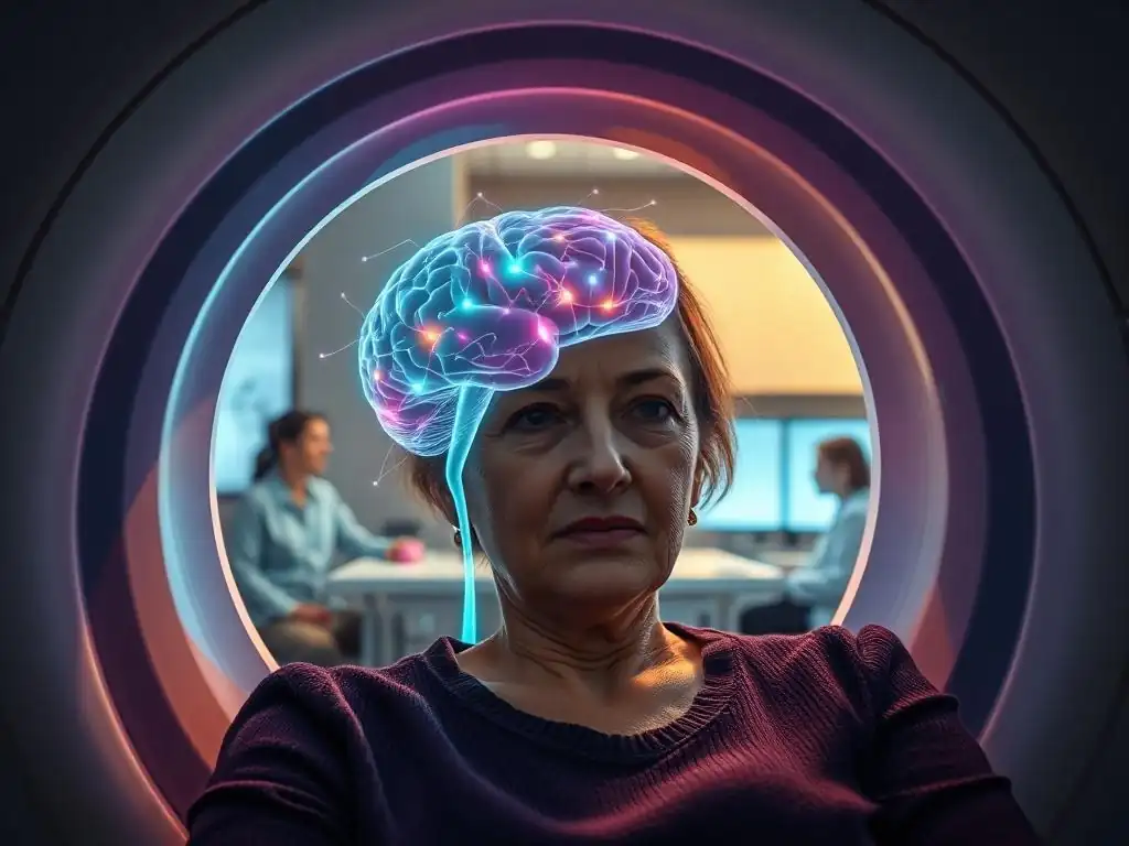
Ever wondered what truly happens within your brain during long-term therapy? This article delves into the fascinating world of neuroscience, exploring the profound and lasting brain therapy longterm effects revealed by the latest brain scan insights. Prepare to uncover the transformative power of therapy and understand how it reshapes your neural pathways, fostering lasting positive change.
- Brain Therapy Longterm Effects: Unveiling Changes with Scan Insights - Mind Care Guide
- How Long-Term Therapy Alters Brain Function: A Neuroscience Perspective
- Brain Scan Technologies Used to Measure Therapy's Impact
- Specific Brain Regions Affected by Long-Term Therapy: Scan Results
- The Benefits of Long-Term Therapy on Brain Health: Evidence from Scans
- Types of Long-Term Therapy and Their Distinct Brain Effects
- Brain Changes and Symptom Reduction: Linking Scan Data to Clinical Outcomes
- Challenges and Future Directions in Brain Scan Research for Therapy
- Conclusion: The Power of Long-Term Therapy to Reshape the Brain
-
Frequently Asked Questions
- What specific brain regions are most affected by long-term therapy, according to recent scans?
- How does long-term therapy change the neural pathways in the brain?
- Can brain scans objectively measure the benefits of long-term therapy?
- What are some potential long-term effects of therapy that are visible in brain scans?
- Is there a link between therapy type (e.g., CBT, psychodynamic) and the observed brain changes?
- Final Reflection: The Enduring Power of Neuroplasticity
- Unlock Your Brain's Potential: Take the Next Step
Brain Therapy Longterm Effects: Unveiling Changes with Scan Insights - Mind Care Guide
The Growing Understanding of Brain Plasticity and Mental Health
The human brain is a remarkably dynamic organ, possessing an inherent ability to change and adapt throughout life. This phenomenon, known as neuroplasticity, is the biological foundation for learning, memory, and recovery from injury.
In the context of mental health, researchers are increasingly understanding that conditions like depression, anxiety, and PTSD involve maladaptive patterns in neural circuits, and that neuroplasticity offers the potential to rewire these circuits towards healthier functioning. Early studies showing how experiences, such as learning a new skill or navigating a complex environment, can alter brain structure and connectivity laid the groundwork for exploring how therapeutic experiences might also induce physical changes.
This growing understanding challenges older views that the adult brain was largely static. Instead, we now know that experiences, including therapeutic interventions, can lead to the formation of new synapses, changes in glial cells, and even alterations in the volume of specific brain regions. This means that the thoughts, feelings, and behaviors explored and practiced in therapy aren't just abstract concepts; they are processes that can actively shape the biological structure and function of the brain, offering a tangible pathway for healing and lasting change.
Importance of Brain Scans in Assessing Long-Term Therapy Outcomes
While subjective reports from individuals undergoing therapy are invaluable for understanding their experience and progress, they offer a limited window into the objective biological changes occurring. This is where brain scanning technologies become crucial. Techniques like fMRI, EEG, and PET scans provide non-invasive ways to peer inside the living brain and measure neural activity, connectivity, and even aspects of its physical structure.
By scanning individuals before, during, and after a course of long-term therapy, researchers can gather objective data on how brain function and structure change over time. This allows for the assessment of therapy's impact at a neural level, providing empirical evidence for its effectiveness beyond just symptom reduction. These scans can help validate therapeutic approaches, reveal the specific brain circuits involved in different mental health conditions, and contribute to a deeper understanding of the brain therapy longterm effects.
How Long-Term Therapy Alters Brain Function: A Neuroscience Perspective
Neuroplasticity and the Rewiring of Neural Pathways During Therapy
Long-term therapy leverages the principles of neuroplasticity to facilitate change. Each therapeutic session, whether it involves processing past trauma, challenging negative thought patterns, or practicing new coping skills, provides repeated experiences that influence neural activity. This consistent engagement strengthens specific neural pathways associated with more adaptive responses while potentially weakening those linked to maladaptive behaviors or emotional reactions.
Think of it like carving a new path through a forest. Initially, it's difficult, but with repeated travel, the path becomes clearer and easier to navigate. Similarly, therapy helps individuals practice navigating challenging thoughts or emotions in new ways, reinforcing the neural connections that support these healthier responses. For instance, learning to reappraise a stressful situation in CBT strengthens pathways between the prefrontal cortex (involved in rational thought) and the amygdala (involved in threat response), allowing the PFC to exert greater regulatory control.
Structural Brain Changes Observed in Long-Term Therapy Patients
Beyond changes in functional activity and connectivity, long-term therapy can also lead to observable structural changes in the brain. These changes might include alterations in gray matter density (reflecting changes in neurons and synapses) or white matter integrity (reflecting changes in the connections between brain regions). These structural adaptations are often considered more enduring indicators of the brain therapy longterm effects.
Studies on individuals with conditions like major depressive disorder or post-traumatic stress disorder who have undergone successful long-term therapy have sometimes reported increases in gray matter volume or density in specific areas, such as parts of the prefrontal cortex or the hippocampus. These regions are often impacted by chronic stress and mood disorders. Conversely, areas associated with heightened threat response might show functional decreases, though significant structural changes in areas like the amygdala are less consistently reported than functional ones. These structural findings provide compelling evidence that therapy can induce tangible, physical changes in the brain.
Brain Scan Technologies Used to Measure Therapy's Impact
Functional Magnetic Resonance Imaging (fMRI) and its Role in Brain Therapy Research
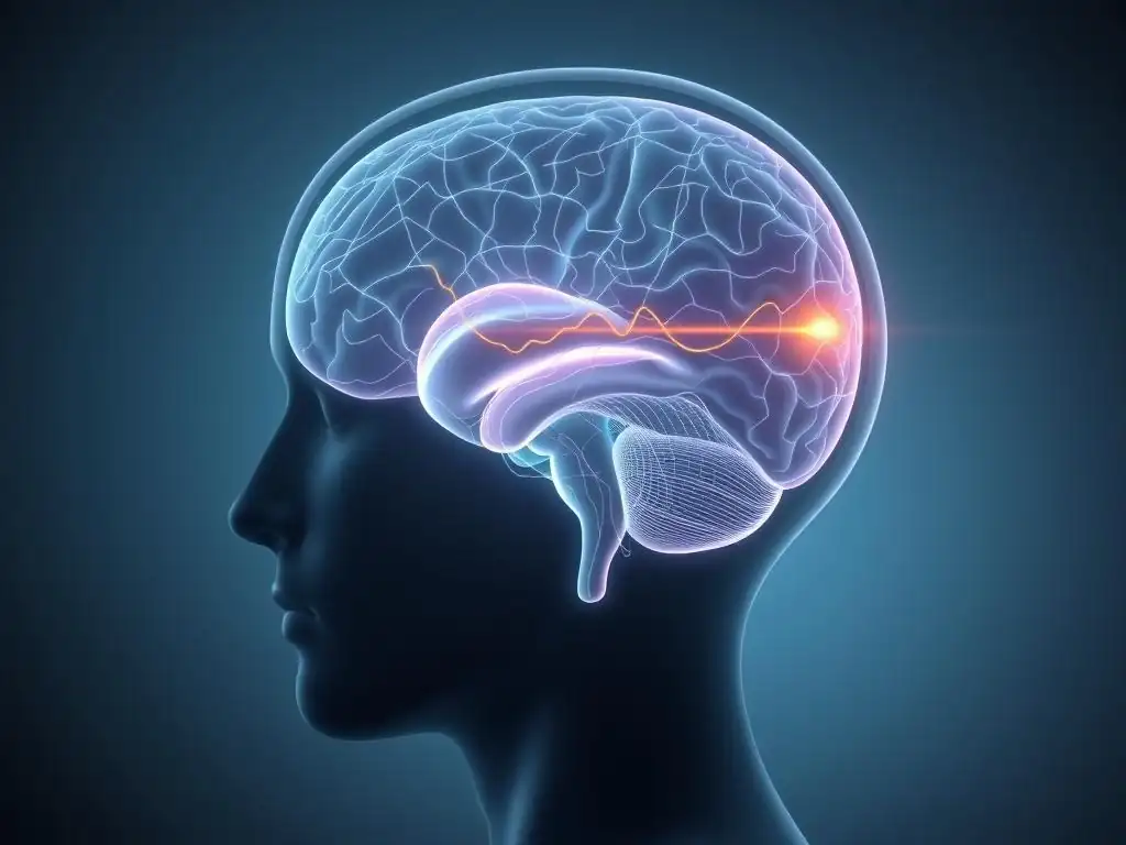
Functional Magnetic Resonance Imaging (fMRI) is a cornerstone technology in studying the brain therapy longterm effects. fMRI measures brain activity indirectly by detecting changes in blood flow. When a brain region is more active, it requires more oxygenated blood, and fMRI can map these changes in blood oxygenation levels (BOLD signal) throughout the brain. This allows researchers to see which brain areas are more or less active during specific tasks (like processing emotional images) or even when the brain is at rest (resting-state fMRI).
By comparing fMRI scans taken before and after a course of therapy, researchers can identify changes in how different brain regions activate or communicate with each other. For example, fMRI studies have shown decreased activity in the amygdala and increased activity in regions of the prefrontal cortex in patients with anxiety disorders or depression after successful CBT. These studies provide visual evidence of the functional neural changes underlying symptom improvement and are invaluable for understanding the brain therapy longterm effects.
Electroencephalography (EEG) and Monitoring Brainwave Activity During Therapy
Electroencephalography (EEG) measures the electrical activity produced by neurons in the brain using electrodes placed on the scalp. While EEG has lower spatial resolution than fMRI (meaning it's less precise at pinpointing activity to very specific deep brain structures), it offers excellent temporal resolution, capable of capturing brain activity changes that occur in milliseconds. This makes EEG particularly useful for studying the timing of neural responses and changes in brainwave patterns associated with different cognitive states or emotional processing.
EEG studies can investigate how therapy influences brainwave frequencies (such as alpha, beta, theta waves), coherence (how synchronized activity is between different brain regions), and event-related potentials (ERPs), which are specific brainwave responses to stimuli. For instance, research on mindfulness-based therapies has used EEG to show changes in alpha and theta power associated with states of relaxed attention and altered processing of emotional information, providing insights into the temporal dynamics of brain therapy longterm effects.
Positron Emission Tomography (PET) Scans: Examining Metabolic Changes in the Brain
Positron Emission Tomography (PET) scans use a small amount of a radioactive tracer injected into the bloodstream to measure metabolic activity, blood flow, or the distribution of specific molecules like neurotransmitter receptors in the brain. Different tracers can target different biological processes, offering a unique perspective on brain function compared to fMRI or EEG.
While less commonly used than fMRI for routine tracking of therapy outcomes due to the use of radiation and higher cost, PET scans can provide valuable insights into the biochemical underpinnings of mental health conditions and how therapy might influence them. For example, PET could be used to study changes in glucose metabolism (an indicator of overall brain activity) or to examine alterations in the density or binding of receptors for neurotransmitters like serotonin or dopamine after therapy, contributing another layer to understanding the comprehensive brain therapy longterm effects.
Specific Brain Regions Affected by Long-Term Therapy: Scan Results
Amygdala and Emotional Regulation: Long-Term Therapy's Influence
The amygdala, a small, almond-shaped structure deep within the brain, is a key player in processing emotions, particularly fear, anxiety, and threat detection. In many mental health conditions characterized by excessive fear or anxiety, such as panic disorder, phobias, or PTSD, the amygdala is often found to be hyperactive, reacting strongly to perceived threats. Long-term therapy, especially exposure-based therapies and Cognitive Behavioral Therapy (CBT), directly targets these fear responses.
Numerous fMRI studies have demonstrated a significant reduction in amygdala activity in response to emotional stimuli after successful long-term therapy for anxiety and trauma-related disorders. For example, research on individuals with social anxiety disorder treated with CBT showed a notable decrease in amygdala activation when they viewed critical faces post-therapy, a change that correlated strongly with their reduced social anxiety symptoms. This dampened amygdala response is a hallmark finding illustrating the brain therapy longterm effects on emotional processing circuits.
Prefrontal Cortex and Cognitive Control: Enhancing Executive Functions
The prefrontal cortex (PFC), located at the front of the brain, is the seat of executive functions, including decision-making, planning, working memory, impulse control, and the ability to regulate emotions. In many mental health conditions, the communication between the PFC and more primitive emotional centers like the amygdala is disrupted, often with reduced top-down control from the PFC.
Long-term therapy is hypothesized to strengthen the PFC's capacity to regulate activity in these lower brain regions. Scan studies using fMRI have frequently shown increased activation in various subregions of the PFC during tasks requiring emotional regulation or cognitive control after therapy for conditions like depression, anxiety, and OCD. Furthermore, analyses of functional connectivity often reveal strengthened pathways between the PFC and the amygdala or other limbic areas, indicating improved regulatory capacity. This enhancement of PFC function is a critical aspect of the brain therapy longterm effects, leading to better emotional management and cognitive abilities.
Hippocampus and Memory Consolidation: Strengthening Emotional Memories
The hippocampus, another structure within the limbic system, is crucial for forming and retrieving declarative memories, including the contextual details surrounding events. It helps us differentiate between past dangers and present safety. In conditions like PTSD and chronic stress, the hippocampus can be affected, sometimes showing reduced volume, and its function in contextualizing traumatic memories may be impaired, leading to intrusive flashbacks.
While the evidence for therapy-induced structural increase in hippocampal volume is less consistent across studies than functional changes in other areas, some research, particularly in the context of trauma-focused therapies for PTSD, suggests potential normalization or improvements in hippocampal function and connectivity. The therapeutic process of re-processing traumatic memories in a safe environment is thought to help the hippocampus re-encode these memories with a more appropriate context, reducing their power to trigger intense fear responses.
Functional scan data showing altered hippocampal activity and connectivity during memory tasks post-therapy support the idea that therapy positively impacts how the brain processes and consolidates emotional memories as part of its brain therapy longterm effects.
The Benefits of Long-Term Therapy on Brain Health: Evidence from Scans
A consistent and significant finding from brain scan research on long-term therapy is the observable reduction in activity within brain regions primarily associated with processing stress, fear, and negative emotions. The amygdala is the most frequently cited example, showing decreased reactivity to emotional or threatening stimuli after successful treatment for anxiety disorders, phobias, and PTSD. Other areas, such as parts of the insula (involved in processing bodily sensations and emotions), may also show modulated activity.
This reduced activity is not merely a passive change but reflects a fundamental shift in how the brain appraises and responds to potentially stressful situations. For instance, a meta-analysis combining data from multiple fMRI studies on anxiety treatment showed a reliable decrease in amygdala activation post-therapy, often correlating with the degree of symptom relief reported by patients. While the percentage of reduction varies depending on the specific disorder and therapy, this pattern of dampened threat response is a robust indicator of the positive brain therapy longterm effects on stress circuitry.
Increased Connectivity Between Brain Areas Associated with Emotional Well-being
Beyond changes within individual brain regions, long-term therapy also influences how different areas of the brain communicate with each other. A common finding is increased functional connectivity between the prefrontal cortex and limbic regions like the amygdala. This strengthened connection is thought to represent enhanced top-down control, where the more rational, executive parts of the brain gain greater influence over emotional responses originating in the limbic system.
Furthermore, research is beginning to explore how therapy impacts broader brain networks. For example, studies have looked at changes in the default mode network (involved in self-referential thought and rumination) and salience network (involved in detecting important stimuli). Changes in the connectivity within and between these networks post-therapy are often observed and are associated with reduced rumination and improved ability to shift attention, contributing to overall emotional regulation and well-being. These complex changes in brain connectivity are key components of the brain therapy longterm effects.
Improved Cognitive Function and Memory Performance After Therapy
Mental health conditions, particularly depression and anxiety, often go hand-in-hand with cognitive difficulties, including problems with concentration, memory, decision-making, and executive functions. As individuals recover through long-term therapy, these cognitive impairments often improve. Brain scans provide a neural basis for these observed cognitive benefits.
Scan studies often show changes in activity patterns in brain regions critical for cognitive processing, such as the prefrontal cortex and parietal cortex, correlating with improved performance on cognitive tests. For instance, individuals recovering from depression often show normalized activity in areas associated with attention and working memory during cognitive tasks after therapy.
While the link between therapy-induced structural hippocampal changes and memory performance is still an area of active research, functional scan data suggests that therapy can improve the efficiency of memory processing and retrieval, particularly in the context of emotional memories. These cognitive enhancements are valuable brain therapy longterm effects that support better daily functioning and quality of life.
Types of Long-Term Therapy and Their Distinct Brain Effects
Cognitive Behavioral Therapy (CBT) and its Impact on Prefrontal Cortex Activity
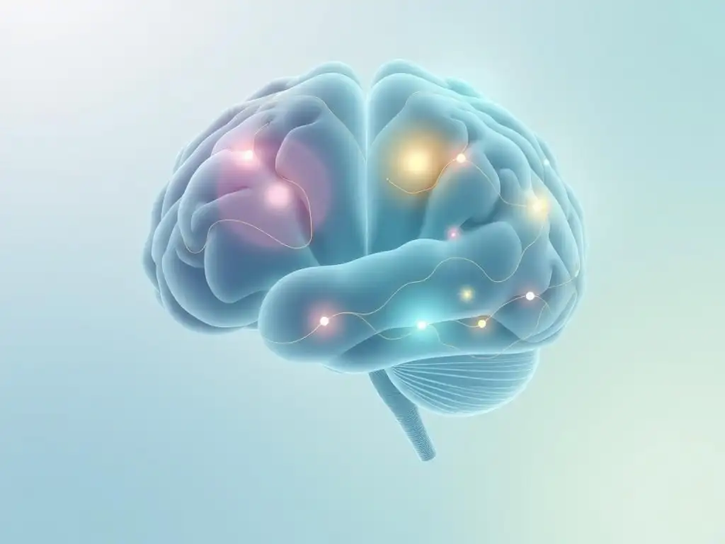
Cognitive Behavioral Therapy (CBT) is a widely researched form of therapy that focuses on identifying and changing negative thought patterns and behaviors that contribute to distress. Because it heavily involves conscious thought processes, reasoning, and behavioral experiments, CBT is strongly associated with changes in the prefrontal cortex (PFC) and its connections to other brain areas.
Numerous fMRI studies have investigated the neural effects of CBT across various conditions. For example, studies on anxiety disorders treated with CBT consistently report decreased activity in the amygdala and increased activity or altered connectivity in specific regions of the PFC, such as the ventromedial or dorsolateral PFC, during emotional tasks.
Research on OCD treated with CBT has shown changes in the cortico-striatal circuits involved in habits and compulsions, including areas like the orbitofrontal cortex and caudate nucleus. These findings underscore how CBT's emphasis on cognitive restructuring and behavioral change translates into measurable brain changes, particularly in areas involved in executive control, as a key brain therapy longterm effect.
Psychodynamic Therapy and Changes in Emotional Processing Centers
Psychodynamic therapy explores the influence of unconscious processes, past experiences, and interpersonal relationships on present difficulties. This type of therapy often involves processing complex emotions, gaining insight into relational patterns, and exploring the roots of psychological distress. While fewer brain scan studies have focused on psychodynamic therapy compared to CBT, existing research suggests it may particularly impact brain regions involved in emotional processing, self-awareness, and interpersonal understanding.
Studies on psychodynamic therapy for depression, for instance, have explored changes in activity within limbic regions and areas involved in self-referential processing, such as the medial prefrontal cortex (mPFC). One study comparing psychodynamic therapy to a control condition in depressed patients found changes in mPFC activity associated with symptom improvement.
Another area of interest is how psychodynamic therapy might affect connectivity between the mPFC (involved in self-reflection) and the amygdala, potentially altering how individuals process emotional information related to the self and others. These findings suggest that psychodynamic therapy's focus on emotional depth and insight can lead to brain therapy longterm effects centered around emotional processing and self-perception circuits.
Mindfulness-Based Therapy and Increased Activity in Attention Networks
Mindfulness-based therapies, such as Mindfulness-Based Stress Reduction (MBSR) and Mindfulness-Based Cognitive Therapy (MBCT), train individuals to cultivate present moment awareness with a non-judgmental attitude. This practice involves intentionally directing and sustaining attention, monitoring internal experiences (thoughts, feelings, bodily sensations), and developing a different relationship with difficult emotions. These processes engage brain networks involved in attention, self-awareness, and emotional regulation.
Brain scan studies on mindfulness practices and mindfulness-based therapies have consistently shown changes in brain regions like the anterior cingulate cortex (ACC) and the insula, both critical for attention, interoception (awareness of internal bodily states), and processing emotions. For example, studies have reported increased gray matter density in these areas after mindfulness training.
Furthermore, fMRI studies show altered functional connectivity, often involving decreased coupling within the default mode network (associated with mind-wandering and rumination) and increased connectivity in networks supporting attention and executive control. These changes reflect the enhanced capacity for focused attention and emotional regulation developed through mindfulness, demonstrating distinct brain therapy longterm effects on attention and self-awareness networks.
Brain Changes and Symptom Reduction: Linking Scan Data to Clinical Outcomes
Correlation Between Amygdala Activity and Anxiety Reduction
One of the most compelling demonstrations of the link between brain changes and clinical outcomes comes from studies showing a strong correlation between the reduction in amygdala activity and the decrease in anxiety symptoms after long-term therapy. As individuals report feeling less anxious and fearful, brain scans reveal a corresponding dampening of the amygdala's response to stimuli that previously triggered fear.
For example, research on panic disorder patients undergoing CBT has shown that the extent to which amygdala reactivity to panic-related cues decreases post-therapy is a significant predictor of how much their panic symptoms improve over time. While the specific correlation coefficients vary between studies and populations, figures in the range of r = -0.5 to -0.7 are sometimes reported, indicating a moderate to strong inverse relationship: the more the amygdala calms down, the more anxiety subsides. This provides powerful objective evidence linking the neural brain therapy longterm effects directly to clinical benefits.
Prefrontal Cortex Activation and Improved Decision-Making Skills
Improvements in cognitive functions, including decision-making and problem-solving, are common benefits of recovery facilitated by long-term therapy, especially for conditions that impair executive functions. Scan data provides insight into the neural basis for this improvement, often showing enhanced activity and efficiency in the prefrontal cortex (PFC), the brain's command center for these processes.
Studies on individuals recovering from depression have shown increased activation in areas like the dorsolateral PFC during cognitive tasks requiring planning or complex decision-making. This increased neural efficiency in the PFC correlates with improved scores on neuropsychological tests assessing executive functions and self-reported improvements in decision-making clarity and problem-solving abilities in daily life. This link between enhanced PFC function observed in scans and real-world cognitive improvements highlights how the brain therapy longterm effects contribute to practical benefits beyond just emotional regulation.
Hippocampal Volume Increase and Enhanced Memory Recall
While therapy-induced structural increases in hippocampal volume are not universally observed, particularly outside of specific trauma contexts, functional changes involving the hippocampus often correlate with improvements in memory processing and recall, especially concerning emotional or traumatic events. The process of therapy can help individuals integrate fragmented or distressing memories into a coherent narrative, a process heavily reliant on the hippocampus.
Functionally, studies have shown altered patterns of hippocampal activity and connectivity with other brain regions involved in memory retrieval and contextualization after therapy. This can lead to improved recall of non-emotional memories and a reduced tendency for traumatic memories to be triggered out of context (e.g., flashbacks), which is a hallmark symptom of PTSD. While the direct link between structural volume changes and memory recall is still being fully elucidated, the functional normalization of hippocampal activity is a plausible mechanism contributing to the positive brain therapy longterm effects on memory and trauma processing.
Challenges and Future Directions in Brain Scan Research for Therapy
Limitations of Current Scanning Techniques
Despite the incredible insights provided by brain scanning technologies, they come with inherent limitations. These techniques are expensive, require specialized equipment and highly trained personnel, which limits their accessibility and scalability for widespread use in clinical practice. Each technology also has its own trade-offs: fMRI offers good spatial detail but is relatively slow, capturing activity changes over seconds; EEG is very fast (milliseconds) but struggles to pinpoint activity deep within the brain; and PET requires the use of radioactive tracers.
Furthermore, interpreting brain scan data is complex. Brain activity is highly dynamic and influenced by many factors beyond the therapeutic intervention. Findings from studies often represent average changes across groups of patients, and there is significant variability in how individual brains respond to therapy. Establishing definitive cause-and-effect relationships based solely on correlational scan data remains challenging, and it is difficult to completely isolate the effects of therapy from spontaneous recovery or other life events.
The Need for Longitudinal Studies to Track Brain Changes Over Time
Most brain scan studies investigating therapy outcomes are relatively short-term, often assessing brain changes only immediately before and after a standard course of therapy, which might last anywhere from a few weeks to several months. While these studies provide valuable snapshots of immediate effects, they offer limited information about the long-term trajectory of brain changes.
To truly understand the sustained impact and durability of the brain therapy longterm effects, there is a critical need for longitudinal studies that follow individuals for extended periods – months or even years – after therapy has concluded. Such studies could reveal whether the observed brain changes are maintained, if they continue to evolve over time, whether booster sessions or continued practice are necessary to preserve these changes, and if early neural markers can predict long-term recovery trajectories. Longitudinal research is essential for building a comprehensive picture of therapy's lasting influence on the brain.
Integrating Brain Scan Data with Clinical Assessments for Personalized Treatment
The future of brain scan research in therapy lies in moving beyond group-level findings to utilize this information for individual patients. Integrating objective brain scan data with detailed clinical assessments, including symptom profiles, individual history, genetic factors, and treatment response patterns, holds immense potential for developing more personalized and effective treatment strategies.
Imagine a scenario where initial brain scan patterns could help clinicians determine which type of therapy is most likely to be effective for a specific individual, based on their unique neural profile. For example, someone with pronounced hyperconnectivity in fear circuits might be guided towards therapies known to effectively modulate the amygdala, while someone with diminished prefrontal activity might benefit more from approaches that strengthen cognitive control. This integration of neural and clinical data could revolutionize the field, leading to truly tailored interventions that optimize the brain therapy longterm effects for each person.
Conclusion: The Power of Long-Term Therapy to Reshape the Brain
Emphasizing the Importance of Understanding Brain Therapy Longterm Effects
The accumulating evidence from brain scan research provides compelling and objective validation for the transformative power of long-term therapy. These studies show unequivocally that therapy is not just about talking; it is an experience that can lead to measurable, physical, and functional changes in the brain. Understanding these biological underpinnings of change is crucial for appreciating therapy as a potent intervention for mental health conditions.
By revealing changes in brain structure, activity, and connectivity, research into the brain therapy longterm effects demystifies the therapeutic process and highlights its capacity to foster profound and lasting neural adaptations. These adaptations support improved emotional regulation, enhanced cognitive function, and a greater capacity for overall well-being. This scientific insight reinforces the value of investing time and effort in therapeutic work.
Mind Care Guide: Your Resource for Brain Health and Mental Well-being
At Mind Care Guide, we are committed to providing accessible, evidence-based information to empower individuals on their journey towards mental wellness. We believe that understanding the science behind mental health treatments, including the remarkable brain therapy longterm effects illuminated by advanced scanning techniques, is vital for making informed decisions about your care.
We aim to be your trusted resource for exploring topics like brain health, the neuroscience of mental well-being, and the tangible outcomes of different therapeutic approaches. Explore our articles and guides to learn more about how your brain functions, how mental health challenges impact it, and how effective interventions like long-term therapy can help reshape your neural landscape for lasting health and resilience.
Frequently Asked Questions
What specific brain regions are most affected by long-term therapy, according to recent scans?
Studies show significant changes in the prefrontal cortex (responsible for executive functions) and the amygdala (involved in emotional processing). These changes correlate with improved emotional regulation and reduced anxiety.
How does long-term therapy change the neural pathways in the brain?
Long-term therapy encourages the formation of new, healthier neural pathways while weakening maladaptive ones. This process, called neuroplasticity, is crucial for lasting change.
Can brain scans objectively measure the benefits of long-term therapy?
Yes, brain scans, such as fMRI, can provide objective evidence of the physiological changes associated with therapy. These scans can reveal increased activity in areas linked to positive emotional regulation.
What are some potential long-term effects of therapy that are visible in brain scans?
Scans often show increased grey matter volume in regions associated with emotional intelligence and self-awareness. Reduced activity in the amygdala can indicate a decrease in fear responses.
Is there a link between therapy type (e.g., CBT, psychodynamic) and the observed brain changes?
While more research is needed, preliminary studies suggest that different therapy types may activate slightly different brain regions. However, all effective therapies ultimately aim to improve overall brain function and mental well-being.
Final Reflection: The Enduring Power of Neuroplasticity
The insights gleaned from brain scans regarding long-term therapy reveal a profound truth: our brains are not static entities, but dynamic landscapes constantly reshaped by experience. This understanding, once a niche scientific concept, is now revolutionizing our approach to mental health, offering tangible hope for lasting change and challenging the limitations of traditional models. The ability to visualize these changes, to literally see the brain rewiring itself, is a powerful testament to the efficacy of sustained therapeutic intervention.
The impact of long-term therapy extends far beyond the individual; it ripples outwards, influencing families, communities, and ultimately, the very fabric of our society. By fostering emotional regulation, resilience, and healthier coping mechanisms, therapy contributes to a more compassionate and understanding world. As Viktor Frankl so eloquently stated, >“Between stimulus and response there is a space.
In that space is our power to choose our response. In our response lies our growth and our freedom.” This space, cultivated through the consistent work of therapy, is where true healing and personal growth blossom. It's a space where the brain, guided by skilled therapeutic intervention, can rewrite its own narrative.
Consider the implications of this for your own life. Are you ready to embrace the transformative power of neuroplasticity? Long-term therapy isn't just about symptom reduction; it's about cultivating a healthier, more resilient you. It's about actively shaping your brain's landscape to foster well-being and unlock your full potential. Take a moment to reflect on your own mental health journey. What steps can you take today to invest in your brain's well-being and create lasting positive change? The journey may be long, but the rewards – a more empowered, resilient, and fulfilling life – are immeasurable.
Unlock Your Brain's Potential: Take the Next Step
Thanks for exploring the fascinating world of long-term therapy and brain scans with us! You've gained valuable insights into the remarkable changes therapy can create. Now, it's time to put that knowledge into action.
- Schedule a consultation: This is the most important step! Taking the first step towards understanding your own brain health and exploring therapy is a powerful act of self-care.
- Share this article: Spread the word and help others discover the transformative power of long-term therapy. Let's break the stigma together!
- Explore Mind Care Guide's resources: We have additional articles and guides on various mental health topics—dive deeper and find what resonates with you.
Ready to embark on your journey toward a healthier, more resilient you? What specific area of your mental well-being are you most excited to explore next?