
The Science of Change: Brain Imaging of Therapy Outcomes


Welcome to Mind Care Guide, a sanctuary for your mental well-being.
Are you grappling with the question of whether therapy is truly working? We understand. It's a valid concern. In this space, we delve into the fascinating world of brain imaging therapy effectiveness research, exploring how cutting-edge neuroscience is helping us understand and validate the impact of mental health treatments. Curious to see what the science reveals about the tangible changes happening within your mind? Let's explore the evidence together...
The Science of Change: Brain Imaging of Therapy Outcomes
- Introduction: Understanding Brain Changes Through Therapy
- Brain Imaging Techniques Used in Therapy Outcome Research
- Brain Imaging Therapy Effectiveness Research: Key Findings
- Long-Term Therapy Outcomes and Brain Changes: Specific Examples
- Limitations and Future Directions in Brain Imaging of Therapy Outcomes
- Conclusion: The Promise of Brain Imaging for Improving Mental Healthcare
-
Frequently Asked Questions
- What is the role of brain imaging in researching the effectiveness of therapy?
- What types of brain imaging techniques are typically used in brain imaging therapy effectiveness research?
- How does brain imaging contribute to understanding the mechanisms of therapeutic change?
- Can brain imaging predict which individuals will benefit most from specific types of therapy?
- What are the current limitations and future directions of brain imaging therapy effectiveness research?
- Final Reflection: The Evolving Landscape of Therapeutic Change
- Unlock Your Brain's Potential: Take Charge of Your Therapy Journey
Introduction: Understanding Brain Changes Through Therapy
Exploring the Neuroscience of Mental Health Treatment
The human brain is a complex and dynamic organ, the seat of our thoughts, emotions, and behaviors. Understanding its intricate workings is fundamental to comprehending mental health conditions and developing effective treatments.
Neuroscience provides the tools to explore the biological underpinnings of disorders such as depression, anxiety, schizophrenia, and PTSD, revealing how disruptions in neural circuits and communication pathways contribute to symptoms. This exploration involves studying everything from the molecular level of neurotransmitters to the macroscopic connectivity of different brain regions, offering insights into why certain individuals are more vulnerable to mental health challenges and how these conditions manifest in distinct patterns of brain activity and structure.
Decades of research in the neuroscience of mental health have illuminated specific brain areas implicated in various disorders. For example, the amygdala, involved in processing fear and emotional responses, often shows heightened activity in anxiety disorders. The prefrontal cortex, critical for executive functions, decision-making, and emotional regulation, may exhibit reduced activity or altered connectivity in depression and schizophrenia.
By mapping these neural correlates, researchers gain a deeper understanding of the biological basis of mental illness, paving the way for targeted therapeutic interventions designed to restore healthier brain function. This foundational knowledge is crucial for evaluating how treatments, including psychotherapy, bring about change at the neural level.
The Role of Brain Imaging in Assessing Therapy Effectiveness
Brain imaging techniques represent a revolutionary approach to observing the living brain in action, offering unprecedented insights into how psychological therapies exert their effects. Before the advent of modern imaging, understanding the impact of therapy on the brain was largely theoretical, based on behavioral observations and patient reports. Now, non-invasive methods allow researchers to visualize changes in brain structure, activity patterns, and connectivity that occur as a direct result of therapeutic interventions. This capability provides empirical evidence that therapy doesn't just change minds; it changes brains, offering a biological validation for the effectiveness of psychological treatments.
Utilizing brain imaging to assess therapy effectiveness allows researchers to move beyond simply measuring symptom reduction. It enables the identification of specific neural mechanisms that underpin successful therapeutic outcomes. For instance, imaging studies can reveal whether therapy leads to decreased activity in hyperactive fear circuits in anxiety or increased activity in prefrontal regions associated with emotional control in depression.
This detailed understanding of how therapy works at the neural level can inform and refine treatment protocols, potentially leading to more efficient and personalized interventions. Such brain imaging therapy effectiveness research is vital for advancing our understanding and improving mental healthcare delivery.
Brain Imaging Techniques Used in Therapy Outcome Research
Functional Magnetic Resonance Imaging (fMRI) for Mapping Brain Activity
Functional Magnetic Resonance Imaging (fMRI) is a widely used technique in brain imaging therapy effectiveness research due to its ability to non-invasively measure brain activity. fMRI detects changes in blood flow and oxygenation that occur when specific brain regions are more active. When neurons fire, they require more oxygen, leading to increased blood flow to that area.
By tracking these hemodynamic responses, fMRI can create detailed maps showing which parts of the brain are engaged during specific tasks, emotional states, or even during rest. This allows researchers to compare patterns of brain activity in individuals before and after therapy to identify neural changes associated with symptom improvement.
In therapy outcome studies, fMRI is often used to examine how treatment alters the brain's response to stimuli related to the disorder. For example, individuals with social anxiety might show excessive amygdala activation when viewing faces expressing negative emotions.
An fMRI study could assess whether a course of cognitive behavioral therapy (CBT) leads to a reduction in this amygdala response and an increase in prefrontal cortex activity related to regulation, potentially showing a correlation with reduced social anxiety symptoms. The spatial resolution of fMRI, typically down to a few millimeters, provides detailed maps of brain regions involved, making it a powerful tool for understanding the neural mechanisms of therapeutic change.
Electroencephalography (EEG) for Tracking Brainwave Patterns
Electroencephalography (EEG) is another crucial brain imaging technique, particularly valuable for its excellent temporal resolution – its ability to track rapid changes in brain activity, measured in milliseconds. EEG works by placing electrodes on the scalp to detect the electrical activity produced by neurons firing in the brain.
This activity is recorded as brainwaves, which have different frequencies (e.g., delta, theta, alpha, beta, gamma) associated with different states of consciousness and cognitive processes. While EEG's spatial resolution (pinpointing exactly where activity originates) is lower than fMRI, its ability to capture the timing and sequence of neural events is unparalleled among non-invasive methods.
In the context of brain imaging therapy effectiveness research, EEG is used to assess how therapy alters brainwave patterns and the dynamic connectivity between brain regions. For instance, studies might examine changes in alpha asymmetry (differences in alpha wave activity between the left and right hemispheres) in depression following psychotherapy, as this pattern has been linked to emotional processing.
EEG can also measure event-related potentials (ERPs), which are characteristic brainwave responses to specific stimuli, allowing researchers to see how therapy changes the brain's immediate processing of emotional or cognitive information. Its high temporal precision makes EEG ideal for studying the rapid neural dynamics affected by therapeutic interventions.
Positron Emission Tomography (PET) for Analyzing Brain Metabolism
Positron Emission Tomography (PET) is a brain imaging technique that provides insights into the metabolic and biochemical activity of the brain, which fMRI and EEG do not directly measure. PET involves injecting a small amount of a radioactive tracer, often a glucose analog, into the bloodstream. Active brain cells consume glucose, so areas with higher metabolic rates will accumulate more tracer, which can then be detected by the PET scanner. This allows researchers to measure regional brain metabolism, blood flow, or the density of specific neurotransmitter receptors or transporters, depending on the tracer used.
PET scans can be used in brain imaging therapy effectiveness research to investigate how therapy influences neurochemical systems or resting metabolic activity associated with a disorder. For example, a PET study might examine changes in serotonin transporter availability in patients with obsessive-compulsive disorder (OCD) undergoing exposure and response prevention therapy, as disruptions in the serotonin system are implicated in OCD.
Another application is measuring glucose metabolism in specific brain regions linked to mood regulation in individuals receiving therapy for depression. While PET has lower spatial and temporal resolution compared to fMRI and EEG, its ability to probe specific neurochemical pathways provides unique and valuable information about the biological changes induced by therapy.
Diffusion Tensor Imaging (DTI) for Assessing White Matter Integrity
Diffusion Tensor Imaging (DTI) is a specialized MRI technique that measures the diffusion of water molecules in the brain. In white matter tracts – bundles of nerve fibers that connect different brain regions – water diffusion is anisotropic, meaning it moves more freely along the direction of the fibers than across them.
DTI exploits this property to map the orientation and integrity of these white matter pathways, providing insights into the structural connectivity of the brain. The fractional anisotropy (FA) value, derived from DTI data, is often used as a measure of white matter integrity; lower FA can indicate damage or abnormalities in the fiber tracts.
In the context of brain imaging therapy effectiveness research, DTI is used to explore whether therapy can induce changes in the structural connections between brain regions. For instance, studies might investigate whether psychotherapy for PTSD leads to improved integrity of white matter tracts connecting the prefrontal cortex (involved in regulation) and the amygdala (involved in fear processing).
Changes in these structural pathways could represent a biological mechanism through which therapy enhances communication and improves emotional regulation. DTI provides a complementary perspective to functional imaging, showing how therapy might reshape the brain's underlying wiring diagram, offering valuable data for brain imaging therapy effectiveness research.
Brain Imaging Therapy Effectiveness Research: Key Findings

Neural Correlates of Successful Cognitive Behavioral Therapy (CBT)
Brain Activity Changes in Anxiety Disorders After CBT
Brain imaging therapy effectiveness research has provided compelling evidence for how Cognitive Behavioral Therapy (CBT) alters brain activity in individuals with anxiety disorders. Studies using fMRI have consistently shown that successful CBT for conditions like social anxiety disorder or specific phobias is associated with reduced activity in brain regions involved in processing fear and threat, such as the amygdala.
For example, research on social anxiety has found that after CBT, patients exhibit significantly less activation in the amygdala and hippocampus when anticipating or experiencing social situations, compared to before therapy. This suggests that CBT helps dampen the brain's alarm system response to perceived threats.
Furthermore, brain imaging studies often reveal increased activity in prefrontal cortical areas, particularly the ventromedial prefrontal cortex (vmPFC), following successful CBT for anxiety. The vmPFC is known to play a crucial role in regulating emotional responses and inhibiting the amygdala. Findings suggest that CBT helps strengthen the top-down control exerted by the prefrontal cortex over subcortical emotional regions.
For instance, one study on specific phobia found that therapy led to increased vmPFC activity during exposure to the feared stimulus, correlating with reduced fear ratings. These brain activity changes provide neural signatures for the mechanisms by which CBT helps individuals manage and overcome their anxiety.
Brain Imaging Evidence for CBT's Impact on Depression
Brain imaging therapy effectiveness research has also shed light on the neural impact of CBT for depression. Major depressive disorder is often associated with altered activity and connectivity in networks related to emotional processing, reward, and cognitive control, including areas like the prefrontal cortex, anterior cingulate cortex (ACC), and striatum.
fMRI studies have shown that effective CBT for depression is linked to a normalization of activity in some of these key regions. For example, research has demonstrated that successful CBT can lead to increased activity in the dorsolateral prefrontal cortex (dlPFC), an area involved in cognitive control and planning, which is often hypoactive in depression.
Moreover, studies have indicated that CBT can alter the connectivity between brain regions in depressed patients. For instance, abnormal connectivity within the default mode network (DMN), a network active when the mind is at rest, is often observed in depression. Some brain imaging therapy effectiveness research suggests that CBT can help normalize this connectivity.
Additionally, altered connectivity between the amygdala and the ACC, involved in conflict monitoring and emotional regulation, has been observed to shift towards patterns seen in healthy individuals after successful CBT. These findings underscore that CBT doesn't just change thought patterns; it appears to reconfigure the functional architecture of the brain involved in mood regulation.
Neuroplasticity and Psychotherapy: Evidence from Brain Scans
How Therapy Shapes Brain Structure and Function
The concept of neuroplasticity – the brain's ability to change and adapt throughout life – is central to understanding how psychotherapy works at a biological level. Brain imaging techniques have provided compelling evidence that therapy can indeed induce measurable changes in both the structure and function of the brain.
Structural MRI studies, for example, have shown that successful therapy can be associated with changes in gray matter volume in specific brain regions. For instance, studies on mindfulness-based therapies have reported increases in gray matter density in areas related to learning, memory, emotional regulation, and self-referential processing, such as the hippocampus and parts of the prefrontal cortex.
Functional imaging, such as fMRI, further illustrates how therapy reshapes brain activity and connectivity patterns. As discussed earlier, psychotherapy can lead to altered activation levels in response to stimuli and changes in the strength or patterns of connections between different brain networks.
These functional changes are thought to reflect the learning and adaptation processes that occur during therapy – for example, learning new coping strategies, re-evaluating negative thought patterns, or developing new emotional regulation skills. This brain imaging therapy effectiveness research provides tangible proof that the therapeutic process is not merely abstract psychological work but a biological process of neural learning and adaptation.
Brain's Adaptive Responses to Therapeutic Interventions
Therapeutic interventions leverage the brain's inherent capacity for adaptation to promote recovery from mental health challenges. Brain imaging allows researchers to observe these adaptive responses in action. For example, in trauma-focused therapies like Prolonged Exposure (PE), individuals learn to confront traumatic memories in a safe environment. Brain imaging studies before and after PE have shown changes consistent with the brain adapting to process fear more effectively, such as reduced amygdala reactivity and increased prefrontal control during trauma recall. This indicates a process of fear extinction and habituation occurring at the neural level, facilitated by the therapeutic process.
Similarly, in therapies targeting social skills deficits, such as those used in schizophrenia, brain imaging might show changes in areas involved in social cognition and understanding others' intentions, like the temporoparietal junction (TPJ) or the medial prefrontal cortex (mPFC). Improvements in social functioning observed clinically can be correlated with changes in activity or connectivity in these specific social brain networks, reflecting the brain's adaptation to new social learning experiences provided by therapy.
The brain's adaptive responses visualized through imaging underscore that successful therapy involves rewiring neural circuits, reinforcing the biological basis of psychological healing and providing powerful evidence for brain imaging therapy effectiveness research.
Brain Imaging and Personalized Treatment Approaches
Identifying Biomarkers for Predicting Therapy Response
One of the exciting frontiers in brain imaging therapy effectiveness research is the potential to identify neural biomarkers that can predict an individual's likely response to a specific type of therapy. Currently, selecting the most effective therapy for a patient often involves trial and error, which can be time-consuming and costly. Brain imaging offers the possibility of using pre-treatment brain characteristics – such as specific patterns of activity, connectivity, or structural features – to predict who is most likely to benefit from a particular therapeutic approach, like CBT versus psychodynamic therapy.
For instance, preliminary brain imaging therapy effectiveness research in depression has explored whether the level of connectivity within certain brain networks or the activity in specific regions (like the subgenual ACC) before treatment can predict remission rates with CBT compared to medication. Some studies have suggested that different baseline brain patterns might predict better outcomes with one treatment modality over another.
While this field is still developing and findings need replication across larger cohorts, the promise of using brain imaging to identify neural signatures that predict therapy response could revolutionize personalized mental healthcare, allowing clinicians to select the optimal treatment from the outset.
Tailoring Therapy Based on Individual Brain Profiles
Beyond predicting general response, brain imaging also holds potential for tailoring therapeutic interventions based on an individual's unique brain profile. Understanding the specific neural dysfunctions underlying a patient's symptoms – as revealed by brain imaging – could allow therapists to customize techniques and focus areas within a therapy. For example, if brain imaging indicates particularly strong and widespread connectivity within fear circuits in an anxiety patient, the therapist might place extra emphasis on exposure techniques designed to weaken these specific connections.
Conversely, if imaging suggests deficits in prefrontal control mechanisms, the therapy might focus more heavily on cognitive restructuring and strengthening executive functions related to emotional regulation. While the direct use of individual patient brain scans to guide therapy in routine clinical practice is not yet standard, brain imaging therapy effectiveness research is building the knowledge base to support such personalized approaches in the future. By understanding the unique neural landscape of each individual's mental health challenge, therapy could potentially become more precise, efficient, and effective, moving towards a truly personalized medicine model for mental health.
Long-Term Therapy Outcomes and Brain Changes: Specific Examples
Brain Imaging of Trauma Therapy Effects
Trauma-focused therapies, such as Eye Movement Desensitization and Reprocessing (EMDR) or Prolonged Exposure (PE), aim to help individuals process traumatic memories and reduce associated symptoms like hyperarousal, avoidance, and negative cognitions. Brain imaging therapy effectiveness research has investigated the neural changes associated with successful treatment of Post-Traumatic Stress Disorder (PTSD). Studies have shown that effective trauma therapy can lead to a decrease in amygdala reactivity when processing trauma-related stimuli, indicating a reduction in the threat response.
Furthermore, imaging studies have reported increased activity or connectivity in brain regions involved in emotional regulation and cognitive control, such as the medial prefrontal cortex (mPFC) and the hippocampus, following successful trauma therapy. For instance, research has found that after PE, veterans with PTSD showed increased hippocampal volume and connectivity with the mPFC, suggesting improved memory processing and regulatory capacity. These findings provide neural evidence that trauma therapy helps the brain integrate traumatic experiences into autobiographical memory in a less threatening way and enhances the brain's ability to manage fear responses.
Neural Changes Following Eye Movement Desensitization and Reprocessing (EMDR)
Eye Movement Desensitization and Reprocessing (EMDR) is a specific trauma therapy that involves bilateral stimulation (often eye movements) while processing traumatic memories. Brain imaging therapy effectiveness research on EMDR is still evolving, but some studies suggest unique neural effects. For example, some fMRI studies have shown that successful EMDR treatment for PTSD is associated with decreased activity in the amygdala and increased activity in the anterior cingulate cortex (ACC) and prefrontal cortex when recalling traumatic memories.
Hypotheses suggest that the bilateral stimulation used in EMDR may help to facilitate the processing and integration of traumatic memories by engaging neural networks involved in memory consolidation and emotional regulation. While the exact mechanisms are still under investigation, brain imaging studies using techniques like fMRI and EEG are providing initial insights into how EMDR might alter brain activity patterns associated with trauma processing, contributing to the growing body of brain imaging therapy effectiveness research in trauma.
Brain Changes in Patients with Schizophrenia Undergoing Therapy
Psychological therapies, particularly cognitive remediation and social skills training, play a crucial role in managing symptoms and improving functional outcomes for individuals with schizophrenia, alongside medication. Brain imaging therapy effectiveness research in schizophrenia has explored how these therapies impact brain structure and function, which are significantly altered in the disorder. Studies using structural MRI have sometimes shown that cognitive training can be associated with increases in gray matter volume in areas related to executive function and memory, such as the prefrontal cortex and hippocampus, although findings can vary.
Functional imaging studies using fMRI have investigated the effects of therapies on brain activity during cognitive tasks or social interactions. For example, cognitive remediation therapy aimed at improving working memory might show increased activity or efficiency in prefrontal-parietal networks involved in working memory tasks after treatment. Social skills training might lead to changes in activity or connectivity within social cognition networks. This brain imaging therapy effectiveness research provides biological support for the benefits of psychological interventions in schizophrenia, demonstrating that these therapies can induce positive changes in brain regions and networks associated with the core deficits of the disorder.
Brain Imaging of Therapy Outcomes in Mood Disorders
Brain imaging therapy effectiveness research in mood disorders, particularly depression and bipolar disorder, has extensively documented the neural impact of various psychotherapies, including CBT, Interpersonal Therapy (IPT), and mindfulness-based therapies. As mentioned earlier, CBT for depression has been linked to normalization of activity in prefrontal-limbic circuits. IPT, which focuses on interpersonal relationships, has also been shown to induce changes in brain regions associated with social processing and emotional regulation, sometimes overlapping with areas affected by CBT but potentially involving distinct pathways related to social cognition.
Mindfulness-based cognitive therapy (MBCT) and mindfulness-based stress reduction (MBSR) have been associated with structural and functional changes in brain areas related to attention, emotion regulation, and self-awareness. Studies have reported increased gray matter in regions like the hippocampus and prefrontal cortex, and altered functional connectivity within the default mode network and between attention and emotion networks. This body of brain imaging therapy effectiveness research demonstrates that diverse psychotherapeutic approaches for mood disorders can lead to measurable and sometimes distinct changes in brain circuitry, reflecting their different mechanisms of action in promoting recovery.
Limitations and Future Directions in Brain Imaging of Therapy Outcomes
Challenges in Interpreting Brain Scan Data
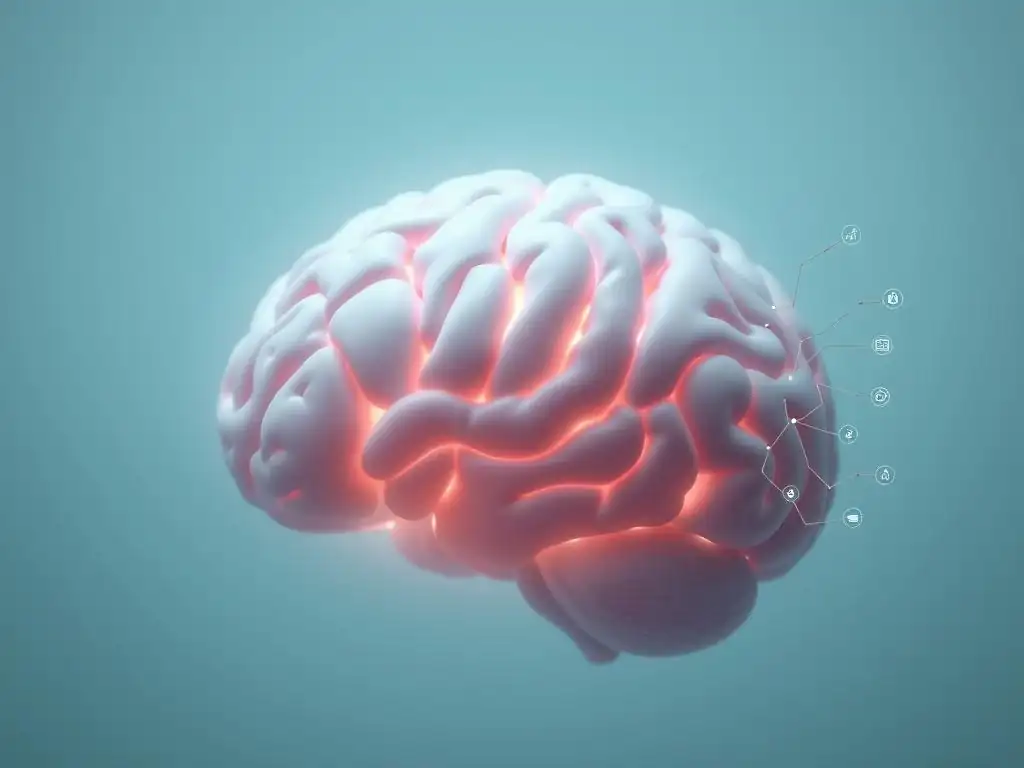
Despite its immense potential, interpreting brain scan data in the context of therapy outcomes presents several challenges. One significant difficulty lies in the complex relationship between brain activity/structure and psychological states or behaviors. A change observed in a specific brain region after therapy doesn't always definitively explain why the therapy was effective; it merely shows a correlation. Pinpointing the causal link requires sophisticated experimental designs. Furthermore, different studies using similar therapies may report changes in slightly different brain areas or networks, potentially due to variations in methodology, patient populations, or the specific aspects of therapy being investigated.
Another challenge is the issue of specificity. Changes seen after therapy might not be unique to that specific treatment or even to therapeutic change itself; they could reflect general effects of learning, reduced stress, or the passage of time. Distinguishing therapy-specific neural changes from these general effects requires careful control groups, including those receiving placebo or alternative treatments.
The field is also grappling with the best ways to analyze complex brain network data, moving beyond focusing on single regions to understanding how entire systems reorganize in response to therapy. These interpretive hurdles highlight the need for continued methodological refinement in brain imaging therapy effectiveness research.
The Need for Larger Sample Sizes and Longitudinal Studies
Current brain imaging therapy effectiveness research, while promising, often suffers from limitations imposed by relatively small sample sizes. Conducting brain imaging studies is expensive and logistically challenging, making it difficult to recruit and scan large numbers of participants. Small sample sizes can lead to findings that are not reproducible and may overestimate the true effect size of therapy on the brain. To draw more robust conclusions and identify reliable biomarkers, future research critically needs to involve larger, multi-site studies that can pool data across different locations.
Furthermore, most studies examine brain changes immediately before and after a course of therapy. While valuable, this cross-sectional approach doesn't fully capture the dynamic process of change or assess the long-term stability of neural alterations. Longitudinal studies that follow participants months or even years after therapy are essential to understand whether the observed brain changes are sustained and how they relate to the maintenance of therapeutic gains over time. Such long-term brain imaging therapy effectiveness research will provide a more complete picture of the enduring impact of psychotherapy on the brain.
Ethical Considerations in Brain Imaging Research
The increasing use of brain imaging in mental health research raises important ethical considerations. One concern relates to the potential for misinterpretation or oversimplification of findings. Presenting complex brain data in a deterministic way could lead to stigma or a reductionist view of mental illness, ignoring the multifaceted interplay of biological, psychological, and social factors. Researchers and communicators must be careful to explain findings accurately and avoid implying that brain scans are definitive diagnostic tools or solely determine a person's identity or potential.
Another ethical aspect involves privacy and data security. Brain imaging data is highly personal and potentially identifiable. Ensuring the robust protection and anonymization of this sensitive information is paramount. As research moves towards potentially using brain imaging for personalized treatment selection or prediction, there are also ethical questions about equity of access to such technologies and the potential for discrimination based on brain profiles. These ethical considerations must be actively addressed as brain imaging therapy effectiveness research continues to advance.
Future Innovations in Brain Imaging Techniques
The field of brain imaging is constantly evolving, with future innovations promising even greater insights into the neural effects of therapy. Advances in MRI technology, such as higher field strength scanners, offer improved spatial resolution and the ability to detect more subtle structural and functional changes. Development of new fMRI paradigms, including naturalistic scanning during therapy sessions or using virtual reality, could provide more ecologically valid data on how the brain responds during real-world therapeutic interactions.
Improvements in EEG technology, such as high-density arrays and mobile EEG systems, allow for more detailed and flexible recordings of brain electrical activity, potentially even outside the lab setting. Future developments in PET tracers could enable researchers to study a wider range of neurotransmitter systems and cellular processes affected by therapy. Integrating data from multiple imaging modalities (e.g., combining fMRI and EEG for better spatiotemporal resolution) and using advanced computational methods like machine learning to analyze complex datasets are also key future directions that will enhance brain imaging therapy effectiveness research.
Conclusion: The Promise of Brain Imaging for Improving Mental Healthcare
Brain Imaging as a Tool for Monitoring Treatment Progress
Brain imaging holds significant promise as a tool for objectively monitoring treatment progress in mental healthcare. While symptom checklists and clinical interviews remain essential, brain scans offer a complementary perspective on how the brain is responding to therapy.
By obtaining brain imaging data at multiple points throughout a course of treatment – for instance, at the start, mid-treatment, and end – clinicians and researchers could potentially track neural changes that correlate with symptom improvement. This could provide early indicators of whether a therapy is having its intended biological effect, potentially allowing for adjustments to the treatment plan if the desired neural changes are not occurring.
For example, if brain imaging therapy effectiveness research shows that successful therapy for depression involves increased connectivity in a specific circuit, monitoring this connectivity during treatment could help assess if the patient's brain is moving towards a healthier state. While routine clinical brain scanning for monitoring progress is not yet standard practice, ongoing brain imaging therapy effectiveness research is building the evidence base and developing the necessary methods to potentially integrate such objective neural markers into future clinical workflows, offering a more data-driven approach to evaluating treatment response.
Towards a More Objective and Personalized Approach to Therapy
The insights gained from brain imaging are paving the way for a more objective and personalized approach to psychological therapy. By providing a window into the biological underpinnings of mental health conditions and the mechanisms by which therapy induces change, brain imaging moves beyond subjective reports to offer empirical evidence of therapeutic impact. This objectivity can help validate the power of psychotherapy and reduce stigma by framing mental health challenges as conditions with tangible biological correlates that can be influenced by psychological interventions.
Crucially, the potential to use brain imaging to predict therapy response and tailor interventions based on individual brain profiles represents a significant step towards personalized medicine in mental health. Imagine a future where a brain scan could help guide the choice between different therapeutic modalities or inform the specific techniques used within a therapy, maximizing the likelihood of a positive outcome for each individual. This vision, supported by ongoing brain imaging therapy effectiveness research, holds the potential to transform mental healthcare delivery, making it more efficient, effective, and truly tailored to the needs of the individual.
The Importance of Continued Brain Imaging Therapy Effectiveness Research
Continued investment in brain imaging therapy effectiveness research is paramount for realizing the full potential of these technologies in improving mental healthcare. There is still much to learn about the complex interplay between brain function, psychological processes, and therapeutic change across the wide spectrum of mental health conditions. Research is needed to replicate findings across diverse populations, establish reliable neural biomarkers, understand the long-term effects of different therapies, and develop standardized protocols for data acquisition and analysis.
Further research is also necessary to translate findings from the lab into practical clinical tools. This involves developing user-friendly methods for analyzing brain data and integrating this information ethically and effectively into clinical decision-making. By continuing to explore the science of change through brain imaging, researchers can unlock new insights into how therapy works, refine existing treatments, develop novel interventions, and ultimately improve the lives of individuals struggling with mental health challenges. The ongoing commitment to brain imaging therapy effectiveness research is an investment in the future of mental well-being.
Frequently Asked Questions
What is the role of brain imaging in researching the effectiveness of therapy?
Brain imaging provides a visual representation of brain activity before and after therapy. This helps researchers understand how therapy impacts brain function and identify neural correlates of improvement.
What types of brain imaging techniques are typically used in brain imaging therapy effectiveness research?
Common techniques include fMRI (functional Magnetic Resonance Imaging) to measure brain activity, PET (Positron Emission Tomography) to track metabolic processes, and EEG (Electroencephalography) to assess brainwave patterns. These techniques provide different insights into brain function.
How does brain imaging contribute to understanding the mechanisms of therapeutic change?
Brain imaging can reveal which brain regions are activated or deactivated during therapy. This allows researchers to pinpoint the specific neural pathways involved in the therapeutic process and understand how change occurs.
Can brain imaging predict which individuals will benefit most from specific types of therapy?
Research aims to identify biomarkers through brain imaging that predict treatment response. This could lead to personalized treatment plans based on an individual's unique brain characteristics and improve therapy outcomes.
What are the current limitations and future directions of brain imaging therapy effectiveness research?
Limitations include the high cost and complexity of imaging techniques, as well as the need for larger sample sizes. Future directions involve developing more precise imaging methods and integrating brain imaging data with other clinical information.
Final Reflection: The Evolving Landscape of Therapeutic Change
The ability to visualize therapeutic change within the brain offers not just a scientific breakthrough, but a profound shift in how we understand and approach mental health. This research, focusing on brain imaging and therapy effectiveness, is no longer a futuristic concept; it's actively shaping the future of mental healthcare, offering personalized treatments and paving the way for more effective interventions.
The impact of this research extends far beyond the laboratory, influencing the very fabric of our understanding of mental illness and recovery. It challenges outdated stigmas and fosters a more nuanced appreciation for the complex interplay between the mind and brain. The ability to objectively measure therapeutic progress, as demonstrated through brain imaging, empowers both patients and clinicians. The greatest discovery of all time is that a person can change his future by merely changing his attitude.
– Oprah Winfrey. This quote resonates deeply with the potential of brain imaging to show tangible evidence of positive change, fostering hope and resilience in the therapeutic journey.
Consider the implications of this research for your own life. Perhaps you know someone struggling with mental health challenges, or perhaps you've faced these challenges yourself. This article offers a glimpse into a future where treatment is not just about managing symptoms, but about actively reshaping the brain towards well-being. Take a moment to reflect on the power of hope and the potential for change. Seek out resources, advocate for better mental health support, and remember that the science of change is on your side.
Unlock Your Brain's Potential: Take Charge of Your Therapy Journey
Thanks for exploring the fascinating science behind therapeutic change with us! We hope you gained valuable insights into how brain imaging is revolutionizing our understanding of mental well-being. Now, it's time to put this knowledge into action.
- Start journaling your therapy progress: Track your emotional shifts and notice how your perspective changes over time. This is a powerful way to personalize the insights from the article and actively engage in your own healing.
- Share this article with someone who might find it helpful: Spread awareness about the incredible power of therapy and the advancements in understanding brain function.
- Consider exploring different therapeutic approaches: Armed with this new understanding, you can research and discuss various methods with your therapist to find what works best for you.
So, what small step will you take today to nurture your brain and enhance your therapy experience?