
Molecular Imaging: Seeing Brain Chemistry in Action with PET and SPECT

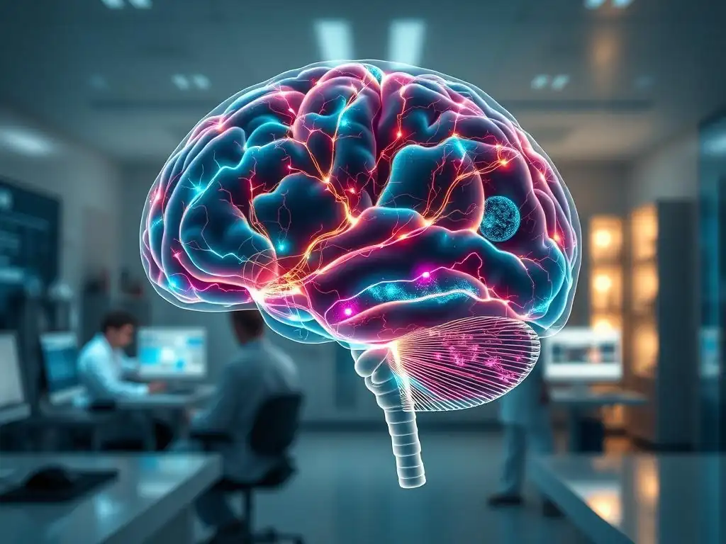
Welcome to Mind Care Guide, your trusted companion on the journey to mental well-being. Feeling lost in the complexities of your mind? We understand. In this article, we delve into the fascinating world of PET SPECT brain scans – imagine seeing the very chemistry of your brain in action! What secrets can these powerful tools unlock about mental health, and how might they pave the way for a brighter future? Let's explore together...
- Molecular Imaging: Unveiling Brain Function with PET and SPECT
- PET Scans: Positron Emission Tomography for Brain Function
- SPECT Scans: Single-Photon Emission Computed Tomography for Brain Blood Flow
- PET and SPECT Brain Scans: A Comparative Analysis
- Molecular Imaging and Brain Function: Key Insights
- Advancements in PET/SPECT Brain Scan Technology
- Ethical Considerations in PET/SPECT Brain Imaging
- The Future of Molecular Imaging in Brain Research
-
Frequently Asked Questions
- What are PET and SPECT scans and how do they relate to mental well-being?
- How do PET and SPECT scans help in diagnosing mental health challenges?
- What kind of information do PET and SPECT scans provide that other imaging techniques like MRI or CT scans do not?
- Are PET and SPECT scans safe, and what are the potential risks involved?
- How can I access PET or SPECT scans for mental health evaluation, and what should I expect during the procedure?
- Final Reflection: Unveiling the Mind's Mysteries Through Molecular Imaging
- Unlock the Mysteries of Your Brain: Explore Imaging's Potential
Molecular Imaging: Unveiling Brain Function with PET and SPECT
Principles of Molecular Imaging
Molecular imaging is a revolutionary field that allows scientists and clinicians to visualize and measure specific biological processes within living organisms, including the human brain, at a molecular and cellular level. Unlike structural imaging techniques like MRI or CT, which primarily show anatomy, molecular imaging focuses on function – how molecules behave in the brain, such as neurotransmitter binding, metabolic activity, or protein aggregation. This is achieved by introducing trace amounts of molecular probes, often labeled with radioactive isotopes, into the body. These probes are designed to target specific molecules or pathways involved in brain function or dysfunction.
Once the probe is administered, it distributes throughout the body, including the brain, binding to its target molecules. Specialized scanners then detect signals emitted by the radioactive label. The distribution and intensity of these signals provide detailed information about the location, quantity, and activity of the target molecules in different brain regions.
This allows researchers to non-invasively study the complex biochemistry of the brain in real-time, offering unprecedented insights into the mechanisms underlying both healthy brain function and various neurological and psychiatric conditions. The insights gained from these advanced PET SPECT brain scans are invaluable for research and potential clinical applications.
Applications in Neuroscience and Psychiatric Research
Molecular imaging has become an indispensable tool in neuroscience and psychiatric research, significantly advancing our understanding of brain disorders. It allows researchers to investigate hypotheses about the molecular basis of conditions like Alzheimer's disease, Parkinson's disease, schizophrenia, depression, and addiction. For instance, researchers can use specific radiotracers to measure dopamine receptor density in individuals with Parkinson's or schizophrenia, or quantify amyloid plaque burden in patients suspected of having Alzheimer's.
Beyond understanding disease mechanisms, molecular imaging plays a crucial role in drug development and evaluation. By visualizing the binding of potential therapeutic compounds to their molecular targets in the brain, researchers can assess drug efficacy, optimize dosing, and predict potential side effects. This capability is particularly important for psychiatric medications, where understanding drug interaction with neurotransmitter systems is key. The ability to directly observe these molecular interactions in vivo using techniques like PET and SPECT brain scans accelerates the development of more targeted and effective treatments.
PET and SPECT: Visualizing Brain Function
Positron Emission Tomography (PET) and Single-Photon Emission Computed Tomography (SPECT) are two primary techniques within the realm of molecular imaging used to visualize brain function. Both methods rely on the administration of a radiotracer that emits detectable radiation, but they differ in the type of radiation detected and the scanning technology used. PET detects pairs of gamma rays produced by the annihilation of positrons emitted by the radiotracer, while SPECT detects single gamma rays.
These differences in detection methodology lead to variations in resolution, sensitivity, and the types of radiotracers that can be used. However, both PET and SPECT brain scans provide functional information that is critical for understanding brain health and disease. They allow us to see things like regional blood flow, metabolic rate (glucose metabolism), and the distribution and binding of neurotransmitters or receptors, offering a dynamic view of brain activity that complements the structural information provided by MRI or CT.
PET Scans: Positron Emission Tomography for Brain Function
PET Scan Methodology: Radiotracers and Positron Detection
PET imaging relies on the use of radiotracers labeled with positron-emitting isotopes, such as Fluorine-18 (¹⁸F), Carbon-11 (¹¹C), Oxygen-15 (¹⁵O), or Nitrogen-13 (¹³N). These isotopes have short half-lives, meaning they decay rapidly, which is important for minimizing radiation dose to the patient. For brain imaging, a common radiotracer is ¹⁸F-fluorodeoxyglucose (¹⁸F-FDG), which is taken up by brain cells in proportion to their metabolic activity (glucose utilization). Other tracers target specific receptors, transporters, or enzymes. Once administered intravenously, the radiotracer travels through the bloodstream and accumulates in brain regions based on the biological process it's designed to measure.
When a positron-emitting isotope decays, it emits a positron. This positron travels a short distance (typically less than a millimeter in tissue) before colliding with an electron. This annihilation event converts the mass of both particles into energy, which is released as a pair of gamma rays traveling in nearly opposite directions (180 degrees apart).
The PET scanner, which consists of a ring of detectors surrounding the patient's head, detects these coincident pairs of gamma rays. By detecting many such pairs and tracing their lines of flight back to their origin, a computer can reconstruct a 3D image showing the distribution and concentration of the radiotracer in different parts of the brain, reflecting the underlying biological activity.
PET Scan Applications in Neurological Disorders
PET scans have become invaluable in the diagnosis and management of various neurological disorders. Perhaps one of the most well-known applications is in the evaluation of Alzheimer's disease and other dementias. ¹⁸F-FDG PET can reveal patterns of reduced glucose metabolism in specific brain regions (like the temporal and parietal lobes) that are characteristic of Alzheimer's pathology, often before significant structural changes are visible on MRI. Furthermore, specific PET tracers like ¹⁸F-florbetapir or ¹¹C-Pittsburgh compound B (PiB) can directly visualize amyloid plaques, a hallmark of Alzheimer's disease, providing crucial diagnostic information, particularly in ambiguous cases.
Beyond dementia, PET is used in epilepsy to locate seizure foci by identifying areas of hypometabolism during the interictal (between seizures) period or hypermetabolism during a seizure. In movement disorders like Parkinson's disease, while SPECT is more commonly used for dopamine transporter imaging, PET can be used with specific tracers (e.g., targeting dopa decarboxylase activity) to assess the integrity of the dopaminergic system. PET scans also play a role in differentiating types of brain tumors, assessing their aggressiveness, and monitoring treatment response by measuring metabolic activity.
Advantages and Disadvantages of PET Imaging
One of the primary advantages of PET imaging is its high sensitivity, allowing detection of very low concentrations of radiotracer and thus enabling the study of molecular processes at trace levels. PET generally offers higher spatial resolution compared to SPECT (often around 4-6 mm), which allows for better visualization of smaller brain structures and more precise localization of activity.
The wide range of available positron-emitting isotopes and the ability to synthesize tracers for various molecular targets make PET highly versatile for studying diverse biological pathways. Furthermore, PET can provide quantitative data, allowing for precise measurement of parameters like receptor density or metabolic rate.
However, PET imaging also has significant disadvantages. The short half-lives of many useful positron emitters (like ¹¹C and ¹⁵O) mean they must be produced on-site or nearby using a cyclotron, which is a very expensive piece of equipment. This limits the availability of many PET tracers to specialized centers.
PET scanners themselves are also costly to acquire and maintain. The total radiation dose from a PET scan, while generally considered safe, is higher than that from a typical CT scan or MRI. Finally, while resolution is better than SPECT, it is still lower than structural MRI, meaning PET images are often co-registered with MRI scans for anatomical context.
PET Scan vs. MRI: A Comparison of Brain Imaging Modalities
Comparing PET scans and MRI is crucial for understanding their respective roles in brain imaging. MRI (Magnetic Resonance Imaging) provides highly detailed structural images of the brain, excellent for visualizing anatomy, detecting lesions, tumors, strokes, and other structural abnormalities with resolutions often down to 1 mm or less. It works by using strong magnetic fields and radio waves to generate signals from water molecules in tissues. MRI is non-ionizing, meaning it does not use radiation.
In contrast, PET scans are a functional imaging technique that visualizes biological processes at the molecular level, such as metabolism, blood flow, or receptor binding, using radioactive tracers. While PET provides functional information critical for understanding disease mechanisms and activity, its spatial resolution is significantly lower than that of structural MRI.
Therefore, PET and MRI are often used complementarily; a PET scan showing metabolic abnormalities might be overlaid onto a high-resolution MRI scan of the same brain to precisely locate the functional changes within the anatomical structure. Neither technique is inherently "better"; they provide different, yet often synergistic, types of information about the brain.
SPECT Scans: Single-Photon Emission Computed Tomography for Brain Blood Flow
SPECT Scan Principles: Radiotracers and Gamma Ray Detection
SPECT is another nuclear medicine technique used for molecular imaging of the brain, primarily focused on visualizing blood flow and, to a lesser extent, receptor binding. Like PET, SPECT requires the administration of a radiotracer, but it uses isotopes that emit single gamma rays, such as Technetium-99m (⁹⁹mTc), Iodine-123 (¹²³I), or Thallium-201 (²⁰¹Tl). These isotopes generally have longer half-lives than those used in PET, making the radiotracers easier to produce and distribute to centers without cyclotrons.
Once the SPECT radiotracer is administered (typically intravenously), it distributes throughout the brain in proportion to regional cerebral blood flow (rCBF) or binds to specific targets like neurotransmitter transporters. The SPECT scanner uses gamma cameras, which are large detectors that rotate around the patient's head.
These cameras detect the single gamma rays emitted by the radiotracer. By detecting gamma rays from multiple angles as the camera rotates, a computer can reconstruct a 3D image showing the distribution and intensity of the radiotracer within the brain, which in the case of blood flow tracers, reflects the metabolic activity of different brain regions.
Clinical Applications of SPECT in Neurology and Psychiatry
SPECT imaging has several important clinical applications in neurology and psychiatry. Its ability to measure regional cerebral blood flow makes it useful in evaluating conditions where blood supply to the brain is compromised or altered. For example, SPECT can help assess the extent of damage after a stroke or identify areas of reduced perfusion in conditions like vascular dementia. It can also be used in the evaluation of persistent symptoms after traumatic brain injury, where blood flow abnormalities may be present.
In psychiatry, SPECT is particularly valuable for assessing dopamine transporter (DAT) density in the striatum, a key structure involved in movement and reward. This is achieved using specific radiotracers like ¹²³I-ioflupane (DaTscan). Reduced DAT uptake is characteristic of neurodegenerative disorders affecting the dopaminergic system, such as Parkinson's disease and Lewy body dementia. While SPECT is not used for routine psychiatric diagnoses like depression or schizophrenia, research applications have explored its use to study blood flow patterns or receptor availability in these conditions, contributing to our understanding of their neurobiological underpinnings.
SPECT Scan for Diagnosis and Treatment Monitoring
SPECT scans play a role in the diagnostic workup for certain conditions, particularly those involving the dopaminergic system or cerebral blood flow. For instance, a DaTscan using SPECT can help differentiate Parkinson's disease and Lewy body dementia from essential tremor, which typically does not involve significant DAT loss. This can be crucial for guiding patient management and prognosis. While not a definitive diagnostic tool in isolation for most psychiatric disorders, abnormal blood flow patterns on SPECT can support clinical suspicion in complex cases or help rule out alternative diagnoses.
Beyond diagnosis, SPECT can occasionally be used to monitor the effects of treatment, although this is less common than in PET imaging. For example, changes in regional cerebral blood flow might be assessed in response to therapies for stroke rehabilitation or certain psychiatric interventions, though this application is more often seen in research settings than routine clinical practice.
The relative ease of use and availability of SPECT compared to PET means it remains a valuable tool in the clinical neuroimaging arsenal, often serving as an initial functional assessment before potentially moving to more complex imaging like PET SPECT brain scans.
SPECT Scan Safety: Radiation Exposure and Risk Mitigation
Like all nuclear medicine procedures, SPECT scans involve exposure to a small amount of ionizing radiation from the administered radiotracer. The dose varies depending on the specific radiotracer used and the patient's body weight, but it is generally comparable to or slightly lower than the radiation dose received from a diagnostic CT scan. Regulatory bodies establish dose limits and guidelines to ensure that the risks associated with radiation exposure are minimized and outweighed by the clinical benefits of the scan.
To mitigate risks, healthcare professionals adhere to the principle of ALARA (As Low As Reasonably Achievable), using the minimum amount of radiotracer necessary to obtain diagnostic quality images. Patient history, including recent radiation exposure from other procedures, is reviewed. Pregnant individuals are typically not scanned unless absolutely necessary, and breastfeeding mothers may be advised to temporarily suspend breastfeeding after the procedure. The radiotracers used in SPECT have relatively short biological half-lives and are eliminated from the body naturally over time, further reducing long-term exposure.
PET and SPECT Brain Scans: A Comparative Analysis
PET vs SPECT: Resolution, Cost, and Clinical Utility
When comparing PET and SPECT brain scans, several factors distinguish them, including image resolution, cost, and their primary clinical utility. Generally, PET offers higher spatial resolution (typically 4-6 mm) than SPECT (often 7-10 mm or more). This allows PET to resolve smaller structures and provide more detailed functional maps. PET is also generally more sensitive than SPECT, capable of detecting lower concentrations of radiotracer.
However, SPECT scans are typically less expensive to perform than PET scans. This is largely because the radiotracers used in SPECT often have longer half-lives and can be shipped from central pharmacies, eliminating the need for an on-site cyclotron required for many common PET tracers. This lower cost and greater accessibility make SPECT more widely available in clinical settings.
In terms of clinical utility, PET excels in quantitative measurement of metabolic rate and specific receptor binding, making it powerful for research and specific diagnostic challenges (like amyloid imaging). SPECT is highly effective and widely used for assessing regional cerebral blood flow and dopamine transporter imaging (e.g., DaTscan), offering practical diagnostic value in conditions like Parkinsonism and stroke.
Complementary Use of PET and SPECT for Comprehensive Brain Imaging
Despite their differences, PET and SPECT brain scans are often viewed as complementary techniques rather than competing ones. They provide different types of functional information that, when combined with structural imaging like MRI, can offer a more comprehensive picture of brain health and disease.
For example, in the evaluation of dementia, a structural MRI might show atrophy, an ¹⁸F-FDG PET might reveal patterns of hypometabolism suggesting Alzheimer's, and an amyloid PET might confirm the presence of amyloid plaques. In movement disorders, a clinical assessment might be supported by a SPECT DaTscan showing reduced dopamine transporters, while a research PET scan might investigate specific dopamine receptor subtypes.
In some cases, the choice between PET and SPECT for a particular application depends on the specific radiotracer available and the clinical question being asked. For example, assessing cerebral blood flow can be done with both modalities, but SPECT is more commonly used clinically for this purpose due to tracer availability and cost. Assessing glucose metabolism is primarily done with PET using ¹⁸F-FDG. The integration of information from different imaging modalities, including both PET and SPECT brain scans, alongside clinical data, is becoming increasingly standard for complex neurological and psychiatric evaluations.
Amyloid Imaging with PET and Dopamine Transporter Imaging with SPECT
Two prominent examples highlight the specific strengths of PET and SPECT brain scans in molecular imaging: amyloid imaging with PET and dopamine transporter (DAT) imaging with SPECT. Amyloid PET imaging uses tracers like ¹⁸F-florbetapir or ¹⁸F-florbetaben to bind to amyloid plaques, a key pathological feature of Alzheimer's disease. Visualizing and quantifying these plaques in vivo provides strong evidence supporting an Alzheimer's diagnosis, particularly in cases of mild cognitive impairment where the cause is uncertain. This application is largely exclusive to PET due to the specific tracer chemistry required.
Conversely, SPECT is the modality of choice for routine clinical imaging of dopamine transporters (DAT) using tracers like ¹²³I-ioflupane (DaTscan). DATs are proteins on nerve terminals that reabsorb dopamine. Loss of dopaminergic neurons in the striatum, characteristic of Parkinson's disease and Lewy body dementia, leads to reduced DAT density.
A SPECT DaTscan showing decreased uptake in the striatum is highly indicative of these conditions and helps differentiate them from other movement disorders that do not involve significant dopaminergic degeneration. While PET can also image aspects of the dopaminergic system, SPECT DaTscan is more widely available and cost-effective for this specific clinical question.
Molecular Imaging and Brain Function: Key Insights
Neurotransmitter Systems and PET/SPECT Imaging
Molecular imaging, particularly PET and SPECT, provides a powerful window into the complex world of neurotransmitter systems in the living brain. Neurotransmitters like dopamine, serotonin, acetylcholine, and glutamate are chemical messengers that play critical roles in regulating mood, cognition, movement, and behavior. Dysregulation of these systems is implicated in numerous psychiatric and neurological disorders, including depression, schizophrenia, addiction, and Parkinson's disease.
Using specific radiotracers that bind to neurotransmitter receptors, transporters, or enzymes, PET and SPECT brain scans can quantify their distribution, density, and binding affinity in different brain regions. For example, PET tracers can measure dopamine D2/D3 receptor availability, which is altered in conditions like schizophrenia and addiction. SPECT tracers, as mentioned, are widely used to assess dopamine transporter density in Parkinsonism. These studies help researchers understand how these systems function in health and how they are altered in disease states, providing crucial insights into the neurobiological basis of mental health challenges.
Receptor Binding Studies with Molecular Imaging
Receptor binding studies are a cornerstone of molecular imaging applications in neuroscience and psychiatry. These studies involve administering a radiotracer that is designed to selectively bind to a specific type of neurotransmitter receptor or transporter in the brain. By measuring the amount of radiotracer bound in different brain regions, researchers can estimate the density and availability of these target molecules. This information is vital because the number and state of receptors can change in response to disease, stress, aging, or pharmacological treatments.
For instance, PET studies using tracers like ¹¹C-raclopride have shown altered dopamine D2 receptor availability in the striatum of individuals with schizophrenia or substance use disorders. Similarly, PET studies targeting serotonin transporters (SERT) have revealed reduced SERT density in patients with major depression. SPECT studies using tracers like ¹²³I-iomazenil can assess benzodiazepine receptor availability, which is related to anxiety disorders. These receptor binding studies provide direct evidence of molecular changes associated with mental health conditions, aiding in diagnosis, understanding pathology, and evaluating the effects of therapeutic interventions.
PET and SPECT in Drug Development and Target Validation
Molecular imaging plays a critical role throughout the drug development lifecycle, from target identification to clinical trials. Before developing a new drug, researchers use PET or SPECT to validate potential drug targets in the brain by visualizing their presence and distribution in healthy and diseased states. For example, if a new drug is being developed to target a specific receptor implicated in depression, a PET scan can confirm that the receptor is indeed present and accessible in the relevant brain regions in human subjects.
During preclinical and clinical trials, PET and SPECT brain scans are used to assess the pharmacokinetics and pharmacodynamics of potential drug candidates. This includes measuring how the drug is absorbed, distributed, metabolized, and excreted (pharmacokinetics) and, crucially, confirming that the drug binds to its intended target in the brain and at what dose (pharmacodynamics).
Receptor occupancy studies using PET, for instance, can determine what percentage of target receptors are occupied by a drug at a given dose, helping optimize dosing regimens and predict therapeutic efficacy and potential side effects. This accelerates the development of safer and more effective treatments for neurological and psychiatric disorders.
Advancements in PET/SPECT Brain Scan Technology
Novel Radiotracers for Specific Brain Targets
Significant advancements in molecular imaging technology are driven by the development of novel radiotracers capable of targeting an ever-expanding array of molecules in the brain. Early tracers focused on basic processes like glucose metabolism or blood flow, but modern radiochemistry allows for the synthesis of complex molecules that bind selectively to specific neurotransmitter receptors, transporters, enzymes, or even pathological proteins like amyloid and tau.
Recent developments include tracers that target neuroinflammation markers (e.g., TSPO ligands), synaptic vesicle glycoprotein 2A (SV2A) density (a marker of synaptic density), and specific subtypes of opioid or cannabinoid receptors. The development of new ¹⁸F-labeled tracers, which have a longer half-life than ¹¹C, has made PET more accessible by allowing tracers to be produced off-site and transported. The ongoing search for highly selective and sensitive tracers is crucial for unlocking new insights into brain function and pathology and developing more targeted therapies for conditions studied using PET SPECT brain scans.
Enhanced Resolution and Image Quality in Molecular Imaging
Technological progress has led to substantial improvements in the resolution and image quality achievable with PET and SPECT scanners. Detector technology has advanced, with smaller, faster, and more efficient crystals and photodetectors enabling higher sensitivity and spatial resolution. Techniques like Time-of-Flight (TOF) PET, which measures the difference in arrival time of the two annihilation photons with picosecond precision, allow for more accurate localization of the annihilation event, significantly improving image quality, signal-to-noise ratio, and effective resolution, often reducing scan times or injected dose.
For SPECT, advancements include the development of multi-head cameras and sophisticated reconstruction algorithms. Dedicated brain SPECT cameras with closer detector geometry can also improve sensitivity and resolution compared to general-purpose systems. While SPECT resolution still lags behind PET, these improvements make SPECT brain scans more informative. Ongoing research aims to further push the boundaries of resolution and sensitivity in both modalities, potentially allowing visualization of even smaller structures and molecular aggregates.
Development of Quantitative Analysis Methods for PET/SPECT Data
Beyond hardware improvements, significant progress has been made in the software and analytical methods used to process and interpret PET and SPECT brain scan data. Quantitative analysis is essential for extracting meaningful biological information, such as absolute metabolic rates, receptor densities, or binding potentials. This involves complex mathematical models that account for factors like radiotracer delivery to the brain, binding kinetics, and metabolism.
Modern quantitative methods include compartmental modeling, reference tissue modeling, and graphical analysis techniques. These methods require accurate kinetic data acquired over time (dynamic scanning) and often arterial blood sampling to measure tracer concentration in the blood. Advancements in image registration, normalization to standard brain atlases, and statistical parametric mapping allow for objective comparison of data between individuals and groups, facilitating the identification of subtle but significant differences associated with disease or treatment effects. Machine learning and artificial intelligence are also increasingly being applied to analyze patterns in PET SPECT brain scans, potentially aiding in automated diagnosis and prediction.
Ethical Considerations in PET/SPECT Brain Imaging
Informed Consent and Data Security
As with any medical procedure involving radiation and sensitive personal health information, ethical considerations are paramount in PET and SPECT brain imaging. Obtaining truly informed consent from participants, particularly in research settings or for individuals with cognitive impairments, requires careful attention. Patients must be fully informed about the procedure, including the administration of a radioactive substance, the potential risks (albeit small), the purpose of the scan, and how their data will be used and protected.
Data security and privacy are also critical concerns. Molecular imaging data, especially when combined with other clinical or genetic information, can be highly sensitive. Robust measures must be in place to protect patient confidentiality, anonymize data when appropriate for research, and secure storage and transmission of images and associated information. Compliance with regulations like HIPAA in the United States or GDPR in Europe is essential to ensure patient trust and protect their rights.
Responsible Use of Molecular Imaging Data
The power of molecular imaging to reveal detailed information about brain function comes with the responsibility to use this data ethically and appropriately. In clinical settings, interpreting PET SPECT brain scans requires expertise to ensure findings are placed in the correct clinical context and not overinterpreted or used to make diagnoses in isolation. Given that molecular markers (like amyloid plaques) can be present in individuals without symptoms, careful consideration is needed when communicating findings that may have significant implications for a patient's future health or insurability.
In research, responsible use includes ensuring that studies are designed with clear scientific objectives, minimizing radiation exposure to participants, and avoiding the use of imaging for non-validated applications. There is also an ethical obligation to disseminate research findings responsibly and avoid sensationalizing or misrepresenting the capabilities of molecular imaging techniques, particularly regarding complex conditions like psychiatric disorders.
Addressing Potential Biases in Image Interpretation
Image interpretation in molecular imaging, like other medical specialties, is susceptible to potential biases. These can arise from various sources, including observer bias, limitations of the imaging technology, variability in radiotracer uptake, and the inherent complexity of brain function. For example, differences in scanner calibration, reconstruction algorithms, or analysis software can influence quantitative results. Furthermore, the interpretation of regional uptake patterns can be subjective without standardized atlases and quantitative methods.
Efforts to address these biases include rigorous quality control procedures for scanners and radiotracers, standardization of acquisition protocols, use of validated quantitative analysis methods, and employing blind interpretation where appropriate in research studies. Training and certification of nuclear medicine physicians and technologists are crucial. Researchers are also working on developing artificial intelligence tools for image analysis that can provide more objective and consistent interpretations of PET SPECT brain scans, potentially reducing human variability.
The Future of Molecular Imaging in Brain Research
Personalized Medicine and PET/SPECT Scans
The future of molecular imaging, particularly PET and SPECT brain scans, is increasingly intertwined with the concept of personalized medicine. By providing insights into the specific molecular alterations occurring in an individual's brain, these techniques can help tailor diagnosis, prognosis, and treatment strategies. For example, identifying the presence and distribution of specific receptor subtypes or metabolic patterns could help predict an individual's response to different psychiatric medications or guide the selection of therapies for neurodegenerative diseases.
Molecular imaging can help determine if a patient is a suitable candidate for a targeted therapy (e.g., anti-amyloid antibodies for Alzheimer's) by confirming the presence of the target pathology. It can also be used to monitor treatment response at a molecular level, providing earlier feedback than clinical symptoms alone. As we gain a deeper understanding of the molecular heterogeneity of brain disorders, the ability of PET and SPECT to visualize these differences in living patients will become even more critical for delivering truly personalized care.
Early Detection and Prevention of Neurological and Psychiatric Disorders
One of the most exciting potential future applications of PET and SPECT brain scans lies in the early detection and even prevention of neurological and psychiatric disorders. By visualizing molecular changes that occur very early in the disease process, perhaps years or even decades before the onset of clinical symptoms, molecular imaging could identify individuals at high risk. For instance, amyloid PET can detect amyloid accumulation long before dementia develops.
Identifying individuals in preclinical or prodromal stages could open windows for preventative interventions or early treatment, potentially altering the disease trajectory. For psychiatric disorders, molecular imaging might identify early markers of vulnerability or predict the likelihood of developing a condition based on subtle alterations in neurotransmitter systems or metabolic activity. While significant research is still needed, the potential for PET and SPECT to contribute to proactive brain health management is substantial.
Integration of Molecular Imaging with Other Neuroimaging Techniques
The future of brain imaging will likely involve the increasing integration of molecular imaging (PET and SPECT) with other neuroimaging techniques, such as structural MRI, functional MRI (fMRI), and electroencephalography (EEG). Each modality provides unique information about the brain – structure from MRI, metabolic/molecular function from PET/SPECT, neural activity dynamics from fMRI and EEG. Combining these different types of data offers a more complete and nuanced understanding of brain function and dysfunction.
Hybrid scanners, such as integrated PET/MRI systems, are already becoming more common, allowing simultaneous acquisition of both structural and molecular data, which improves co-registration accuracy and can reduce scan time. Advanced data analysis techniques are being developed to fuse and analyze multimodal datasets, uncovering relationships between structural changes, molecular alterations, and patterns of neural activity. This integrated approach, potentially incorporating data from PET SPECT brain scans with other modalities, holds immense promise for advancing our understanding of complex brain disorders and developing more effective diagnostic and therapeutic strategies.
Frequently Asked Questions
What are PET and SPECT scans and how do they relate to mental well-being?
PET (Positron Emission Tomography) and SPECT (Single-Photon Emission Computed Tomography) scans are molecular imaging techniques used to visualize brain activity. They offer insights into brain chemistry and function, helping diagnose and understand certain mental health conditions.
How do PET and SPECT scans help in diagnosing mental health challenges?
These scans can reveal abnormalities in brain metabolism, neurotransmitter activity, and receptor binding. This information can aid in diagnosing conditions like depression, anxiety disorders, and neurodegenerative diseases affecting mental well-being, offering a more objective assessment.
What kind of information do PET and SPECT scans provide that other imaging techniques like MRI or CT scans do not?
While MRI and CT scans focus on brain structure, PET and SPECT scans provide information about brain function and biochemical processes. They visualize how the brain is actually working, not just its physical appearance.
Are PET and SPECT scans safe, and what are the potential risks involved?
The scans involve injecting small amounts of radioactive tracers, which carry a minimal radiation risk. Allergic reactions are rare but possible. The benefits of improved diagnosis often outweigh the low radiation exposure.
How can I access PET or SPECT scans for mental health evaluation, and what should I expect during the procedure?
Consult with a psychiatrist or neurologist who can determine if a PET or SPECT scan is appropriate for your condition. The procedure typically involves a short injection followed by lying still in a scanner while the images are acquired, usually a painless and non-invasive experience.
Final Reflection: Unveiling the Mind's Mysteries Through Molecular Imaging
The ability to visualize brain chemistry in real-time, using techniques like PET and SPECT, represents a monumental leap forward in our understanding of neurological and psychiatric disorders. This isn't just about technological advancement; it's about unlocking the doors to personalized medicine and revolutionizing how we diagnose, treat, and ultimately, prevent debilitating brain conditions. The implications for improving patient outcomes are profound and far-reaching.
The impact of PET and SPECT on our understanding of the brain is undeniable, reshaping diagnostic practices and driving research into new therapeutic avenues. These technologies have moved beyond simply identifying structural abnormalities; they allow us to witness the intricate dance of neurotransmitters and receptors, providing crucial insights into the biochemical underpinnings of disease. This shift in perspective is transformative. The greatest advances in medicine are not made by chance, but by the deliberate search for knowledge.
This quote highlights the importance of continued investment in research and development within the field of molecular imaging.
Consider the possibilities: earlier and more accurate diagnoses, tailored treatment plans based on individual brain chemistry, and the development of novel drugs targeting specific molecular pathways. This isn't a distant future; it's happening now. You, the reader, are empowered to learn more about these groundbreaking technologies and advocate for their wider accessibility. Engage with the scientific community, support research initiatives, and demand better healthcare options based on the advancements in molecular imaging. The future of brain health depends on our collective commitment to understanding and harnessing the power of PET and SPECT.
Unlock the Mysteries of Your Brain: Explore Imaging's Potential
Thanks for diving into the fascinating world of molecular brain imaging with us! You've now gained a valuable understanding of how PET and SPECT scans reveal brain chemistry and function. Ready to take the next step?
- Schedule a consultation with your doctor to discuss if molecular imaging is right for you. This is your first step towards potentially gaining crucial insights into your brain health.
- Share this article with someone who might find it helpful. Let's spread awareness about these powerful diagnostic tools!
- Explore reputable sources online for more information on brain health and imaging technologies. This can help deepen your understanding and empower you to have more informed conversations with healthcare professionals.
What questions do you have about utilizing molecular imaging to understand your brain better? Let's discuss!