
The Neurobiology of Trauma: Imaging the Brain in PTSD

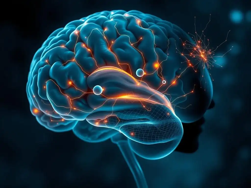
Welcome to Mind Care Guide, a safe space to explore the complexities of mental well-being.
Feeling lost in the aftermath of trauma? You're not alone. We understand the profound impact PTSD can have on your life. This article delves into the fascinating world of PTSD brain imaging techniques, offering a glimpse into the neurobiological changes associated with trauma. Join us as we unravel the science behind the suffering, shedding light on potential pathways to healing and recovery. Curious to understand what's happening inside the brain? Let's begin this journey together...
- The Neurobiology of Trauma: Imaging the Brain in PTSD
- Understanding PTSD Through Brain Imaging
- Brain Regions Affected by Trauma: A Neurobiological Overview
- PTSD Brain Imaging Techniques
- Research Findings: What Brain Imaging Reveals About PTSD
- Clinical Applications of PTSD Brain Imaging
- Challenges and Future Directions in PTSD Brain Imaging Research
- PTSD Brain Imaging: Implications for Mental Well-being
-
Frequently Asked Questions
- How do PTSD brain imaging techniques help us understand the condition?
- What are some common PTSD brain imaging techniques used in research?
- Can PTSD brain imaging techniques diagnose PTSD with certainty?
- How can PTSD brain imaging techniques improve treatment outcomes?
- Are there any risks associated with undergoing PTSD brain imaging techniques?
- Final Reflection: Unmasking the Trauma-Shaped Brain
- Unlock Your Understanding: Explore the Brain's Response to Trauma
The Neurobiology of Trauma: Imaging the Brain in PTSD
Trauma can leave indelible marks, not just on a person's psyche, but also on the very structure and function of their brain. Post-Traumatic Stress Disorder (PTSD) is a complex condition, and understanding its neurobiological underpinnings is crucial for developing effective treatments. Research into the brain of individuals with PTSD has significantly advanced thanks to sophisticated neuroimaging techniques. These PTSD brain imaging techniques allow scientists and clinicians to peer inside the living brain, observing how trauma might reshape its networks and activity patterns.
This exploration delves into how different parts of the brain are affected by trauma and examines the various PTSD brain imaging techniques employed in research and potentially clinical settings. By visualizing the brain's response to traumatic stress, we gain valuable insights into the mechanisms driving PTSD symptoms, paving the way for more targeted interventions and a deeper, more empathetic understanding of this challenging disorder.
Understanding PTSD Through Brain Imaging
Investigating the brain in the context of trauma and PTSD provides a biological window into the disorder. While PTSD is diagnosed based on behavioral and psychological symptoms, these symptoms are ultimately rooted in altered brain function. PTSD brain imaging techniques offer a non-invasive way to study these alterations directly.
The Role of Brain Imaging in Understanding PTSD
Brain imaging plays a pivotal role in shifting our understanding of PTSD from a purely psychological condition to one with significant neurobiological correlates. Techniques like MRI and fMRI reveal structural changes and functional patterns that differentiate individuals with PTSD from those who have experienced trauma but did not develop the disorder, or from those with no trauma history. This helps in identifying potential biomarkers.
Furthermore, brain imaging aids in understanding the heterogeneity of PTSD. Not everyone experiences or responds to trauma in the same way, and imaging can help identify different neurobiological profiles associated with varying symptom clusters, such as hyperarousal versus dissociation. This detailed insight is vital for tailoring interventions.
Why Study the Brain in PTSD?
Studying the brain in PTSD is essential for several reasons. Firstly, it helps validate the subjective experiences of individuals suffering from the disorder by providing objective evidence of neurological changes. This can reduce stigma and increase empathy. Secondly, identifying specific brain regions or networks involved provides targets for therapeutic interventions, including psychotherapy, pharmacology, or even neuromodulation techniques like TMS.
Moreover, understanding the neurobiological pathways implicated in PTSD can shed light on why certain treatments are effective for some individuals but not others. By observing how brain activity or structure changes in response to therapy using PTSD brain imaging techniques, researchers can refine existing treatments and develop novel approaches that directly address the underlying brain dysfunction.
Brain Regions Affected by Trauma: A Neurobiological Overview
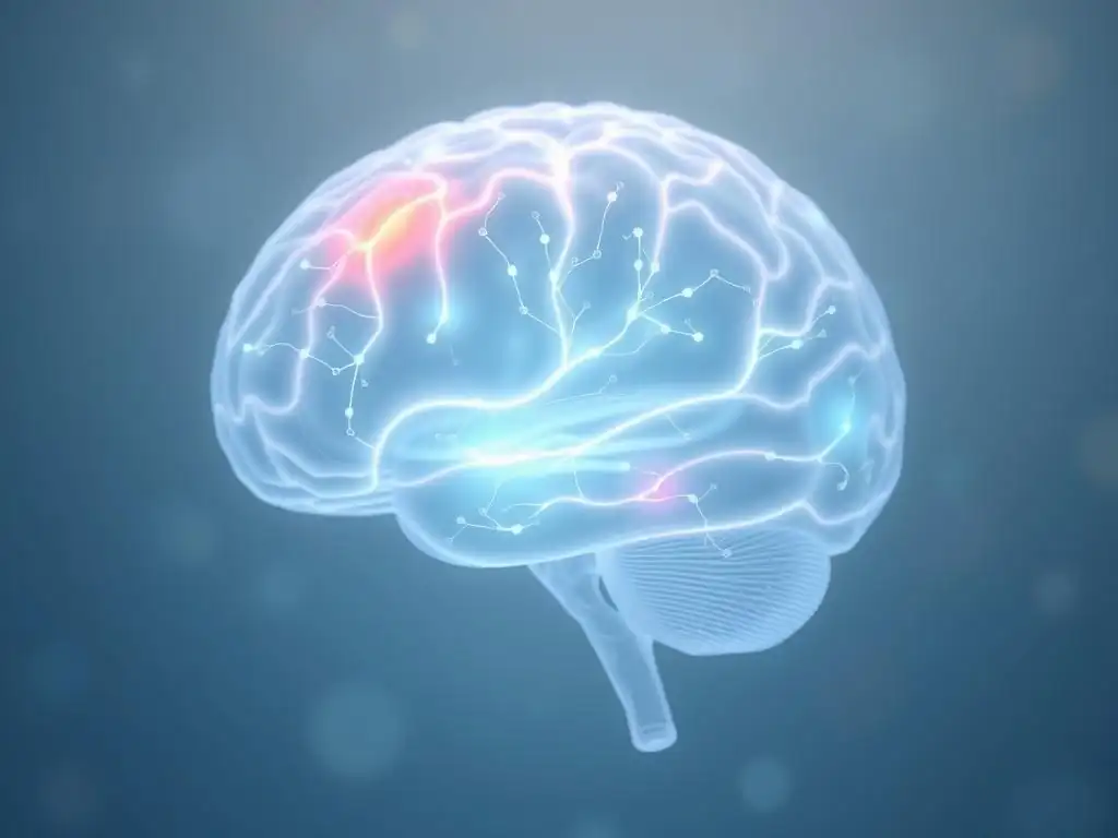
Trauma doesn't impact the brain uniformly; certain regions appear particularly vulnerable or central to the development and maintenance of PTSD symptoms. Key areas include those involved in processing fear, memory, and emotional regulation.
The Amygdala and Fear Processing in PTSD
The amygdala, a small almond-shaped structure deep within the temporal lobe, is a cornerstone of the brain's fear response system. In individuals with PTSD, PTSD brain imaging techniques, particularly fMRI, often reveal exaggerated activity in the amygdala when exposed to trauma-related cues or even general threat stimuli. This hyperresponsiveness is thought to underpin the heightened fear, anxiety, and hypervigilance characteristic of the disorder.
Studies have demonstrated that the amygdala in PTSD can show increased volume or altered connectivity with other brain regions. For example, research using structural MRI has sometimes indicated larger amygdala volumes in certain PTSD subgroups, while functional studies consistently show its hyperactivation during fear conditioning and extinction tasks, suggesting a deficit in inhibiting fear responses.
The Hippocampus and Memory Formation in PTSD
The hippocampus, located near the amygdala, is critical for forming and retrieving declarative memories, including the context surrounding traumatic events. In PTSD, memory disturbances are common, ranging from intrusive flashbacks to difficulties recalling details of the trauma or other life events. PTSD brain imaging techniques like structural MRI have frequently shown reduced hippocampal volume in individuals with chronic PTSD, sometimes cited as being 10-15% smaller on average compared to controls.
This volume reduction and altered hippocampal function are hypothesized to contribute to the fragmented, decontextualized nature of traumatic memories in PTSD. The inability to properly contextualize the memory (e.g., remembering that the event is in the past and not currently happening) is believed to drive symptoms like flashbacks and nightmares. Functional studies also show altered hippocampal activity during memory tasks in this population.
The Prefrontal Cortex and Emotional Regulation in PTSD
The prefrontal cortex (PFC), especially the medial prefrontal cortex (mPFC) and its sub-regions like the ventromedial prefrontal cortex (vmPFC), plays a crucial role in executive functions, decision-making, and regulating emotions, including inhibiting the amygdala's fear response. In PTSD, there is often evidence of reduced activity or volume in certain parts of the PFC, observed through structural and functional PTSD brain imaging techniques.
This impaired prefrontal control is thought to contribute significantly to the difficulty individuals with PTSD have in regulating their emotional responses to trauma reminders. A less active or smaller vmPFC may be less effective at dampening the hyperactive amygdala, leading to persistent feelings of threat and difficulty recovering from stress. Connectivity studies often show weakened connections between the PFC and amygdala in individuals with PTSD.
PTSD Brain Imaging Techniques
A variety of advanced neuroimaging methods are employed to study the brain in PTSD, each offering unique insights into different aspects of brain structure, function, and chemistry. These PTSD brain imaging techniques can be broadly categorized as structural or functional.
Structural Brain Imaging Techniques for PTSD
Structural imaging provides detailed pictures of the brain's anatomy, allowing researchers to measure the size of different regions or assess the integrity of white matter tracts.
Magnetic Resonance Imaging (MRI) and PTSD Brain Structure
Magnetic Resonance Imaging ( MRI) uses strong magnetic fields and radio waves to create detailed anatomical images of the brain. For studying PTSD, structural MRI is primarily used to measure the volume and shape of specific brain regions, such as the hippocampus, amygdala, and prefrontal cortex. As noted, numerous studies have reported findings like reduced hippocampal volume using this technique.
While structural MRI provides clear images of brain anatomy, it does not directly measure brain activity. However, identifying structural differences can point to areas where function might also be altered or which may be more vulnerable to the effects of trauma.
Diffusion Tensor Imaging (DTI) and White Matter Integrity in PTSD
Diffusion Tensor Imaging ( DTI) is a specialized MRI technique that measures the diffusion of water molecules in brain tissue. Since water diffuses differently along nerve fibers (axons) compared to across them, DTI can provide information about the orientation and integrity of white matter tracts, which are the brain's communication highways.
Research using DTI in PTSD has explored potential disruptions in the connectivity between different brain regions. Findings have sometimes indicated altered white matter integrity in tracts connecting the amygdala, hippocampus, and prefrontal cortex, suggesting impaired communication between these key regions involved in fear and memory processing.
Functional Brain Imaging Techniques for PTSD
Functional imaging techniques measure brain activity or metabolic processes, offering insights into how different brain regions work and interact in real-time or under specific conditions.
Functional Magnetic Resonance Imaging (fMRI) and Neural Activity in PTSD
Functional Magnetic Resonance Imaging ( fMRI) measures brain activity by detecting changes in blood flow. When a brain region is more active, it requires more oxygen, leading to changes in local blood oxygenation levels that fMRI can detect. This is perhaps the most widely used of the functional PTSD brain imaging techniques.
fMRI is used to study brain responses to trauma cues, emotional stimuli, or during cognitive tasks in individuals with PTSD. As mentioned earlier, fMRI studies frequently show heightened amygdala activity and reduced PFC activity during fear processing, providing dynamic evidence of altered neural circuit function.
Positron Emission Tomography (PET) and Neurotransmitter Function in PTSD
Positron Emission Tomography ( PET) uses radioactive tracers injected into the bloodstream to measure metabolic activity, blood flow, or the distribution of specific molecules like neurotransmitter receptors or transporters. PET offers a window into the brain's neurochemistry, which is critical for understanding conditions like PTSD.
PET studies in PTSD have investigated various neurotransmitter systems, including serotonin, dopamine, and opioid systems. Findings have suggested potential dysregulation in these systems, which are involved in mood, reward, stress response, and pain perception, all relevant to PTSD symptomatology. For example, some studies have indicated altered serotonin transporter availability in certain brain regions.
Electroencephalography (EEG) and Brainwave Patterns in PTSD
Electroencephalography ( EEG) measures electrical activity in the brain using electrodes placed on the scalp. EEG has excellent temporal resolution (measuring activity changes very quickly), making it useful for studying rapid brain responses to stimuli. It captures brainwave patterns associated with different states (e.g., sleep, wakefulness) and cognitive processes.
In PTSD research, EEG can reveal alterations in brainwave oscillations (e.g., alpha, beta, theta waves) and event-related potentials (ERPs), which are specific brain responses time-locked to a stimulus. Studies have sometimes shown altered patterns of brain oscillations, particularly in response to emotional stimuli, reflecting differences in attention, processing, and emotional reactivity in individuals with PTSD.
Comparing Different PTSD Brain Imaging Methods
Each of these PTSD brain imaging techniques offers complementary information.
- MRI/DTI: Provide structural detail and white matter integrity.
- fMRI: Offers good spatial resolution for functional activity, showing where activity changes occur.
- PET: Provides insight into neurochemistry and metabolic processes.
- EEG: Offers excellent temporal resolution, showing when activity changes occur very rapidly.
Often, researchers use multimodal imaging, combining techniques like fMRI and DTI, or fMRI and PET, to get a more comprehensive picture of the structural, functional, and chemical alterations in the PTSD brain.
Comparing Techniques:
- [x] Structural vs. Functional
- [x] Spatial Resolution
- [x] Temporal Resolution
- [x] Information Type (Anatomy, Activity, Chemistry, Electrical)
Research Findings: What Brain Imaging Reveals About PTSD
Decades of research using various PTSD brain imaging techniques have provided significant insights into the neurobiological signatures of trauma. These studies have not only confirmed hypotheses derived from clinical observations but also uncovered novel aspects of how trauma impacts the brain.
Brain Imaging Studies on PTSD
Numerous studies have consistently reported findings across different imaging modalities. For instance, a large meta-analysis pooling data from many structural MRI studies estimated an average hippocampal volume reduction of around 10% in individuals with PTSD compared to trauma-exposed controls without PTSD. Functional fMRI studies frequently highlight a pattern of increased amygdala activity and decreased vmPFC activity during fear processing tasks in PTSD.
These imaging findings provide empirical support for the idea that PTSD is associated with measurable alterations in brain structure and function, particularly within circuits involved in fear, memory, and emotion regulation.
Further research using DTI has suggested altered connectivity, potentially indicating disrupted communication pathways. For example, reduced fractional anisotropy (a measure of white matter integrity) has been reported in fiber tracts connecting limbic regions and the prefrontal cortex in some PTSD populations.
Neurobiological Markers of PTSD
The consistent findings across various PTSD brain imaging techniques suggest potential neurobiological markers for the disorder. While no single imaging marker is currently definitive for diagnosis, patterns of altered volume, activity, and connectivity in regions like the amygdala, hippocampus, and PFC are considered strong correlates.
Research is ongoing to determine if these markers can predict who will develop PTSD after trauma, distinguish PTSD from other conditions like depression, or indicate the severity of symptoms. For example, some studies have explored whether baseline amygdala reactivity measured by fMRI could predict symptom severity later.
Brain Imaging to Predict PTSD Treatment Response
An exciting area of research involves using PTSD brain imaging techniques to predict how well an individual might respond to specific treatments. The idea is that different neurobiological profiles might be more or less amenable to certain therapies, such as trauma-focused psychotherapy (e.g., Prolonged Exposure, EMDR) or pharmacological interventions (e.g., SSRIs).
For example, some preliminary studies have suggested that baseline connectivity patterns between the amygdala and PFC, as measured by fMRI, might predict response to psychotherapy. Individuals with stronger baseline regulatory connections might benefit more readily from therapies that require emotional processing and cognitive restructuring. Using imaging in this way could help personalize treatment selection.
Clinical Applications of PTSD Brain Imaging
While primarily a research tool, PTSD brain imaging techniques hold potential for future clinical applications, although they are not yet standard for routine diagnosis or treatment planning.
Diagnosing PTSD with Brain Imaging
Currently, PTSD is diagnosed based on clinical interviews and symptom criteria outlined in diagnostic manuals like the DSM-5. Brain imaging is not used for routine clinical diagnosis. The observed brain differences in PTSD are group-level findings (averages across many individuals) and show significant variability at the individual level.
However, ongoing research aims to develop imaging-based biomarkers that could potentially aid diagnosis in complex cases, differentiate PTSD from conditions with overlapping symptoms, or provide objective support for a diagnosis. This would require highly reliable and specific imaging signatures that are not yet established.
Personalized Treatment Approaches Based on Brain Imaging Data
The potential to use PTSD brain imaging techniques to personalize treatment is a significant clinical goal. If specific brain profiles predict response to different therapies, clinicians could potentially use imaging data to guide treatment decisions, moving away from trial-and-error approaches.
For instance, a patient showing a specific pattern of amygdala-PFC connectivity might be directed towards a therapy known to be effective for that profile. This personalized medicine approach, while still in the research phase for PTSD, is a major driving force in current neuroimaging studies.
Monitoring Treatment Effectiveness Using Brain Imaging
Beyond diagnosis and initial treatment selection, PTSD brain imaging techniques could potentially be used to monitor the effectiveness of interventions. Observing whether structural or functional brain abnormalities normalize or improve following therapy could provide objective evidence of treatment success before symptom resolution is fully apparent.
Studies have used imaging to track brain changes throughout the course of psychotherapy or pharmacological treatment. For example, some studies have shown changes in amygdala reactivity or hippocampal volume after successful trauma-focused therapy, suggesting that effective treatment can lead to measurable neurobiological changes.
Challenges and Future Directions in PTSD Brain Imaging Research
Despite the significant progress made using PTSD brain imaging techniques, several challenges remain, and the field is actively exploring new directions to overcome these hurdles and deepen our understanding.
Ethical Considerations in Brain Imaging Research on Trauma
Research involving individuals with a history of trauma requires careful ethical consideration. Participants may experience distress when discussing or being exposed to trauma-related stimuli during imaging sessions, especially in functional studies.
- Ensuring informed consent is robust and participants understand the potential risks.
- Providing adequate psychological support before, during, and after scanning.
- Protecting participant privacy and the confidentiality of sensitive brain data.
- Avoiding the potential for imaging findings to be misinterpreted or misused in legal or social contexts.
Researchers must adhere to strict ethical guidelines and prioritize the well-being of participants.
Improving the Accuracy and Reliability of PTSD Brain Imaging
A key challenge is the variability in findings across different studies. This can be due to differences in sample characteristics (age, trauma type, comorbidity), imaging protocols, data analysis methods, and scanner technology. Improving the accuracy and reliability of PTSD brain imaging techniques is crucial for translating research findings into clinical tools.
Efforts are underway to increase sample sizes through multi-site collaborations, standardize imaging protocols and analysis pipelines, and develop more sophisticated computational methods to analyze complex brain network data. The goal is to identify robust, reproducible neurobiological signatures of PTSD.
The Future of PTSD Research
The future of PTSD brain imaging research is promising, focusing on several key areas:
- Longitudinal Studies: Tracking brain changes over time from immediately after trauma to months or years later to understand the trajectory of PTSD development and recovery.
- Predictive Modeling: Using machine learning and artificial intelligence to integrate imaging data with other biological and clinical information to better predict risk, diagnosis, and treatment response.
- Connectivity and Network Analysis: Moving beyond analyzing individual brain regions to understanding how entire brain networks are disrupted in PTSD.
- Integration with Other Data: Combining imaging data with genetics, epigenetics, and molecular data for a more holistic understanding of PTSD.
- Novel Imaging Techniques: Exploring newer techniques or contrasts that might reveal different aspects of brain pathology.
These efforts aim to refine our understanding of the neurobiological basis of PTSD and accelerate the development of more effective, personalized interventions.
PTSD Brain Imaging: Implications for Mental Well-being
The insights gained from PTSD brain imaging techniques have profound implications for mental well-being. By providing objective evidence of the biological impact of trauma, imaging helps to destigmatize PTSD, validating the experiences of those affected. It underscores that PTSD is not simply a psychological weakness but a disorder with tangible roots in brain alterations.
Understanding how trauma affects the brain offers hope. It points researchers and clinicians towards specific targets for intervention, driving the development of therapies designed to help the brain heal and restore more adaptive functioning. As imaging techniques become more refined and findings more consistent, they hold the potential to revolutionize how PTSD is diagnosed, understood, and treated, ultimately improving the lives of millions affected by trauma.
Frequently Asked Questions
How do PTSD brain imaging techniques help us understand the condition?
These techniques allow researchers and clinicians to visualize structural and functional changes in the brain associated with PTSD. This helps in understanding the neurological basis of trauma and informing treatment strategies. They can highlight areas affected like the amygdala and hippocampus.
What are some common PTSD brain imaging techniques used in research?
Common techniques include functional Magnetic Resonance Imaging (fMRI), which shows brain activity, and Positron Emission Tomography (PET) scans, which can measure glucose metabolism and neurotransmitter function. Magnetic Resonance Spectroscopy (MRS) is also used to analyze brain chemistry.
Can PTSD brain imaging techniques diagnose PTSD with certainty?
Currently, brain imaging cannot definitively diagnose PTSD on its own. They are used in conjunction with clinical assessments and patient history. Imaging provides valuable insights but is not a standalone diagnostic tool.
How can PTSD brain imaging techniques improve treatment outcomes?
By identifying specific brain regions affected in individuals with PTSD, treatment can be tailored to target those areas. This personalized approach may lead to more effective therapies and better symptom management. It also allows for monitoring the impact of treatment on brain function.
Are there any risks associated with undergoing PTSD brain imaging techniques?
Generally, MRI and fMRI are considered safe, though there might be contraindications for some individuals (e.g., metal implants). PET scans involve exposure to a small amount of radiation, which is usually considered minimal risk. Always discuss potential risks with your doctor.
Final Reflection: Unmasking the Trauma-Shaped Brain
The intricate neural pathways disrupted by trauma are not merely abstract concepts; they are tangible realities, visible through the lens of advanced brain imaging techniques. Understanding these visible changes is crucial not only for diagnosing PTSD but also for developing more effective and targeted treatments. The implications of this research extend far beyond the clinic, impacting our societal understanding of mental health and the need for compassionate support.
The journey through this article has highlighted the profound impact of trauma on the brain, a landscape forever altered by the experience of overwhelming adversity. The images presented, while clinical in nature, reveal a deeply human story of resilience and vulnerability. The human brain is the most complex thing we know of in the universe. It's also the most fragile.
– This quote underscores the delicate balance within the brain and the importance of understanding how trauma disrupts this delicate equilibrium. The ongoing research into PTSD brain imaging techniques offers a path towards a more empathetic and effective approach to healing.
Now, consider the implications of this knowledge. How can we, as individuals and as a society, use this understanding of the neurobiology of trauma to foster greater compassion, reduce stigma, and advocate for improved mental healthcare access? You have the power to share this knowledge, to advocate for better support systems, and to challenge the pervasive misunderstandings surrounding PTSD. Let the images and insights presented here inspire you to become a champion for those struggling with the invisible wounds of trauma.
Unlock Your Understanding: Explore the Brain's Response to Trauma
Thanks for delving into the fascinating, yet complex, world of trauma and its impact on the brain! We hope you gained valuable insights into the neurobiological mechanisms behind PTSD. Now, let's put this knowledge into action:
- Share your thoughts and experiences: What resonated most with you from this exploration of trauma's neurological effects? Start a conversation in the comments below – your perspective is valuable!
- Deepen your knowledge: Research related resources on trauma-informed care and support networks in your community. You might be surprised by the availability of local help.
- Consider this: What small step can you take today to promote understanding and empathy surrounding trauma in your own life, or within your community?
Ready to take that first step towards increased awareness and support? Let's start the discussion!