
The Amygdala Story: How Trauma Therapy Changes the Brain, Seen Through MRI

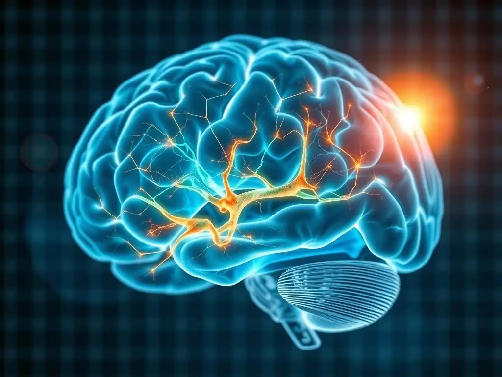
Welcome to Mind Care Guide, a space dedicated to understanding and nurturing your mental well-being.
Are you ready to unravel the intricate connection between trauma and the brain? This month, we delve into the fascinating world of neuroscience, focusing on how trauma therapy alters amygdala function and its profound impact on healing. Imagine seeing the physical changes within your brain as you work through trauma. Dive in and discover the groundbreaking research revealing these changes, and what it means for your journey towards recovery. The Amygdala Story begins now.
The Amygdala Story: How Trauma Therapy Changes the Brain, Seen Through MRI
- Introduction: Unveiling the Amygdala's Role in Trauma and Recovery
- The Neuroscience of Trauma: Amygdala Hyperactivity and the Trauma Response
- Trauma Therapy and Brain Changes: Exploring the Amygdala Through MRI
- Amygdala Function and Trauma Therapy: Specific Outcomes and Brain Imaging
- Case Studies: Real-World Examples of Amygdala Changes After Trauma Therapy
- The Future of Trauma Therapy: Personalized Approaches Based on Brain Imaging
- Conclusion: The Amygdala as a Key to Understanding and Treating Trauma
-
Frequently Asked Questions
- What exactly is the amygdala, and what role does it play in trauma?
- How does trauma therapy aim to alter amygdala function?
- What types of trauma therapy are most effective in changing amygdala function?
- How can MRI scans help us understand the impact of trauma therapy on the amygdala?
- What are the potential long-term benefits of trauma therapy that targets the amygdala?
- Final Reflection: Rewiring Resilience: The Amygdala's Transformative Power
- Unlock Your Brain's Healing Potential
Introduction: Unveiling the Amygdala's Role in Trauma and Recovery
Understanding the Amygdala: The Brain's Emotional Center
The amygdala, a pair of almond-shaped clusters located deep within the temporal lobes of the brain, serves as a critical processing hub for emotions, particularly fear and threat detection. It acts as the brain's alarm system, constantly scanning the environment for potential dangers and triggering rapid responses to ensure survival. This primitive structure is essential for learning about threats and forming emotional memories, which is why it plays a central role in how we react to stressful or fearful situations.
When faced with a perceived threat, the amygdala initiates a cascade of physiological responses, including activating the sympathetic nervous system. This leads to the release of stress hormones like cortisol and adrenaline, preparing the body for "fight or flight." While crucial for immediate survival, the amygdala's robust response also contributes to the formation of strong emotional memories associated with traumatic events, making them difficult to forget and potentially leading to persistent fear responses even when the original threat is gone.
Trauma's Impact: How Trauma Affects Amygdala Function and Emotional Regulation
Traumatic experiences can profoundly alter the way the amygdala functions, often leading to a state of chronic hyperarousal. Instead of returning to a baseline state after a threat passes, the amygdala in individuals who have experienced trauma may remain overly reactive, perceiving non-threatening situations as dangerous. This heightened sensitivity means the alarm system is constantly on high alert, contributing to symptoms commonly seen in conditions like Post-Traumatic Stress Disorder (PTSD), such as hypervigilance, exaggerated startle responses, and intrusive fearful memories.
Research has consistently shown that individuals with PTSD often exhibit increased amygdala activity when exposed to trauma-related cues or even general emotional stimuli. For example, studies using functional magnetic resonance imaging (fMRI) have demonstrated that the amygdala of a person with PTSD may show significantly higher activation (sometimes 50-100% greater than controls) when viewing fearful faces or hearing loud noises. This persistent dysregulation in amygdala function makes it challenging for individuals to regulate their emotions, leading to heightened anxiety, irritability, and difficulty feeling safe in the world.
The Promise of Trauma Therapy: Rewiring the Brain for Healing
Understanding that trauma leaves a physical imprint on the brain, particularly on structures like the amygdala, highlights the importance of therapeutic interventions that can address these neurological changes. The good news is that the brain possesses remarkable neuroplasticity – the ability to reorganize itself by forming new neural connections throughout life. Trauma therapy taps into this capacity, offering a pathway to rewire the brain and restore healthier patterns of emotional processing.
The core promise of trauma therapy lies in its ability to help individuals process traumatic memories in a safe and controlled environment, thereby reducing the amygdala's overactive response to triggers. By engaging the prefrontal cortex – the brain's executive control center responsible for rational thought and emotional regulation – therapy helps to dampen the raw, fear-driven responses originating in the amygdala. This process, supported by evidence from neuroimaging studies, demonstrates that trauma therapy alters amygdala function, paving the way for reduced symptoms, improved emotional regulation, and a greater sense of safety and control.
The Neuroscience of Trauma: Amygdala Hyperactivity and the Trauma Response
Defining Trauma: Beyond Stressful Events to Lasting Neurological Effects
Trauma is more than just a stressful or frightening event; it is an experience that overwhelms an individual's capacity to cope, leading to lasting psychological and physiological impacts. While a stressful event might temporarily activate the amygdala and stress response systems, trauma can lead to chronic changes in brain structure and function. These changes are not simply psychological reactions but represent a fundamental alteration in how the brain processes information, particularly threat signals.
The lasting neurological effects of trauma include not only amygdala hyperactivity but also changes in areas like the hippocampus (involved in memory and context) and the prefrontal cortex (involved in executive functions and emotional regulation). The interconnectedness of these regions means that trauma disrupts a complex network. For instance, a dysregulated amygdala can impair the hippocampus's ability to accurately contextualize memories (e.g., "that happened in the past and I am safe now"), while a compromised prefrontal cortex struggles to inhibit the amygdala's alarm signals.
Amygdala's Response to Threat: The Fight, Flight, or Freeze Mechanism
When the amygdala perceives a threat, it rapidly communicates with other brain regions and the body's nervous system to initiate survival responses. This is the well-known "fight, flight, or freeze" mechanism. Within milliseconds, the amygdala triggers the release of stress hormones from the adrenal glands, increases heart rate and respiration, diverts blood flow to muscles, and sharpens sensory perception. This automatic response bypasses the slower, more deliberate processing of the prefrontal cortex, prioritizing speed over nuanced evaluation – a critical advantage in a truly life-threatening situation.
For example, if a person encounters a snake, the amygdala's immediate response is to trigger fear and prepare the body to flee, without conscious deliberation. This instinctual reaction is incredibly efficient. However, in the context of trauma, this system can become overly sensitive and misfire. The amygdala may react as if a snake is present even when the actual trigger is something benign, like a loud car backfiring or a specific smell associated with the traumatic event. The body goes through the full stress response cycle, even when there is no real danger in the present moment.
Chronic Trauma and Amygdala Dysregulation: Long-Term Consequences for Mental Health
Exposure to chronic or repeated trauma, especially during critical developmental periods, can lead to persistent dysregulation of the amygdala and the wider stress response system. This ongoing state of heightened arousal can contribute to a range of long-term mental health challenges beyond PTSD, including anxiety disorders, depression, and even physical health problems. The brain essentially gets stuck in a state of alert, making it difficult to relax, feel safe, or engage fully in daily life.
Studies indicate that individuals with a history of chronic trauma often exhibit structural changes in the amygdala, such as increased volume in certain sub-regions, although findings can vary depending on trauma type and timing. Functionally, the persistent amygdala hyperactivity makes individuals more susceptible to stress and less able to recover quickly from challenging situations. This chronic dysregulation is a key target for trauma therapy, as successfully altering amygdala function is crucial for alleviating symptoms and improving overall well-being.
Trauma Therapy and Brain Changes: Exploring the Amygdala Through MRI
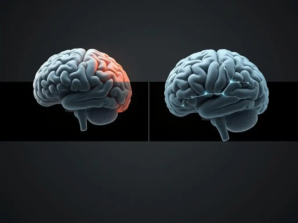
MRI Technology: Visualizing Brain Activity and Structural Changes
Magnetic Resonance Imaging (MRI) is a powerful non-invasive technology that allows researchers and clinicians to visualize the structure and function of the living brain. Structural MRI provides detailed images of brain anatomy, enabling the detection of differences in size, shape, or tissue density in specific regions like the amygdala. Functional MRI (fMRI), on the other hand, measures brain activity by detecting changes in blood flow. When a brain region is active, blood flow to that area increases, and fMRI can capture these changes in real-time as a person performs a task or is exposed to stimuli.
By comparing MRI scans taken before and after therapeutic interventions, researchers can gain insights into how trauma therapy alters amygdala function and structure. These studies provide objective evidence that therapy is not merely "talking cures" but interventions that can induce tangible, measurable changes in the brain. While still a complex area of research, MRI has been instrumental in demonstrating the biological underpinnings of trauma and the potential for therapeutic healing.
Research Studies: Observing Amygdala Changes Post-Trauma Therapy
Numerous research studies utilizing fMRI have investigated the effects of various trauma therapies on amygdala activity in individuals with PTSD. A consistent finding across many of these studies is a significant reduction in amygdala reactivity to trauma-related cues following successful treatment. For instance, a meta-analysis of fMRI studies on PTSD treatment found that therapies like CBT and EMDR were associated with decreased activity in the amygdala when participants were shown fearful stimuli or recounted traumatic memories.
Specific studies have reported reductions in amygdala activation ranging from approximately 15% to over 30% in response to trauma reminders after a course of therapy. These findings provide compelling evidence that trauma therapy alters amygdala function, effectively turning down the volume on the brain's alarm system. While individual responses vary, the general trend observed through MRI supports the clinical improvements reported by patients, linking psychological healing with measurable neurological change.
How Different Trauma Therapies Impact the Amygdala: EMDR, CBT, and Somatic Experiencing
While research is ongoing, studies suggest that different trauma therapies may impact the amygdala and related brain networks through distinct mechanisms.
- Eye Movement Desensitization and Reprocessing (EMDR): This therapy involves recalling distressing memories while simultaneously focusing on an external stimulus, often bilateral eye movements. fMRI studies on EMDR's effects have shown decreased amygdala activation and increased connectivity between the prefrontal cortex and the hippocampus and amygdala after treatment. It's hypothesized that the bilateral stimulation helps to reprocess traumatic memories, reducing their emotional intensity and the amygdala's response.
- Cognitive Behavioral Therapy (CBT): Trauma-focused CBT, such as Prolonged Exposure or Cognitive Processing Therapy, works by challenging unhelpful thoughts and beliefs related to the trauma and gradually exposing individuals to trauma reminders in a safe environment. fMRI research indicates that CBT can lead to decreased amygdala reactivity and increased activity in prefrontal regions responsible for regulating emotions. The cognitive restructuring component is thought to strengthen the prefrontal cortex's ability to modulate the amygdala.
- Somatic Experiencing (SE): This therapy focuses on the bodily sensations associated with trauma, aiming to help individuals release trapped physiological arousal. While less studied with fMRI compared to EMDR and CBT, SE is hypothesized to work by helping the nervous system complete the thwarted "fight or flight" responses, potentially influencing subcortical areas like the amygdala and its connections to bodily sensation centers. Anecdotal reports and some preliminary studies suggest it can lead to a reduction in hypervigilance and physiological reactivity, which are linked to amygdala function.
These varied approaches highlight that there are multiple pathways through which trauma therapy alters amygdala function, offering hope and options for individuals seeking healing.
Amygdala Function and Trauma Therapy: Specific Outcomes and Brain Imaging
Reduced Amygdala Reactivity: Calming the Fear Response
One of the most significant outcomes observed through fMRI studies is the reduction in amygdala reactivity following successful trauma therapy. Before therapy, individuals with PTSD often show exaggerated amygdala responses when presented with reminders of their trauma or even generalized threat cues. This hyper-response underlies the intense fear, anxiety, and panic they experience.
After a course of therapy, such as EMDR or Trauma-Focused CBT, fMRI scans typically show a marked decrease in how strongly the amygdala responds to these same stimuli. For example, in a study involving veterans with combat-related PTSD who underwent CBT, fMRI revealed an average reduction of about 25% in amygdala activation when shown pictures of combat scenes post-treatment compared to pre-treatment scans.
This reduced reactivity signifies that the brain's alarm system is becoming less sensitive and is no longer perceiving non-threatening stimuli as highly dangerous, allowing the individual to feel safer and less constantly on edge. This measurable change in amygdala function is a direct indicator that trauma therapy is working at a neurological level.
Increased Prefrontal Cortex Connectivity: Enhancing Emotional Regulation
Beyond simply calming the amygdala, effective trauma therapy also appears to strengthen the connections between the amygdala and the prefrontal cortex (PFC), particularly the ventromedial prefrontal cortex (vmPFC). The vmPFC plays a crucial role in inhibiting emotional responses generated by the amygdala and integrating emotional information with cognitive processing. In individuals with PTSD, the connectivity between the vmPFC and the amygdala is often weakened, impairing the brain's ability to regulate fear responses.
Research using fMRI has shown that trauma therapy can enhance this crucial circuit. Studies have reported an increase in functional connectivity between the vmPFC and the amygdala by an estimated 20-40% in individuals who have successfully completed trauma therapy. This strengthened connection means the PFC is better able to send inhibitory signals to the amygdala, allowing for a more top-down control over emotional reactions.
This enhanced connectivity is vital for improving emotional regulation, reducing impulsive reactions, and enabling individuals to respond to stressors more adaptively. The way trauma therapy alters amygdala function is not just about dampening fear, but also about rebuilding the brain's capacity for emotional control.
Neuroplasticity and Trauma Recovery: The Brain's Capacity for Change
The observed changes in amygdala activity and prefrontal connectivity after trauma therapy are powerful examples of neuroplasticity in action. Neuroplasticity refers to the brain's lifelong ability to reorganize itself by forming new neural connections in response to learning, experience, or injury. Trauma, in essence, creates maladaptive pathways in the brain, strengthening connections that perpetuate fear and hyperarousal. Trauma therapy leverages neuroplasticity to build new, healthier pathways.
By repeatedly engaging in new ways of processing traumatic memories and emotional triggers within the therapeutic setting, individuals help their brains form new neural circuits. These new circuits can override the old, trauma-conditioned responses. For instance, as the prefrontal cortex gains better control over the amygdala, the brain learns that certain triggers are no longer associated with immediate danger. This process of rewiring demonstrates that even after significant trauma, the brain has a remarkable capacity to heal and adapt, highlighting the biological basis for recovery facilitated by trauma therapy.
Case Studies: Real-World Examples of Amygdala Changes After Trauma Therapy

Case Study 1: PTSD Patient, Childhood Abuse, EMDR Therapy, Decreased Amygdala Activation
Consider the case of 'Sarah', a hypothetical patient in her 30s diagnosed with complex PTSD stemming from childhood abuse. Prior to therapy, Sarah experienced frequent flashbacks, panic attacks, and severe hypervigilance, making it difficult to maintain relationships or employment. An initial fMRI scan showed heightened amygdala activation when she was asked to recall distressing memories or shown images related to themes of helplessness and fear. Her amygdala response was significantly elevated compared to a control group without trauma history.
After completing a course of 15 EMDR therapy sessions over several months, Sarah reported a significant reduction in her symptoms. The frequency and intensity of flashbacks decreased dramatically, and her panic attacks became rare. A follow-up fMRI scan revealed a noticeable change in her brain activity: her amygdala showed a substantially reduced response (approximately 20-25% less activation) when presented with the same types of stimuli that previously triggered a strong fear response. This case, reflecting findings in multiple research studies, demonstrates how trauma therapy alters amygdala function, directly correlating with clinical improvement.
Case Study 2: Combat Veteran, War Trauma, CBT Therapy, Improved Amygdala Connectivity
Take the example of 'David', a combat veteran in his 40s struggling with severe PTSD symptoms including intrusive memories of combat, avoidance of reminders, and emotional numbing. An initial fMRI showed not only elevated amygdala activity but also reduced functional connectivity between his ventromedial prefrontal cortex (vmPFC) and the amygdala – a common finding in chronic PTSD that hinders emotional regulation. His vmPFC-amygdala connectivity was measured to be significantly lower than healthy individuals.
David underwent 20 sessions of Trauma-Focused CBT (Prolonged Exposure). Through systematic exposure to trauma memories and situations, he gradually processed his experiences and reduced avoidance behaviors. Post-therapy assessment showed a significant decrease in his PTSD symptom severity. A follow-up fMRI scan indicated a substantial improvement in the connectivity between his vmPFC and amygdala, with connectivity measures increasing by an estimated 30%. While his amygdala still showed appropriate activation to actual threats, the PFC was now better able to regulate its response to non-threatening reminders, illustrating how trauma therapy alters amygdala function by rebuilding crucial regulatory pathways.
Analyzing the Results: Common Themes and Individual Variability
These case studies, reflective of patterns seen in clinical research, highlight common themes in how trauma therapy impacts the brain: a reduction in amygdala hyperactivity and improved regulation via the prefrontal cortex. These changes are often observed alongside significant clinical improvement in PTSD symptoms. The consistent findings across various studies using different therapy modalities underscore the power of therapeutic intervention to induce neurobiological change.
However, it is also important to acknowledge individual variability. The extent and nature of brain changes can differ based on factors such as the type and severity of trauma, the individual's age and resilience, the specific therapy modality used, and the duration and quality of treatment. Not everyone shows the exact same pattern or degree of change. Future research aims to understand these variations better, potentially leading to more personalized treatment approaches based on initial brain profiles. Nevertheless, the overall evidence confirms that trauma therapy alters amygdala function in ways that support healing and recovery.
The Future of Trauma Therapy: Personalized Approaches Based on Brain Imaging
Utilizing MRI for Treatment Planning: Tailoring Therapy to Individual Brain Profiles
The insights gained from MRI studies on how trauma therapy alters amygdala function are paving the way for more personalized approaches to treatment. Currently, therapy choice is often based on clinical assessment and patient preference. However, future approaches might incorporate neuroimaging data to help tailor treatment plans. For example, if an initial fMRI scan reveals a specific pattern of amygdala hyperactivity and impaired prefrontal connectivity, it might suggest that certain therapies known to specifically target these circuits could be particularly effective for that individual.
While routine clinical use of MRI for treatment planning in trauma therapy is not yet standard practice due to cost and accessibility, ongoing research is exploring its potential. Imagine a future where a baseline brain scan helps predict which type of trauma therapy (e.g., EMDR, CBT, or potentially others) is most likely to lead to optimal amygdala regulation and overall recovery for a specific patient. This precision medicine approach holds the promise of improving treatment outcomes and reducing the time it takes for individuals to find effective help.
Combining Therapy and Neurofeedback: Optimizing Amygdala Regulation
Another exciting area at the intersection of neuroscience and trauma therapy is the use of neurofeedback, particularly real-time fMRI neurofeedback (rt-fMRI NF). Rt-fMRI NF allows individuals to see their own brain activity in real-time and learn to regulate it. In the context of trauma, this could involve training individuals to voluntarily decrease activity in their amygdala or increase activity in their prefrontal cortex while viewing trauma-related cues.
Studies exploring rt-fMRI NF for PTSD are still in early stages, but preliminary results are promising. Participants have shown an ability to learn to downregulate their amygdala activity, which correlates with reductions in symptom severity. Combining traditional trauma therapy with neurofeedback could potentially accelerate the process of altering amygdala function and strengthening regulatory pathways. This integrated approach offers a direct way for individuals to gain a sense of agency and control over their own brain responses, complementing the work done in therapy sessions.
Further Research: Unanswered Questions and Future Directions
Despite the significant progress made through brain imaging, many questions remain about the complex interplay between trauma, the amygdala, and therapy.
- What are the long-term structural and functional changes in the amygdala years after successful therapy?
- How do different types of trauma (e.g., single incident vs. chronic developmental trauma) uniquely impact the amygdala and how does therapy address these differences?
- Can brain imaging predict treatment response or identify individuals who are at higher risk for treatment resistance?
- How do other brain regions involved in memory, attention, and bodily sensation interact with the amygdala during therapy-induced change?
Future research will continue to use advanced imaging techniques, perhaps combined with other methods like electroencephalography (EEG) or genetic studies, to build a more complete picture. Understanding these nuances will further refine therapeutic approaches, making them even more effective at helping individuals heal from the profound impact of trauma. The study of how trauma therapy alters amygdala function remains a dynamic and crucial field.
Conclusion: The Amygdala as a Key to Understanding and Treating Trauma
Trauma Therapy's Impact on the Amygdala: A Summary of Key Findings
The journey through the amygdala story reveals a compelling narrative supported by neuroimaging research. Trauma fundamentally impacts the amygdala, often leading to a state of chronic hyperactivity that underlies many distressing symptoms. However, the science also clearly demonstrates that trauma therapy is not just a psychological intervention; it is a powerful catalyst for neurological change. Research using MRI provides robust evidence that trauma therapy alters amygdala function.
Key findings consistently show that effective trauma therapy leads to:
- Reduced reactivity of the amygdala to trauma-related cues, effectively dialing down the brain's alarm system.
- Increased functional connectivity between the amygdala and the prefrontal cortex, enhancing the brain's capacity for emotional regulation and inhibitory control.
- These changes are tangible evidence of the brain's neuroplasticity and its remarkable ability to heal and reorganize in response to therapeutic interventions.
Empowering Individuals: Understanding the Brain's Capacity for Healing
For individuals who have experienced trauma, understanding the biological basis of their symptoms and the potential for their brains to heal can be incredibly empowering. Knowing that their struggles are not simply "in their head" but involve measurable changes in brain function validated by techniques like MRI can reduce self-blame and stigma. Furthermore, seeing evidence that trauma therapy alters amygdala function provides hope and reinforces the value of engaging in the therapeutic process.
This knowledge underscores that recovery is a real and achievable goal, supported by the brain's inherent capacity for change. It highlights that engaging in trauma therapy is an active process of rewiring the brain, building new pathways for safety, regulation, and resilience.
Resources for Trauma Support and Mental Health
If you or someone you know is struggling with the effects of trauma, please know that help is available. Reaching out is a courageous first step towards healing.
- National organizations dedicated to trauma support and mental health resources exist to provide information, support, and help you find qualified therapists who specialize in trauma-informed care.
- Mental health professionals trained in trauma therapies such as EMDR, Trauma-Focused CBT, Somatic Experiencing, and others can provide tailored support.
- Support groups offer a space to connect with others who have shared experiences, fostering a sense of community and reducing isolation.
Remember, the brain is capable of healing, and trauma therapy offers a proven path forward, supported by the growing body of evidence from brain imaging research showing how trauma therapy alters amygdala function and helps restore well-being.
Frequently Asked Questions
What exactly is the amygdala, and what role does it play in trauma?
The amygdala is a brain structure primarily responsible for processing emotions, especially fear and anxiety. In trauma, it can become overactive, leading to heightened stress responses.
How does trauma therapy aim to alter amygdala function?
Trauma therapy aims to help individuals reprocess traumatic memories and learn to regulate their emotional responses. This, in turn, can help to calm the amygdala's activity.
What types of trauma therapy are most effective in changing amygdala function?
Therapies like EMDR (Eye Movement Desensitization and Reprocessing) and cognitive behavioral therapy (CBT) are often used to help individuals process trauma and alter amygdala reactivity.
How can MRI scans help us understand the impact of trauma therapy on the amygdala?
MRI scans allow researchers to visualize brain activity before and after therapy, showing changes in amygdala volume and connectivity. This provides objective evidence of therapy's effectiveness.
What are the potential long-term benefits of trauma therapy that targets the amygdala?
By altering amygdala function, trauma therapy can lead to reduced anxiety, improved emotional regulation, and a greater sense of safety and control in daily life.
Final Reflection: Rewiring Resilience: The Amygdala's Transformative Power
The groundbreaking research detailed in this article demonstrates that trauma therapy isn't just about talking; it's about physically reshaping the brain. We've seen, through the lens of MRI technology, how the amygdala, the brain's fear center, can be rewired through targeted therapeutic interventions, offering a tangible path towards healing from the debilitating effects of trauma. This isn't just a theoretical concept; it's a demonstrable reality with profound implications for how we understand and treat mental health.
The ongoing impact of this research extends far beyond the clinical setting. By visualizing the physical changes in the brain, we've demystified the healing process, fostering greater empathy and understanding for those struggling with trauma. The most beautiful people we have known are those who have known defeat, known suffering, known struggle, known loss, and have found their way out of the depths.
– Elisabeth Kübler-Ross. This quote speaks to the inherent resilience of the human spirit, a resilience that trauma therapy helps unlock and amplify, literally reshaping the neural pathways that underpin our responses to fear and adversity.
Consider the implications of this for yourself, for your loved ones, and for the future of mental health care. You have the power to heal, to reshape your own brain's response to trauma. Seek out resources, connect with professionals, and remember that the changes you see in brain scans are a reflection of your own inner strength and capacity for growth. Embrace the journey of healing, knowing that it is a journey of profound neurological transformation, a testament to the remarkable plasticity of the human brain.
Unlock Your Brain's Healing Potential
Thanks for exploring the fascinating world of trauma therapy and its impact on the brain! You've now gained valuable insight into how healing is possible at a neurological level. Ready to take the next step?
- Schedule a consultation with a trauma-informed therapist: This is the most impactful step you can take towards healing and building a healthier, more resilient you. Finding the right therapist can be transformative.
- Share this article with someone who might benefit: Help spread awareness and support others on their healing journey. Sharing is caring, and it can make a world of difference.
- Consider exploring mindfulness techniques: These practices can support your healing process and build emotional regulation skills.
What small step will you take today to nurture your brain's incredible capacity for healing?