
LongTerm Brain Benefits: What Scans Show After Successful Therapy


Welcome to Mind Care Guide, a sanctuary dedicated to your mental well-being.
Ever wondered what truly happens within your mind when therapy unlocks its potential? We understand the journey to mental wellness can feel like navigating uncharted territory. In this article, we delve into the fascinating world of neuroscience, revealing the long-term brain benefits observable through brain scans post-successful therapy. Prepare to be amazed as we uncover the tangible changes and lasting impact of healing on your brain's architecture. Intrigued to see how therapy reshapes your neural pathways? Let's explore!
- Long-Term Brain Benefits: What Brain Scans Show After Successful Therapy
- Neuroscience of Mental Health: Linking Therapy to Brain Changes
- Brain Scans Post-Successful Therapy: A Visual Journey
- Analyzing Brain Scan Results: Key Findings After Therapy
- The Significance of Brain Scan Findings for Mental Health Treatment
- Conclusion: The Future of Brain Scans in Mental Healthcare.
-
Frequently Asked Questions
- What specific brain changes can brain scans reveal after successful therapy?
- How do brain scans help us understand the long-term benefits of successful therapy?
- Are brain scans typically recommended for all patients after successful therapy?
- What types of brain scans are most commonly used to assess the impact of successful therapy?
- Can brain scans predict the likelihood of relapse after successful therapy?
- Final Reflection: The Enduring Power of Healing, Visible in the Brain
- Unlock Your Brain's Potential: Start Your Journey Today!
Long-Term Brain Benefits: What Brain Scans Show After Successful Therapy
Introduction: Unveiling the Lasting Impact of Therapy on the Brain
The journey through mental health challenges can be profound, and while the subjective experience of healing is paramount, modern neuroscience is beginning to provide tangible evidence of therapy's long-term effects. Brain scans postsuccessful therapy offer a fascinating glimpse into the neural changes that accompany recovery. These advanced imaging techniques allow researchers and clinicians to visualize shifts in brain structure, activity, and connectivity, moving beyond anecdotal evidence to demonstrate that therapy doesn't just change how we think or feel – it can fundamentally alter the physical landscape of the brain.
Understanding the long-term brain benefits revealed by scanning after effective treatment provides powerful validation for the impact of psychotherapy. It underscores that mental health interventions are not merely coping strategies but potent tools capable of fostering enduring positive change at the most fundamental level: our neural circuitry. These insights are transforming how we perceive mental health treatment, highlighting its capacity for deep, physiological healing.
Neuroscience of Mental Health: Linking Therapy to Brain Changes
Exploring the Brain's Plasticity: How Therapy Reshapes Neural Pathways
The human brain is remarkably adaptable, a property known as neuroplasticity. This means that the brain's structure and function can change throughout life in response to experiences, learning, and even therapeutic interventions. Therapy, particularly types like Cognitive Behavioral Therapy (CBT), Dialectical Behavior Therapy (DBT), or psychodynamic therapy, provides structured experiences that encourage new ways of thinking, feeling, and behaving. Over time, these new patterns of engagement can strengthen certain neural pathways and weaken others, effectively rewiring the brain.
This reshaping isn't just theoretical; brain scans postsuccessful therapy visually demonstrate these changes. For instance, learning new coping mechanisms in therapy can strengthen connections between the prefrontal cortex, involved in executive functions and regulation, and areas like the amygdala, which processes fear and emotion. This enhanced connectivity can lead to better emotional control and reduced reactivity to stressors, a tangible outcome of neuroplasticity guided by therapeutic processes.
Long-Term Therapy Outcomes and Brain Changes: The Connection
Successful long-term therapy outcomes are intrinsically linked to durable changes within the brain. When therapy helps someone manage anxiety effectively over months or years, it's often because the neural circuits associated with fear and worry have been modulated, and those associated with calm and rational thought have been reinforced. These enduring behavioral and emotional shifts are mirrored by observable changes in brain structure and function, detectable through various imaging techniques.
For example, individuals who have successfully overcome depression through long-term therapy may show increases in gray matter volume in specific brain regions, such as the hippocampus, compared to their state before treatment. This area is crucial for memory and emotional regulation and is often reduced in volume in chronic depression. Brain scanning after effective treatment provides compelling evidence that therapeutic success is underpinned by significant, lasting adaptations within the brain's architecture.
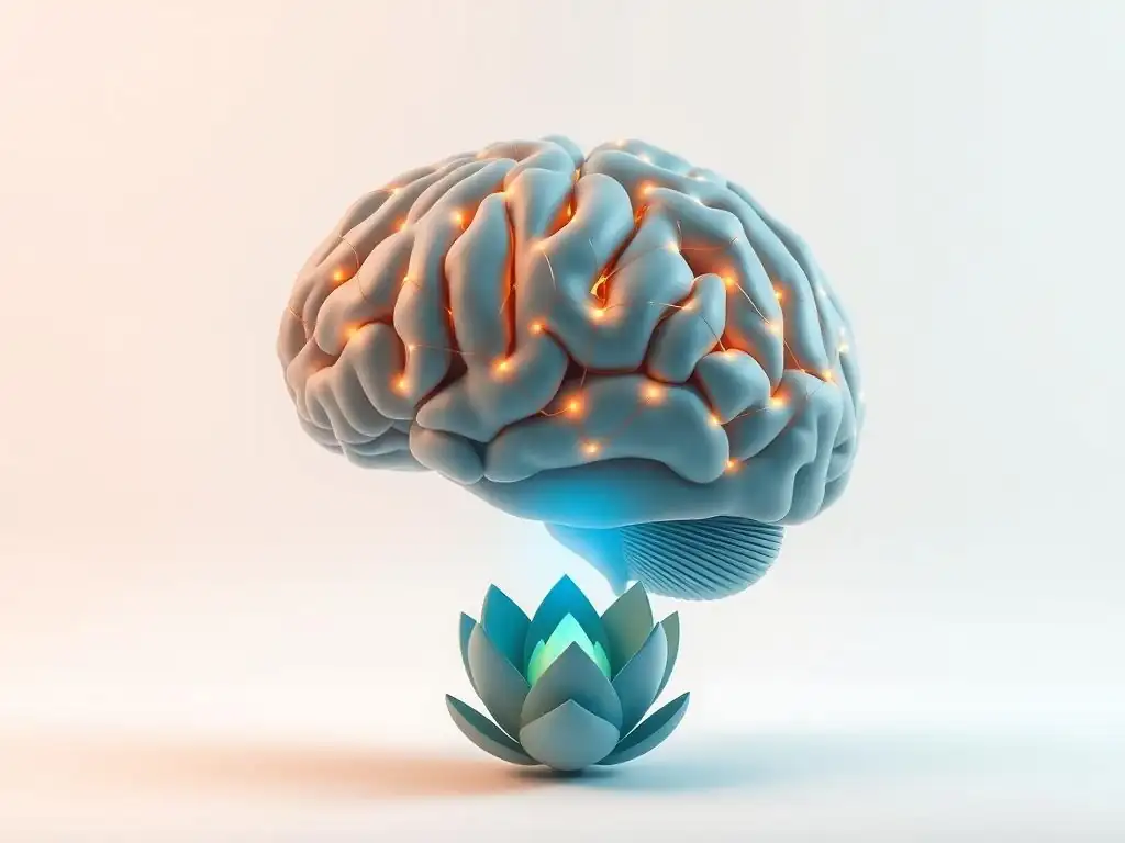
Brain Scans Post-Successful Therapy: A Visual Journey
Understanding Different Brain Scanning Techniques
Exploring the brain's response to therapy relies on sophisticated imaging technologies, each offering a unique perspective on neural structure and activity. Understanding these techniques is key to interpreting what brain scans postsuccessful therapy reveal about the healing process.
-
MRI (Magnetic Resonance Imaging): Visualizing Brain Structure
MRI uses strong magnetic fields and radio waves to create detailed images of brain structures. It's excellent for identifying anatomical features, measuring the volume of different brain regions (like gray matter or white matter), and detecting structural abnormalities. In the context of mental health, MRI can help researchers see if conditions are associated with structural differences or if therapy leads to changes in brain volume or tissue integrity over time.
Studies using structural MRI have sometimes shown differences in the size of regions like the amygdala or hippocampus in individuals with certain conditions compared to healthy controls. Following successful therapy, some research has indicated that these structural differences may normalize or show positive changes, such as increases in hippocampal volume in some cases of recovery from depression or PTSD, suggesting a potential reversal of stress-induced structural changes.
-
fMRI (Functional Magnetic Resonance Imaging): Mapping Brain Activity
fMRI measures brain activity by detecting changes in blood flow. When a brain region is more active, it uses more oxygen, leading to increased blood flow to that area. fMRI captures these changes, providing a dynamic map of which parts of the brain are engaged during specific tasks, rest, or emotional states. It's invaluable for understanding functional connectivity – how different brain regions communicate with each other.
Brain scans postsuccessful therapy using fMRI frequently show altered activity patterns. For instance, fMRI studies on anxiety have demonstrated reduced activity in the amygdala and increased activity in prefrontal regulatory regions when individuals are exposed to anxiety triggers after completing therapy. Similarly, studies on depression recovery have shown normalized connectivity within brain networks involved in mood regulation and self-referential thought.
-
PET (Positron Emission Tomography): Assessing Brain Metabolism
PET scans use a small amount of radioactive tracer injected into the bloodstream to measure metabolic activity, blood flow, or receptor binding in the brain. Different tracers can target specific molecules, providing insights into neurochemical processes. While less common for routine outcome assessment than MRI or fMRI, PET can reveal unique aspects of brain function related to neurotransmitter systems or glucose metabolism, which are often implicated in mental health conditions.
PET studies have sometimes revealed differences in glucose metabolism (an indicator of neural activity) in specific brain areas in conditions like depression or OCD. Research using PET has also explored changes in neurotransmitter receptor availability after therapy, such as serotonin or dopamine receptors, although findings in this area are complex and often part of broader pharmacological studies. These scans contribute to a fuller picture of the biological underpinnings of mental illness and recovery.
Analyzing Brain Scan Results: Key Findings After Therapy
Brain Scan Evidence of Reduced Anxiety Symptoms
Studies employing brain scans postsuccessful therapy for anxiety disorders, such as Generalized Anxiety Disorder (GAD) or Social Anxiety Disorder, consistently point towards observable neural changes correlating with symptom reduction. Functional MRI (fMRI) is particularly useful here, often showing decreased activity in the amygdala – the brain's primary fear processing center – when individuals are exposed to anxiety-provoking stimuli after therapy. Prior to treatment, this area might show heightened reactivity; post-therapy, this response is often significantly dampened.
Beyond reduced amygdala activity, brain scanning after effective treatment for anxiety also reveals changes in connectivity. Research has demonstrated strengthened functional connections between the prefrontal cortex (PFC), responsible for executive control and emotion regulation, and the amygdala. This enhanced "top-down" control from the PFC is believed to help individuals better manage their fear responses, allowing them to appraise situations more rationally and experience less intense anxiety, a change vividly captured by fMRI data.
Brain Scan Evidence of Improved Mood and Emotional Regulation
For individuals recovering from depression, brain scans postsuccessful therapy frequently highlight improvements in mood and emotional regulation circuits. Structural MRI studies have occasionally reported increases in gray matter volume in regions like the hippocampus and prefrontal cortex, areas often observed to have reduced volume in chronic depression. These structural changes suggest a potential reversal or amelioration of some of the physical impacts of long-term depressive states on the brain.
Functionally, fMRI scans after successful depression therapy often show normalized activity in networks involved in processing emotions and self-referential thought, such as the default mode network (DMN) and the salience network. Overactivity or abnormal connectivity within these networks is often seen in depression. Post-treatment brain scans postsuccessful therapy may show a shift towards healthier, more balanced activity patterns, reflecting improved emotional processing and reduced rumination.
Brain Scan Evidence of Enhanced Cognitive Function
Mental health conditions like depression, anxiety, and PTSD can significantly impair cognitive functions, including attention, memory, and executive function. Encouragingly, brain scans postsuccessful therapy often indicate improvements in these areas as well. This is particularly evident in fMRI studies showing increased activity or connectivity in brain regions associated with cognitive processes, such as the dorsolateral prefrontal cortex (involved in planning and working memory) and parietal regions (involved in attention).
For instance, individuals with depression who undergo successful therapy may show improved performance on cognitive tasks, paralleled by increased activation in relevant brain areas compared to their pre-treatment scans. This suggests that therapy not only alleviates mood symptoms but can also help restore cognitive abilities that were hindered by the condition. Brain scanning after effective treatment provides objective data supporting these cognitive gains.
Brain Scans and Trauma Recovery: Healing the Brain After Trauma
Trauma, particularly complex or chronic trauma, can have profound and lasting effects on brain structure and function. Conditions like Post-Traumatic Stress Disorder (PTSD) are associated with changes in the amygdala, hippocampus, and prefrontal cortex. Brain scans postsuccessful therapy targeting trauma, such as Trauma-Focused CBT or EMDR, have provided compelling visual evidence of the brain's capacity for healing after trauma.
Studies using fMRI on individuals with PTSD after effective therapy have shown decreased amygdala hyperreactivity to trauma-related cues and increased activation in areas of the prefrontal cortex involved in regulating fear responses. Structural MRI has sometimes indicated increases in hippocampal volume, which is often reduced in chronic PTSD. These brain scans postsuccessful therapy underscore that therapeutic interventions can help normalize brain function and structure, supporting recovery from the debilitating effects of trauma.
The Significance of Brain Scan Findings for Mental Health Treatment

Informing Personalized Treatment Approaches
The insights gained from brain scans postsuccessful therapy are beginning to influence how mental health treatment is approached. By understanding the specific neural correlates of different conditions and how they respond to various therapies, clinicians may eventually be able to use imaging data to help personalize treatment plans. For example, if a particular brain network imbalance is identified, therapies known to target that network might be prioritized.
While still largely a research tool, the long-term goal is that brain scanning after effective treatment could provide biomarkers that help predict which therapy is most likely to be effective for an individual, or to tailor specific therapeutic techniques to a person's unique neural profile. This move towards precision mental health care could significantly improve treatment outcomes and reduce the trial-and-error often involved in finding the right therapeutic fit.
Monitoring Treatment Progress and Effectiveness
Brain scans offer a potential objective measure for monitoring the progress and effectiveness of therapy, complementing subjective reports from patients and clinical observations. While not yet standard clinical practice, future applications could involve periodic brain scans to assess whether therapy is inducing the expected neural changes associated with recovery. Observing positive shifts in brain activity or structure could provide tangible feedback for both the therapist and the patient.
Using brain scanning after effective treatment as a monitoring tool could help identify early on if a therapy is working at a neural level or if adjustments are needed. For instance, if expected decreases in amygdala activity aren't occurring in someone undergoing anxiety therapy, it might signal a need to modify the therapeutic approach or consider alternative interventions.
Validating the Effectiveness of Psychotherapy
Perhaps one of the most significant contributions of brain scans postsuccessful therapy is the objective validation they provide for the power of psychotherapy. For decades, therapy's effectiveness was primarily measured by symptom reduction and improved quality of life based on self-report and clinical assessment. While invaluable, these measures are subjective. Brain imaging offers concrete, biological evidence that talking therapy, without medication, can induce measurable physical changes in the brain.
This scientific validation helps destigmatize mental health treatment by demonstrating that conditions are associated with real, observable brain differences and that therapy is a powerful intervention capable of addressing these at a biological level. Brain scanning after effective treatment reinforces the message that mental health is brain health and that investing in therapeutic care is investing in the physical well-being of the brain.
Conclusion: The Future of Brain Scans in Mental Healthcare.
Frequently Asked Questions
What specific brain changes can brain scans reveal after successful therapy?
Brain scans, like fMRI, can show changes in neural activity and connectivity, indicating improved emotional regulation and cognitive function after successful therapy. These changes suggest the brain is adapting positively.
How do brain scans help us understand the long-term benefits of successful therapy?
Brain scans provide visual evidence of structural and functional changes in the brain that correlate with improved mental well-being. This helps us understand how therapy reshapes the brain over time.
Are brain scans typically recommended for all patients after successful therapy?
No, brain scans are not routinely recommended. They are primarily used in research settings to understand the neurological impact of therapy or in specific cases where further investigation is needed.
What types of brain scans are most commonly used to assess the impact of successful therapy?
fMRI (functional magnetic resonance imaging) is commonly used to measure brain activity, while structural MRI can reveal changes in brain volume and connectivity after successful therapy.
Can brain scans predict the likelihood of relapse after successful therapy?
While research is ongoing, some studies suggest that brain scans may help identify neural patterns associated with a higher risk of relapse. However, they are not a definitive predictor, and clinical assessment remains crucial.
Final Reflection: The Enduring Power of Healing, Visible in the Brain
The transformative power of successful therapy isn't just felt; it's now demonstrably seen in the brain. Brain scans are increasingly revealing the tangible, structural and functional changes that occur after successful therapeutic interventions, offering compelling visual evidence of the mind's remarkable plasticity and capacity for healing. This isn't just about scientific curiosity; it's about validating the lived experiences of individuals who have undergone therapy and demonstrating the profound, lasting impact of mental health treatment.
The implications of this research extend far beyond the individual, reshaping our understanding of mental illness and its treatment. It challenges the stigma surrounding mental health struggles by providing concrete evidence of positive change. This visible evidence can empower individuals seeking help, offering hope and reassurance that therapy can lead to real, measurable improvements in brain function and overall well-being. The brain is not a static organ; it is constantly changing and adapting in response to our experiences.
This quote perfectly encapsulates the dynamic nature of the brain and the potential for positive transformation through therapeutic intervention.
Consider the implications of these findings. What does this mean for your own understanding of mental health and healing? You are capable of profound change. If you are struggling, know that help is available, and that the positive changes you experience can be reflected in the very structure of your brain. Take the time to research therapy options, find a qualified professional, and embark on a journey of self-discovery and healing. The potential for positive transformation is within you, and now, we have the scientific evidence to prove it.
Unlock Your Brain's Potential: Start Your Journey Today!
Thanks for taking the time to explore the long-term brain benefits of successful therapy. You've gained valuable insights into how healing impacts your mind – now let's put that knowledge into action!
- Schedule a consultation: Take the first step towards unlocking your brain's potential by contacting a therapist near you. This is the key to building a brighter, healthier future.
- Share this article: Inspire others to explore the transformative power of therapy by sharing this article with friends and family who could benefit.
- Explore our resources: Dive deeper into the topic by checking out other articles on our website covering mental health strategies and well-being.
Ready to take control of your mental well-being and experience lasting change? What positive step will you take this week to prioritize your brain health?