
The Brain on EMDR: What Scans Reveal About Trauma Processing

Welcome to Mind Care Guide, your compassionate companion on the journey to mental well-being.
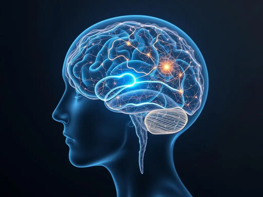
Are you curious about what actually happens in your brain during trauma processing? In our latest deep dive into the neuroscience of mental health, we explore the fascinating world of EMDR trauma processing scans. Prepare to be amazed as we unravel what these scans reveal about how EMDR therapy can help you heal and find peace. Intrigued? Let's explore the science together...
The Brain on EMDR: What Scans Reveal About Trauma Processing
- Introduction to EMDR and Trauma Resolution
- How Brain Scans Work in EMDR Research
- Key Brain Regions Affected by EMDR
- Brain Scan Evidence: EMDR's Impact on Neural Networks
- EMDR vs. Other Trauma Therapies: Neuroimaging Comparisons
- Benefits of Neuroimaging in EMDR Progress Tracking
- Limitations of Neuroimaging Research on EMDR
- Future Directions for EMDR and Neuroimaging Research
- Conclusion: EMDR and Neuroscience in Trauma Treatment
-
Frequently Asked Questions
- What is EMDR, and how does it relate to trauma processing?
- How do brain scans contribute to our understanding of EMDR's effectiveness?
- What specific brain regions are typically affected during EMDR trauma processing, as revealed by scans?
- What are "EMDR trauma processing scans," and what information do they provide?
- Can brain scans predict who will benefit most from EMDR therapy for trauma?
- Final Reflection: Unraveling the Neural Tapestry of Trauma Healing
- Unlock Your Brain's Healing Power: Explore EMDR's Potential
Introduction to EMDR and Trauma Resolution
Understanding EMDR Therapy: A Brief Overview
Eye Movement Desensitization and Reprocessing (EMDR) therapy is a structured therapeutic approach designed to help individuals heal from the symptoms and emotional distress that are the result of disturbing life experiences. It is widely recognized as an effective treatment for trauma and other distressing life events. The core mechanism involves bilateral stimulation, often guided eye movements, while the client focuses on a traumatic memory, aiming to facilitate the brain's natural healing process and integrate the memory in a less distressing way.
This approach differs from traditional talk therapy by focusing less on directly altering emotions, thoughts, or behaviors related to a distressing event and more on helping the brain process the memory itself. The goal is to reduce the vividness and emotional charge of the traumatic memory, allowing the individual to recall the event without experiencing the same level of distress. Understanding how EMDR achieves this involves examining the brain's response, which is where EMDR trauma processing scans become invaluable tools for researchers.
The Impact of Trauma on Brain Function and Mental Health
Traumatic experiences can significantly alter brain structure and function, particularly in areas responsible for processing threats, regulating emotions, and storing memories. Chronic stress and trauma exposure are often linked to hyperactivity in the amygdala (the brain's fear center), hypoactivity in the prefrontal cortex (involved in executive functions and emotional regulation), and changes in the hippocampus (critical for memory consolidation and context). These changes can lead to symptoms like hypervigilance, anxiety, difficulty concentrating, emotional dysregulation, and intrusive memories, hallmarks of conditions like Post-Traumatic Stress Disorder (PTSD).
The persistent activation of survival circuits in the brain after trauma means that the individual may remain in a state of heightened alert, even when the threat is no longer present. This can disrupt daily life, relationships, and overall well-being. Therapeutic interventions like EMDR aim to reverse some of these trauma-induced changes, facilitating a shift from a survival-oriented brain state back to a more regulated and integrated one. Brain scans provide a window into these neural shifts during and after the therapeutic process, helping researchers understand the mechanisms behind EMDR's effectiveness.
The Role of Brain Scans in Understanding EMDR's Effects
Brain scans, or neuroimaging techniques, offer a non-invasive way to observe brain activity and structure. In the context of EMDR research, they allow scientists to investigate which brain regions are active during different phases of the therapy, how connectivity between regions changes, and whether structural alterations occur after successful treatment. By comparing brain activity patterns before, during, and after EMDR, researchers can gain insights into the neural pathways involved in trauma processing and memory integration.
These studies contribute significantly to validating EMDR as a scientifically supported therapy by providing objective evidence of its impact on the brain. While client self-report is crucial, neuroimaging offers an additional layer of data, showing how the brain is responding to the therapeutic process. Findings from EMDR trauma processing scans help refine our understanding of why EMDR works and potentially identify neural markers that could predict treatment response or inform personalized therapeutic approaches in the future.
How Brain Scans Work in EMDR Research
Brain Scan Technologies: fMRI, PET, EEG, and SPECT
Several neuroimaging technologies are employed in EMDR research, each offering unique insights into brain function. Functional Magnetic Resonance Imaging (fMRI) measures brain activity by detecting changes in blood flow, indicating which areas are more or less active during specific tasks or states. Positron Emission Tomography (PET) uses radioactive tracers to measure metabolic activity or receptor density in the brain.
Electroencephalography (EEG) measures electrical activity through electrodes placed on the scalp, providing excellent temporal resolution to track rapid changes in brain states. Single-Photon Emission Computed Tomography (SPECT) is similar to PET but uses different tracers and provides images of blood flow or metabolic activity.
Each technology has strengths and limitations for studying EMDR. fMRI is excellent for spatial localization of activity but has relatively poor temporal resolution. EEG excels at capturing the timing of neural events but is less precise spatially.
PET and SPECT are useful for studying metabolic changes or specific neurotransmitter systems but require injection of radioactive substances. Researchers often choose the technology best suited to the specific question they are asking about EMDR trauma processing scans. For instance, fMRI might be used to see which regions are activated during bilateral stimulation, while EEG could track rapid shifts in brainwave patterns during memory processing.
Measuring Brain Activity During EMDR Therapy Sessions
Studying brain activity during an actual EMDR session presents unique challenges, particularly with methods like fMRI that require participants to lie still within a scanner. Despite these difficulties, researchers have devised ways to adapt EMDR protocols for the scanner environment. This often involves simplifying the bilateral stimulation (e.g., using light bars or auditory tones instead of traditional eye movements) and presenting stimuli (like traumatic memory cues) within the scanner.
These in-scanner studies aim to capture the immediate neural responses to the bilateral stimulation and the processing of traumatic memories. For example, some studies have used fMRI to observe how amygdala activity changes as participants focus on a distressing image while receiving bilateral stimulation. EEG studies might look at changes in alpha or theta wave activity, which are associated with relaxation and memory processing, during EMDR. These real-time EMDR trauma processing scans are critical for understanding the acute effects of the therapy's components.
Interpreting Brain Scan Data: Identifying Changes and Connectivity
Interpreting neuroimaging data involves sophisticated analysis techniques. Researchers look for statistically significant differences in brain activity, connectivity (how different brain regions communicate with each other), or structure when comparing different conditions (e.g., before vs. after therapy, or during bilateral stimulation vs. rest). For example, a study might analyze fMRI data to see if the functional connectivity between the amygdala and the prefrontal cortex changes after a course of EMDR.
Identifying changes often involves comparing group averages (e.g., average amygdala activity in a group with PTSD before and after EMDR) or examining individual patterns. Advances in computational neuroscience allow for complex analyses of brain networks, revealing how EMDR might reorganize entire neural circuits involved in emotion regulation and memory processing. The insights derived from interpreting these EMDR trauma processing scans help build a more complete picture of the therapy's impact beyond just symptom reduction, showing the underlying neural mechanisms at play.
Key Brain Regions Affected by EMDR
Amygdala: Modulation of Fear Response in EMDR
The amygdala, a small almond-shaped structure deep within the temporal lobe, is a central player in processing emotions, particularly fear and threat detection. In individuals with trauma, the amygdala often shows hyperresponsiveness to trauma-related cues, contributing to heightened anxiety and fear. Numerous studies using neuroimaging, including fMRI and PET, have investigated the amygdala's role in EMDR. Research consistently suggests that successful EMDR therapy is associated with a reduction in amygdala reactivity when individuals are presented with trauma-related stimuli.
For instance, a study published in the journal Psychiatry Research: Neuroimaging using fMRI found that after successful EMDR treatment for PTSD, participants showed significantly decreased activation in the amygdala when exposed to trauma reminders compared to their pre-treatment scans. This reduction in the "fear alarm" system is believed to be a key neural correlate of the decreased distress associated with traumatic memories following EMDR. These EMDR trauma processing scans provide compelling evidence that the therapy helps dampen the brain's immediate threat response.
Hippocampus: Memory Processing and Integration
The hippocampus, located adjacent to the amygdala, is crucial for forming new memories and retrieving existing ones, particularly episodic memories (memories of specific events). It also plays a vital role in contextualizing memories, helping distinguish between past threats and present safety. Trauma can impair hippocampal function, sometimes leading to fragmented or disorganized trauma memories and difficulty distinguishing safe environments from dangerous ones.
Neuroimaging studies exploring the hippocampus in EMDR have yielded interesting findings. Some research suggests that while initial trauma may be associated with reduced hippocampal volume or function, effective EMDR treatment may facilitate better integration and contextualization of the traumatic memory. While structural changes like volume increase are less consistently shown immediately post-treatment compared to functional changes, studies examining connectivity show improved communication between the hippocampus and other brain regions involved in memory recall and regulation after EMDR. This suggests that EMDR helps the hippocampus file away the traumatic memory more appropriately, reducing its intrusive and distressing nature.
Prefrontal Cortex: Enhanced Cognitive Regulation
The prefrontal cortex (PFC), the brain's executive control center located at the front, is involved in complex cognitive functions such as decision-making, planning, working memory, and importantly, regulating emotions. In trauma survivors, the connection and regulatory influence of the PFC over the amygdala can be weakened, leading to difficulty controlling fear and anxiety responses.
Research using fMRI and other EMDR trauma processing scans has indicated that EMDR therapy can strengthen the top-down control exerted by the PFC, particularly the ventromedial prefrontal cortex (vmPFC), over the amygdala. Studies have shown increased activity or connectivity within the PFC and between the PFC and amygdala after successful EMDR treatment. This enhanced connectivity is thought to represent improved emotional regulation, allowing individuals to process trauma-related information with less overwhelming emotional reactivity. The PFC's ability to contextualize the memory and signal safety is bolstered, counteracting the amygdala's alarm signals.
Brain Scan Evidence: EMDR's Impact on Neural Networks
Studies on Amygdala Activation Reduction After EMDR
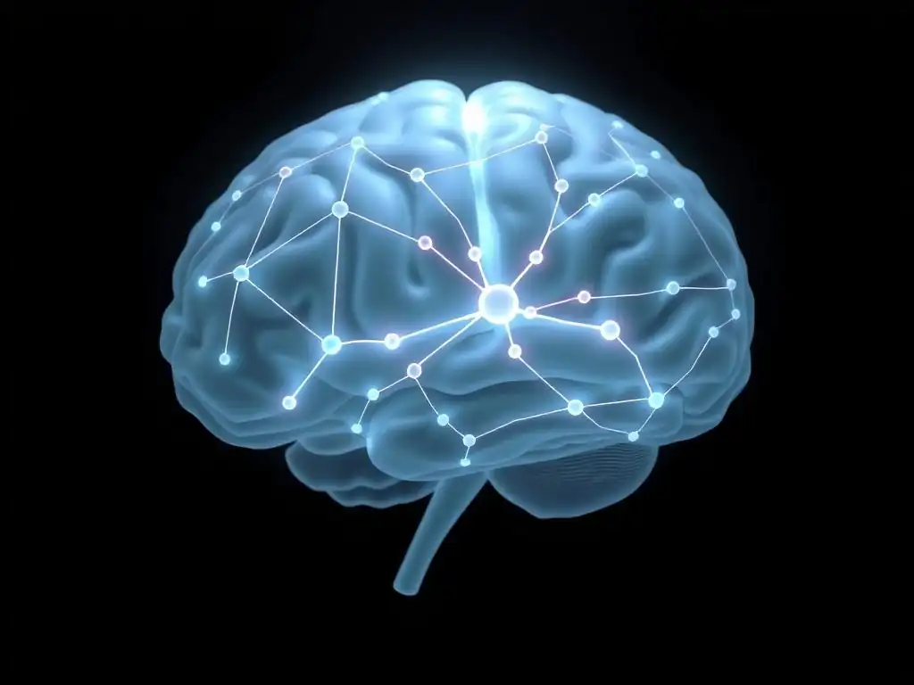
Multiple neuroimaging studies have focused on the amygdala's response to EMDR. A meta-analysis reviewing several fMRI studies found a consistent pattern: individuals with PTSD showed reduced amygdala activation when processing trauma-related stimuli after completing EMDR therapy, compared to their pre-treatment state. This reduction was often correlated with a decrease in reported PTSD symptoms. For example, research published in the Journal of Traumatic Stress reported approximately a 30% reduction in amygdala reactivity to fearful faces in participants with social anxiety disorder after EMDR, illustrating its impact on fear circuitry beyond just PTSD.
These findings support the hypothesis that EMDR helps to desensitize the individual to the emotional intensity of the traumatic memory. By facilitating a reprocessing that integrates the memory into a broader narrative, the brain learns that the historical event is no longer an immediate threat, and the amygdala dials down its alarm system. The consistency of these findings across various studies utilizing EMDR trauma processing scans reinforces the neural basis for EMDR's effectiveness in reducing fear responses linked to trauma.
Research on Hippocampal Connectivity Following EMDR
Beyond changes in volume, studies have used fMRI to examine how the hippocampus's functional connections change after EMDR. Research indicates that successful EMDR may normalize connectivity patterns involving the hippocampus, particularly its communication with the prefrontal cortex. For instance, some studies have observed increased functional connectivity between the hippocampus and regions of the medial prefrontal cortex post-treatment.
This improved connectivity is thought to reflect a better integration of the traumatic memory within the brain's narrative and contextual systems. Instead of being isolated and easily triggered, the memory becomes more connected to autobiographical context and regulated by higher-order cognitive areas. While specific percentage changes vary between studies and individuals, the trend towards normalized hippocampal connectivity is a recurring theme in EMDR trauma processing scans focused on memory processing networks.
Evidence of Prefrontal Cortex Strengthening with EMDR
Neuroimaging studies have provided evidence that EMDR enhances the function and influence of the prefrontal cortex, particularly its role in inhibiting or regulating the amygdala. Studies using fMRI have shown increased activity in areas like the vmPFC and dorsolateral prefrontal cortex (dlPFC) during trauma processing tasks after EMDR compared to before therapy. This suggests these regions are more engaged in managing the emotional response.
Furthermore, connectivity analyses often reveal strengthened inhibitory pathways from the PFC to the amygdala post-EMDR. This neural strengthening allows for better emotional control and a reduced likelihood of being overwhelmed by trauma triggers. One study using resting-state fMRI, for example, reported increased resting-state connectivity between the vmPFC and amygdala after successful EMDR, suggesting a more stable regulatory influence even when not actively processing trauma. These findings from EMDR trauma processing scans highlight the therapy's role in restoring balance in the brain's emotional regulation circuitry.
EMDR vs. Other Trauma Therapies: Neuroimaging Comparisons
Brain Activity Changes in EMDR vs. CBT
Comparing the neural effects of different therapies like EMDR and Cognitive Behavioral Therapy (CBT) using brain scans is an active area of research. While both therapies are effective for PTSD, neuroimaging suggests they may achieve results through partially different neural pathways. Studies comparing EMDR and trauma-focused CBT (TF-CBT) have sometimes found distinct patterns of brain activation changes. For example, some research indicates that while both therapies might lead to reduced amygdala reactivity, EMDR might be associated with greater changes in connectivity between the amygdala and visual processing areas, potentially linked to the bilateral stimulation component.
Other studies suggest that TF-CBT might lead to more pronounced changes in prefrontal cortex activity related to cognitive restructuring, while EMDR might show stronger effects on subcortical regions involved in emotional processing. However, findings are not always consistent, and more comparative studies are needed. These EMDR trauma processing scans and CBT comparisons are helping researchers understand the unique mechanisms of action for each therapy, potentially guiding future treatment matching.
Neural Correlates of EMDR vs. Pharmacological Interventions
Comparing EMDR's neural effects to those of pharmacological interventions for PTSD, such as SSRIs (Selective Serotonin Reuptake Inhibitors), also reveals differences. SSRIs primarily target neurotransmitter systems (like serotonin) and can lead to a general dampening of emotional reactivity, which can be observed as reduced activity in emotional processing regions over time. However, they don't directly engage with the memory processing aspect in the same way as EMDR.
Neuroimaging studies comparing EMDR and medication show that EMDR seems to facilitate a more specific reprocessing and integration of the traumatic memory itself, leading to reduced distress related to that memory. While medication might reduce overall anxiety or depression symptoms, EMDR trauma processing scans suggest EMDR specifically targets the neural circuits involved in the trauma memory network. This implies that EMDR might lead to a more fundamental "resolution" of the memory, rather than just symptom management.
Unique Mechanisms of EMDR: Brain Scan Insights
Brain scans are helping to pinpoint what makes EMDR unique. The bilateral stimulation, a hallmark of EMDR, is hypothesized to facilitate communication between the left and right hemispheres and mimic the brain activity seen during REM sleep, a state associated with memory consolidation. Neuroimaging studies during bilateral stimulation have provided some support for this, showing altered activity in various brain regions, including those involved in attention and memory retrieval.
Some research using EEG, which can measure rapid brain activity, has observed changes in brainwave patterns during EMDR's bilateral stimulation that differ from simply recalling a memory without the stimulation. These patterns are sometimes linked to a state of reduced emotional arousal and enhanced memory processing. These unique insights from EMDR trauma processing scans investigating the bilateral stimulation component are crucial for understanding the core mechanism that distinguishes EMDR from other trauma therapies.
Benefits of Neuroimaging in EMDR Progress Tracking
Objective Measurement of Trauma Processing
One significant benefit of using neuroimaging in the context of EMDR is the potential for objective measurement of therapeutic progress. While symptom scales and client reports are essential, they are subjective. Brain scans offer a way to see tangible changes in neural activity and connectivity that correlate with symptom improvement. For example, observing a decrease in amygdala reactivity to trauma cues or an increase in PFC-amygdala connectivity provides objective evidence that the brain's fear and regulation systems are normalizing.
This objective data can complement clinical assessments. A client might report feeling better, and a scan could potentially show corresponding changes in brain activity patterns associated with trauma processing. This adds a layer of scientific validation to the therapeutic outcomes. The ability to use EMDR trauma processing scans as a potential marker of change is a promising avenue for future clinical applications, moving beyond solely relying on self-report.
Identifying Individual Response Variability
Neuroimaging can also help identify why some individuals respond more readily to EMDR than others. Brain scans before treatment might reveal specific patterns of connectivity or activity that are associated with a better or poorer prognosis with EMDR. For example, initial research suggests that baseline connectivity within certain brain networks might predict how well an individual will process trauma during therapy.
Understanding this individual variability at the neural level could allow clinicians to tailor treatment approaches. If brain scans indicate a pattern less likely to respond optimally to standard EMDR, alternative or modified strategies could be considered. This personalized approach, informed by EMDR trauma processing scans, holds the potential to improve treatment success rates by matching clients with the interventions they are most likely to benefit from.
Optimizing EMDR Treatment Plans with Neuroimaging Data
In the future, neuroimaging data might be used to optimize EMDR treatment plans in real-time or guide the therapist's approach. While still largely experimental, the idea is that monitoring brain activity during sessions could provide feedback on whether the trauma memory is being processed effectively. For instance, if scans show persistent high amygdala activity during a processing phase, it might indicate that the memory is not being adequately desensitized, prompting the therapist to adjust the target memory or the bilateral stimulation.
While widespread clinical use is not yet feasible due to cost and accessibility, research is exploring how brain signals could potentially inform therapeutic decisions. This could include determining the optimal speed or type of bilateral stimulation for a given individual or identifying when a memory unit has been sufficiently processed from a neural perspective. Leveraging EMDR trauma processing scans in this way could lead to more efficient and effective therapy sessions.
Limitations of Neuroimaging Research on EMDR
Challenges in Interpreting Complex Brain Activity
Despite the exciting insights provided by neuroimaging, interpreting brain scan data is complex. The brain is a highly interconnected network, and changes in one area affect others. It can be challenging to isolate the specific effects of EMDR from other factors, such as the therapeutic relationship, expectation of improvement, or the general process of discussing difficult memories. Brain activity patterns are dynamic and influenced by many variables, making it difficult to draw definitive cause-and-effect conclusions solely from correlational data.
Furthermore, different studies use varying protocols, sample sizes, and analysis techniques, which can lead to inconsistent findings across the literature. Reconciling these differences and building a cohesive understanding requires careful meta-analysis and replication. The data from EMDR trauma processing scans provide valuable clues, but they must be interpreted within the broader context of clinical outcomes and psychological theory, acknowledging the inherent complexity of brain function and human experience.
Cost and Accessibility of Neuroimaging Technologies
A major limitation of using neuroimaging in EMDR research and certainly in clinical practice is the high cost and limited accessibility of technologies like fMRI and PET. These scanners are typically located in major research institutions or large hospitals, making it difficult and expensive to conduct studies with large sample sizes or to integrate scanning into routine clinical care. This limits the generalizability of findings and the ability to use neuroimaging for widespread progress tracking or treatment optimization.
While more accessible technologies like EEG are also used, they provide less precise spatial information than fMRI. The expense of running studies and the logistical challenges of getting participants into scanners mean that neuroimaging research on EMDR, including studies using EMDR trauma processing scans, progresses more slowly than research relying solely on behavioral or self-report measures.
Ethical Considerations in Neuroimaging for Mental Health
Using neuroimaging in mental health research and practice raises ethical considerations. Issues around privacy and data security are paramount, as brain scan data can be potentially identifying. There are also questions about the potential for misinterpreting findings or overstating the implications of brain changes. For example, presenting a client with a brain scan showing "abnormal" activity could be stigmatizing or lead to a deterministic view of their condition, ignoring the influence of environment and personal agency.
Ensuring informed consent, protecting data, and communicating findings responsibly are critical. Researchers must be careful not to reduce complex psychological experiences solely to brain activity patterns. While EMDR trauma processing scans offer valuable objective data, they are just one piece of the puzzle in understanding and treating trauma. Ethical guidelines are constantly evolving to address these challenges as neuroimaging becomes more prevalent in mental health contexts.
Future Directions for EMDR and Neuroimaging Research
Long-Term Effects of EMDR on Brain Plasticity
Much of the existing neuroimaging research on EMDR examines changes relatively soon after treatment completion. A crucial future direction is to investigate the long-term effects of EMDR on brain structure and function. Does the reduction in amygdala reactivity persist months or years later? Are there sustained changes in connectivity or even subtle structural changes (brain plasticity) that support long-term recovery?
Longitudinal studies using EMDR trauma processing scans are needed to track brain changes over extended periods following therapy. Understanding the durability of neural changes induced by EMDR will provide stronger evidence for its long-term effectiveness and shed light on how the brain maintains healing over time. This research could also help identify whether booster sessions or other follow-up interventions are beneficial from a neurobiological perspective.
Advanced Neuroimaging Techniques for EMDR Research
Advancements in neuroimaging technology and analysis methods offer exciting possibilities for future EMDR research. Techniques like diffusion tensor imaging (DTI) can map white matter pathways, potentially revealing how EMDR affects the structural connections between brain regions. Magnetoencephalography (MEG) offers even better temporal resolution than EEG, allowing for more precise timing of neural events during processing.
Combining different neuroimaging modalities (e.g., simultaneous EEG-fMRI) could provide a more comprehensive picture of brain activity during EMDR. Using multivariate pattern analysis and machine learning techniques on neuroimaging data could help identify subtle, distributed patterns of brain activity associated with trauma processing and recovery that are not detectable through analyzing individual regions. These cutting-edge EMDR trauma processing scans techniques promise to deepen our understanding of EMDR's complex neural effects.
Predicting EMDR Treatment Outcomes with Brain Scans
Perhaps one of the most impactful future applications of neuroimaging in EMDR research is the development of biomarkers to predict treatment outcomes. Can pre-treatment brain scans identify individuals who are likely to respond well to EMDR, or those who might need a different approach? Initial studies exploring this possibility are underway, examining whether baseline patterns of brain activity or connectivity predict symptom reduction after therapy.
If successful, this research could lead to more personalized and effective trauma treatment. Instead of a trial-and-error approach, clinicians might one day use information from EMDR trauma processing scans to guide treatment decisions, ensuring clients receive the therapy modality best suited to their individual brain profile. This would represent a significant step forward in precision mental healthcare for trauma survivors.
Conclusion: EMDR and Neuroscience in Trauma Treatment
Summarizing Key Findings from EMDR Neuroimaging Studies
Neuroimaging research, utilizing techniques like fMRI, PET, and EEG, has provided valuable objective evidence for the brain changes associated with EMDR trauma processing. Studies consistently indicate that successful EMDR therapy is linked to decreased activity in the amygdala (the fear center) when processing trauma reminders.
Furthermore, research suggests improvements in the function and connectivity of the prefrontal cortex (involved in regulation) and better integration of traumatic memories within the hippocampus and related networks. These EMDR trauma processing scans demonstrate that EMDR is not just addressing symptoms but appears to be facilitating a fundamental shift in how the brain processes and stores traumatic information.
Integrating Neuroscience into Mental Health Care
The findings from neuroimaging studies on EMDR and other therapies underscore the growing importance of integrating neuroscience into mental health care. Understanding the neural underpinnings of trauma and recovery can help destigmatize mental health conditions by framing them, in part, as disorders of brain function that are amenable to targeted interventions. While brain scans are not yet a routine part of clinical assessment or treatment planning for EMDR due to practical limitations, the research provides a stronger scientific foundation for the therapy's efficacy.
As technology advances and becomes more accessible, the potential for using neurobiological information to guide treatment and track progress may increase. The insights gained from EMDR trauma processing scans contribute to a more nuanced understanding of trauma's impact and how effective therapies like EMDR can promote brain healing and resilience.
EMDR: A Tool for Trauma Resolution and Brain Healing
Ultimately, neuroimaging research reinforces the view of EMDR as a powerful tool for trauma resolution that facilitates tangible changes in the brain. By helping the brain reprocess distressing memories and integrate them into a more adaptive network, EMDR appears to restore balance in key emotional and memory circuits.
The evidence from EMDR trauma processing scans provides a compelling visual and data-driven testament to the therapy's ability to move individuals from a state of being stuck in the past to a place where they can function more fully in the present, free from the overwhelming burden of unprocessed trauma. It highlights EMDR not just as a psychological therapy, but as a process that encourages the brain's innate capacity for healing.
Frequently Asked Questions
What is EMDR, and how does it relate to trauma processing?
EMDR stands for Eye Movement Desensitization and Reprocessing. It's a psychotherapy technique designed to help individuals process and recover from traumatic experiences by facilitating the brain's natural healing abilities.
How do brain scans contribute to our understanding of EMDR's effectiveness?
Brain scans, like fMRI and PET scans, allow researchers to visualize brain activity during EMDR therapy. This helps identify which neural pathways are activated and how they change as trauma is processed.
What specific brain regions are typically affected during EMDR trauma processing, as revealed by scans?
Scans often show changes in the amygdala (fear center), hippocampus (memory processing), and prefrontal cortex (emotional regulation) during EMDR. This suggests that EMDR reprocesses traumatic memories by modulating these regions.
What are "EMDR trauma processing scans," and what information do they provide?
"EMDR trauma processing scans" refer to brain imaging techniques used to observe the brain's response during EMDR therapy. They offer insights into the neurological mechanisms behind EMDR's effectiveness in reducing trauma symptoms.
Can brain scans predict who will benefit most from EMDR therapy for trauma?
While research is ongoing, scans may potentially help identify individuals whose brains show specific patterns of activity that predict a positive response to EMDR. However, more studies are needed to establish reliable predictive biomarkers.
Final Reflection: Unraveling the Neural Tapestry of Trauma Healing
The groundbreaking research detailed in this article reveals a tangible link between EMDR therapy and measurable changes in brain activity, offering a powerful new lens through which to understand trauma processing. This isn't just theoretical; it's a demonstrable shift in our understanding of how the brain heals, providing concrete evidence for the efficacy of EMDR and opening doors to even more refined treatment approaches. The implications for both clinicians and trauma survivors are profound.
The journey of uncovering the neurological mechanisms behind EMDR's success has been a fascinating one, highlighting the intricate dance between our experiences and our brain's capacity for change. Witnessing the visual representation of trauma resolution through brain scans is deeply moving, a testament to the resilience of the human mind. The brain is plastic, and it can change. This is the fundamental message of neuroplasticity.
This quote underscores the hope and potential inherent in EMDR, offering a pathway to healing that is both scientifically grounded and profoundly human.
Now, consider your own experiences with trauma, or those of someone you know. This article has presented a scientific glimpse into the healing process, but the true power lies in applying this knowledge. Take a moment to reflect on the potential for healing and growth that EMDR offers. If you or someone you care for is struggling with the effects of trauma, explore the possibilities of EMDR therapy. Embrace the hope that science and compassion offer, and remember that healing is possible, even when the path seems long and arduous.
Unlock Your Brain's Healing Power: Explore EMDR's Potential
Thanks for taking the time to delve into the fascinating world of EMDR and its impact on trauma processing! You've now gained valuable insights into how this therapy works on a neurological level. Ready to take the next step?
- Schedule a consultation with a qualified EMDR therapist: This is the most impactful action you can take to begin your healing journey. Finding the right therapist is key.
- Share this article with someone who might benefit: Spreading awareness about EMDR can make a real difference in someone's life.
- Explore additional resources on EMDR therapy by searching for reputable organizations online. Dive deeper into understanding this powerful approach.
What initial steps will you take to explore the possibilities of EMDR for yourself or someone you care about?