
Seeing the Brain Heal: An Imaging Journey Through Therapy Outcomes

Welcome to Mind Care Guide, a safe space dedicated to your mental well-being.
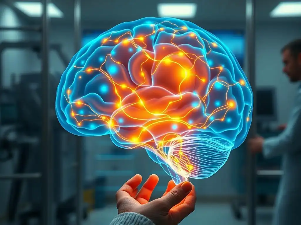
Ever wondered what actually happens inside your brain during therapy? In this article, "Seeing the Brain Heal: An Imaging Journey Through Therapy Outcomes," we delve into the fascinating world of brain imaging therapy outcomes, revealing the tangible changes that occur as you heal. Prepare to witness the power of neuroscience and gain a deeper understanding of your own mental health journey. What secrets do the scans reveal? Let's explore together...
Seeing the Brain Heal: An Imaging Journey Through Therapy Outcomes
- Understanding Brain Imaging in Mental Health Treatment
- Brain Imaging Techniques Used to Track Therapeutic Progress
- Case Studies: Brain Imaging Demonstrating Therapy Success
- How Different Therapies Affect the Brain: A Visual Guide
- Interpreting Brain Scans: What Patients Need to Know
- Limitations and Ethical Considerations of Brain Imaging in Therapy
- Brain Changes and Long-Term Therapy Outcomes: A Deeper Look
- Resources for Learning More About Brain Imaging and Mental Health
-
Frequently Asked Questions
- How does brain imaging help us understand therapy outcomes?
- What types of brain imaging are used to assess therapy outcomes?
- What mental health conditions can benefit from brain imaging to track therapy progress?
- Are there any risks associated with using brain imaging for therapy outcome assessment?
- How does understanding "Brain imaging therapy outcomes" improve patient care?
- Final Reflection: The Evolving Landscape of Brain Healing
- Unveil Your Brain's Healing Power: Take the Next Step
Understanding Brain Imaging in Mental Health Treatment
The landscape of mental health treatment is constantly evolving, moving beyond symptom checklists to explore the biological underpinnings of conditions like depression, anxiety, and PTSD. Brain imaging techniques offer a unique window into the living brain, providing insights into how mental health challenges manifest neurologically and, crucially, how interventions like therapy and medication can lead to observable changes. This journey through brain imaging therapy outcomes reveals the brain's remarkable capacity for change and healing.
The Role of Neuroimaging in Assessing Therapy Effectiveness
Neuroimaging plays a pivotal role in moving towards a more objective assessment of how well mental health treatments are working. While patient self-reports and clinical observations remain fundamental, techniques like fMRI and PET scans allow researchers and clinicians to see where and how the brain is changing in response to therapy.
This isn't just academic; understanding these biological shifts can help refine treatments, personalize approaches, and provide tangible evidence of recovery processes. For example, studies using fMRI have shown that successful cognitive behavioral therapy (CBT) for anxiety can lead to reduced activity in the amygdala, a brain region associated with fear processing, offering a biological correlate to symptom improvement.
Furthermore, tracking brain imaging therapy outcomes provides valuable data for validating different therapeutic modalities. By comparing brain scan data before and after different types of therapy (e.g., CBT vs. mindfulness-based therapy), researchers can identify distinct patterns of neural change associated with each approach.
This helps us understand how these therapies work at a neurological level and why certain therapies might be more effective for specific conditions or individuals. The ability to visualize these changes through brain scans reinforces the idea that mental health conditions are not just "in your head" in a metaphorical sense, but involve real, measurable alterations in brain function and structure.
Why Brain Scans Matter for Long-Term Therapy Outcomes
Understanding brain changes through imaging is particularly crucial when considering the long-term efficacy of therapy. While someone might feel better immediately after starting treatment, brain imaging can help reveal whether the intervention is fostering sustainable changes in neural circuits associated with resilience and emotional regulation.
For instance, research on depression treatment outcomes using brain imaging has looked at connectivity changes in networks like the default mode network (DMN) and the central executive network (CEN). Long-term successful outcomes are often correlated with a normalization of activity and connectivity within and between these networks, suggesting a more fundamental shift in brain function rather than just temporary symptom suppression.
Moreover, brain scans can potentially offer insights into predicting which individuals are likely to benefit most from a particular therapy, although this area is still under active research. By analyzing baseline brain activity patterns or how the brain responds to initial therapy sessions, future applications of brain imaging therapy outcomes might help clinicians tailor treatment plans from the outset, improving the chances of long-term success. This personalized approach, guided by objective neural data, holds significant promise for optimizing care and ensuring that individuals receive the most effective treatment for their specific needs and brain profile.
Brain Imaging Techniques Used to Track Therapeutic Progress
Several advanced neuroimaging techniques are employed to monitor the brain's response to mental health interventions. Each method offers a unique perspective on brain function and structure, contributing to our understanding of brain imaging therapy outcomes. These tools allow researchers and clinicians to observe changes at different levels, from overall activity patterns to specific neurochemical shifts.
fMRI (Functional Magnetic Resonance Imaging): Detecting Brain Activity Changes
Functional Magnetic Resonance Imaging (fMRI) is a widely used technique that measures brain activity by detecting changes in blood flow. When a brain region is more active, it requires more oxygenated blood, and fMRI can pick up on these subtle differences.
In the context of mental health, fMRI is invaluable for observing how different brain circuits involved in emotion, cognition, and behavior respond to therapeutic interventions. For example, studies tracking brain imaging therapy outcomes using fMRI have shown that after successful psychotherapy for social anxiety, there is often reduced activity in the amygdala when individuals are exposed to social stimuli, correlating directly with a decrease in anxiety symptoms.
Another common finding in fMRI studies of therapy outcomes is changes in functional connectivity – how different brain regions communicate with each other. For instance, in individuals with depression, there might be altered connectivity within the reward circuit or between areas involved in rumination. Successful treatment, whether therapy or medication, can lead to a normalization of these connectivity patterns. This offers a dynamic view of how therapy rewires the brain's communication pathways, providing compelling evidence of the biological reality behind positive brain imaging therapy outcomes. Observing these changes helps validate the effectiveness of treatments beyond just symptom reduction.
PET Scans (Positron Emission Tomography): Visualizing Neurochemical Shifts
Positron Emission Tomography (PET) scans offer a different, yet complementary, view of the brain compared to fMRI. PET uses radioactive tracers that bind to specific molecules in the brain, such as neurotransmitter receptors or metabolic markers like glucose.
This allows researchers to visualize and quantify the distribution and activity of these molecules, providing insights into the neurochemical basis of mental health conditions and how they respond to treatment. For example, PET scans have been crucial in understanding how antidepressant medications, particularly SSRIs (Selective Serotonin Reuptake Inhibitors), affect serotonin transporter availability in the brain, directly demonstrating the drug's mechanism of action at a molecular level.
Tracking brain imaging therapy outcomes with PET scans is particularly useful for interventions that directly target neurochemical systems, such as psychotropic medications. Studies using PET have shown changes in dopamine receptor binding in individuals with addiction disorders following treatment, or alterations in serotonin receptor density in patients with obsessive-compulsive disorder (OCD) after successful therapy or medication. These scans provide objective evidence that treatments are reaching their targets and inducing measurable changes in brain chemistry, further solidifying the understanding of how biological processes underpin mental health and recovery.
EEG (Electroencephalography): Monitoring Brainwave Patterns and Therapy Response
Electroencephalography (EEG) measures the electrical activity produced by the brain using electrodes placed on the scalp. While it doesn't provide the detailed spatial resolution of fMRI or PET, EEG offers excellent temporal resolution, meaning it can capture rapid changes in brain activity.
Different mental states and conditions are associated with characteristic brainwave patterns (e.g., alpha, beta, theta waves), and EEG can track how these patterns shift in response to therapy. For instance, studies have used EEG to monitor changes in brainwave coherence or power in specific frequencies before and after treatments for conditions like ADHD or sleep disorders, which are often comorbid with other mental health challenges.
EEG is also being explored for its potential to predict therapy response. By analyzing baseline EEG patterns or early changes during the first few sessions, researchers are investigating whether EEG signatures can indicate which patients are more likely to benefit from a particular intervention.
While still largely a research tool for tracking brain imaging therapy outcomes in this context, advancements in quantitative EEG (qEEG) analysis are making it a promising area for providing insights into the brain's electrical dynamics and how they are modulated by therapeutic processes. The ability to see these electrical patterns change offers another layer of evidence for the brain's adaptability.
Case Studies: Brain Imaging Demonstrating Therapy Success
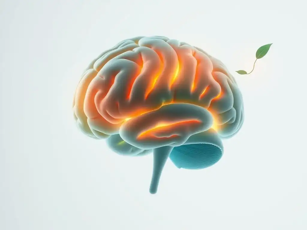
Examining specific case studies provides powerful illustrations of how brain imaging can visually demonstrate the impact of successful mental health treatment. These examples, often drawn from research literature, highlight the tangible changes that occur in the brain as symptoms improve. They offer a compelling narrative of healing, supported by objective neural data, reinforcing the concept of positive brain imaging therapy outcomes.
Case Study 1: Major Depressive Disorder and Brain Scan Evidence of Improvement with SSRIs
Consider a case study involving individuals with Major Depressive Disorder (MDD) treated with Selective Serotonin Reuptake Inhibitors (SSRIs). Before treatment, fMRI scans of these individuals might show increased connectivity within the Default Mode Network (DMN), a network associated with self-referential thought and rumination, and decreased connectivity between the DMN and areas involved in cognitive control.
Additionally, PET scans might reveal lower levels of serotonin transporters in certain brain regions compared to healthy controls. After several weeks or months of successful SSRI treatment, as symptoms of depression lessen, follow-up fMRI scans often show a normalization of DMN connectivity – reduced activity within the network and improved connections with cognitive control areas.
Simultaneously, follow-up PET scans can demonstrate an increase in the binding of the SSRI to serotonin transporters in the brain, confirming the drug's intended action and correlating with the observed clinical improvement. While specific percentage changes vary between studies and individuals, research consistently shows measurable shifts in these markers.
For example, one meta-analysis of fMRI studies on antidepressant treatment outcomes indicated reduced amygdala reactivity to negative stimuli, often showing effect sizes considered medium to large in neuroimaging research (e.g., Cohen's d values around 0.5-0.8), alongside the observed connectivity changes. These brain imaging therapy outcomes provide a biological basis for the relief experienced by patients, showing that medication facilitates tangible neural adjustments.
Case Study 2: Post-Traumatic Stress Disorder Showing Positive Changes on fMRI After Trauma-Focused Therapy
Another compelling example comes from studies on Post-Traumatic Stress Disorder (PTSD) treated with trauma-focused therapies like Prolonged Exposure (PE) or Eye Movement Desensitization and Reprocessing (EMDR). Individuals with PTSD often exhibit hyper-responsivity in the amygdala (the brain's fear center) and hypo-responsivity in the prefrontal cortex (PFC), particularly areas involved in emotional regulation, when processing trauma-related stimuli. This imbalance contributes to symptoms like heightened anxiety, flashbacks, and difficulty regulating emotions.
After successful trauma-focused therapy, fMRI scans often reveal a significant shift in this pattern. Studies have consistently shown decreased activity in the amygdala and increased activity in the medial prefrontal cortex (mPFC) when individuals are exposed to trauma cues. For instance, research has reported reductions in amygdala activation ranging from 20% to 40% in response to fearful faces or trauma scripts following effective therapy.
This suggests that therapy helps the brain process traumatic memories in a safer, more regulated way. These positive brain imaging therapy outcomes illustrate how psychological interventions can lead to measurable, adaptive changes in the neural circuits responsible for fear and emotional processing, offering a visual testament to recovery.
Analyzing Brain Imaging Data to Understand the Impact of Therapy
Analyzing brain imaging data to understand the impact of therapy is a complex process that involves sophisticated statistical methods and computational tools. Researchers compare brain scans taken before and after treatment, often including control groups (e.g., individuals receiving a placebo, waitlist controls, or healthy individuals), to isolate the effects specifically attributable to the therapy. This involves preprocessing the raw imaging data to correct for motion and other artifacts, followed by various analytical approaches depending on the imaging modality and the research question.
For fMRI, this might involve analyzing average activity levels in specific regions of interest (ROIs) or examining functional connectivity patterns across networks. For PET, it involves quantifying tracer binding in different brain areas.
Interpreting these changes requires expertise in neuroscience, statistics, and the specific mental health condition being studied. Researchers look for statistically significant differences in brain activity, connectivity, or neurochemistry that correlate with improvements in clinical symptoms. For example, a study might find a significant increase in mPFC activity (p < 0.05, with appropriate multiple comparison correction) that is positively correlated with a decrease in symptom severity scores (e.g., a correlation coefficient r = -0.6).
These analyses help build a detailed picture of how therapy works in the brain, identifying the neural pathways involved in recovery. Understanding these patterns through rigorous analysis of brain imaging therapy outcomes is key to refining treatment strategies and developing new interventions.
How Different Therapies Affect the Brain: A Visual Guide
Different therapeutic approaches, whether talk therapy, medication, or newer modalities like mindfulness, target different aspects of brain function and structure. Brain imaging provides a visual guide to these diverse impacts, illustrating how various interventions lead to distinct patterns of neural change. Understanding these differences through brain imaging therapy outcomes helps clarify the mechanisms by which therapies exert their beneficial effects.
Cognitive Behavioral Therapy (CBT) and its Impact on Brain Circuits
Cognitive Behavioral Therapy (CBT) is a structured talk therapy that focuses on identifying and changing negative thought patterns and behaviors. Brain imaging studies have shown that CBT primarily impacts the brain circuits involved in emotional regulation, executive control, and threat processing.
For instance, research using fMRI on patients with anxiety disorders before and after successful CBT often shows reduced activity in the amygdala and increased activity in the prefrontal cortex (PFC), particularly the ventromedial PFC (vmPFC), which is involved in extinguishing fear responses. This suggests that CBT helps strengthen the brain's capacity to regulate emotional reactions and reappraise fearful situations.
Furthermore, CBT has been shown to alter connectivity patterns. Studies have observed increased functional connectivity between the vmPFC and the amygdala after CBT for anxiety, indicating improved top-down control over emotional responses. For depression, CBT has been linked to changes in connectivity within the default mode network and between the DMN and areas related to cognitive control, supporting a shift away from excessive self-focus and rumination. These brain imaging therapy outcomes visually demonstrate how learning new ways of thinking and behaving can literally reshape the brain's functional architecture, enhancing its ability to manage challenging emotions and thoughts.
Medication and Its Observable Effects on Neurotransmitter Activity via PET Scans
Psychiatric medications, such as antidepressants, antipsychotics, and mood stabilizers, exert their effects primarily by modulating neurotransmitter systems in the brain. PET scans are particularly well-suited to visualize these effects. As discussed earlier, PET can measure the density or availability of neurotransmitter receptors or transporters. For example, antipsychotic medications used to treat schizophrenia primarily target dopamine receptors (D2 receptors). PET scans can show that these medications occupy a significant percentage of D2 receptors in areas like the striatum, directly demonstrating their pharmacological action in the living brain.
Similarly, antidepressants targeting serotonin or norepinephrine reuptake can be visualized affecting the transporters for these neurotransmitters using specific PET tracers. Studies tracking brain imaging therapy outcomes with PET have shown, for example, that successful antidepressant treatment is associated with changes in serotonin transporter binding potential in regions like the raphe nuclei and cortical areas.
While the precise percentage change varies depending on the medication and study, observable shifts in neurochemistry are consistently detected. These PET findings provide objective evidence that medications are indeed changing brain chemistry in ways believed to alleviate symptoms, offering a neurobiological explanation for their therapeutic effects.
Mindfulness-Based Therapy and Long-Term Changes in Brain Connectivity
Mindfulness-based therapies, such as Mindfulness-Based Stress Reduction (MBSR), focus on cultivating present-moment awareness and non-judgmental observation. Brain imaging studies suggest that these practices can lead to significant long-term changes in brain structure and function, particularly in areas related to attention, self-awareness, and emotional regulation. fMRI studies have shown that mindfulness training can increase activity in the prefrontal cortex and insula, regions associated with attention and interoception (awareness of internal bodily states).
Furthermore, mindfulness practices have been linked to changes in brain connectivity. Research has shown increased functional connectivity within the salience network (involved in detecting important stimuli) and between the salience network and areas of the prefrontal cortex after mindfulness training. Studies have also reported changes in the connectivity of the default mode network, potentially reflecting a reduced tendency for mind-wandering and rumination. These brain imaging therapy outcomes suggest that regular mindfulness practice enhances the brain's capacity for focused attention, self-awareness, and emotional balance, illustrating how cultivating a specific mental state can lead to enduring neural adaptations.
Interpreting Brain Scans: What Patients Need to Know
While brain imaging is a powerful tool in mental health research and increasingly in clinical settings, interpreting scan results requires careful consideration. For patients, understanding what brain scans can and cannot tell us about their mental health and therapy progress is crucial. This section aims to demystify the process and empower patients to engage in informed conversations with their healthcare providers about brain imaging therapy outcomes.
Understanding Your Brain Imaging Report: A Patient's Guide
A brain imaging report, whether from an fMRI, PET, or EEG scan, can seem complex and filled with technical jargon. Typically, a report will describe the findings in various brain regions, noting any observed abnormalities or significant patterns of activity or connectivity compared to what is expected. It might refer to specific brain areas like the amygdala, hippocampus, prefrontal cortex, or networks like the DMN. For PET scans, it might mention tracer uptake or binding potential in certain regions, often in relation to specific neurotransmitter systems.
It's important for patients to understand that these reports describe biological data, not a direct diagnosis of a mental health condition. Mental health diagnoses are clinical diagnoses based on symptoms, history, and clinical evaluation. Brain imaging can provide supporting evidence or insights into potential underlying neural mechanisms, but it's not a standalone diagnostic test for most conditions currently.
When reviewing a report showing changes related to brain imaging therapy outcomes, focus on understanding what the scan measured (e.g., blood flow, metabolic activity, electrical signals) and where changes were observed. Don't hesitate to ask your doctor for clarification on any terms or findings you don't understand.
Discussing Brain Scan Results with Your Therapist or Psychiatrist
If brain imaging has been part of your mental health journey, discussing the results with your therapist or psychiatrist is essential. These professionals can interpret the technical findings in the context of your specific symptoms, history, and treatment plan. They can explain how the observed brain patterns relate to your condition and, if applicable, how changes seen in follow-up scans reflect the impact of your therapy or medication – the brain imaging therapy outcomes.
Prepare questions beforehand. You might ask:
- What specific areas of my brain were assessed?
- What did the scan show before treatment?
- What changes were observed in the follow-up scan after therapy?
- How do these changes relate to my symptoms?
- Does this information change our treatment plan?
- Are these brain changes expected for the type of therapy I received?
Remember that brain scans are just one piece of the puzzle. Your subjective experience, symptom severity, and overall well-being are equally, if not more, important indicators of therapy success. The conversation should integrate the objective data from the scan with your personal journey and clinical progress, providing a holistic view of your brain imaging therapy outcomes.
The Future of Brain Imaging in Personalized Mental Healthcare
The field of brain imaging in mental healthcare is rapidly advancing, with significant potential for personalizing treatment. Researchers are actively exploring whether brain imaging markers can help predict which individuals are most likely to respond to specific therapies or medications. For instance, some preliminary studies suggest that baseline activity or connectivity patterns in certain brain regions might predict response to SSRIs or specific types of psychotherapy for depression or anxiety.
While this is still largely in the research phase, the goal is to move towards a future where brain imaging could help clinicians make more informed decisions about initial treatment choices, potentially saving patients time and distress spent on ineffective therapies. Advanced techniques like machine learning are being applied to large datasets of brain scans and clinical outcomes to identify complex patterns that predict treatment response. The promise of personalized brain imaging therapy outcomes lies in tailoring interventions to an individual's unique neurobiological profile, leading to more efficient and effective mental healthcare.
Limitations and Ethical Considerations of Brain Imaging in Therapy

Despite the immense potential of brain imaging in understanding and tracking mental health treatment, it's important to acknowledge its current limitations and the ethical considerations associated with its use. Brain imaging is not a panacea, and its application in clinical practice requires careful thought and adherence to ethical guidelines.
The Cost and Accessibility of Brain Imaging Technologies
One of the most significant limitations of brain imaging, particularly advanced techniques like fMRI and PET, is their cost and limited accessibility. MRI and PET scanners are expensive to purchase and operate, and the procedures themselves can be costly for patients, often requiring specialized facilities and trained personnel. This means that access to these technologies for routine clinical assessment of brain imaging therapy outcomes is not universally available and may be limited by insurance coverage or geographical location.
While research continues to explore the clinical utility of brain imaging, the current cost often restricts its use primarily to research settings or complex cases in specialized clinics. More accessible and affordable technologies, like EEG, are also being explored for clinical applications, but they offer different types of information. For brain imaging to become a routine tool in assessing therapy progress, costs need to decrease, and availability needs to increase significantly, ensuring equitable access for all who could potentially benefit from the insights it provides into brain imaging therapy outcomes.
Ethical Implications of Using Brain Scans to Predict Therapy Response
The potential use of brain imaging to predict therapy response raises complex ethical questions. While the idea of tailoring treatment based on a patient's brain profile is appealing, concerns exist about the potential for discrimination or stigmatization. If brain scans are used to determine who receives certain types of therapy, particularly expensive or limited ones, it could create new inequalities in access to care.
Furthermore, predicting non-response based on a brain scan could be disheartening for patients and might lead to them being denied potentially beneficial treatments. The brain is plastic and capable of change, and a baseline scan is just a snapshot. Ethical guidelines emphasize that any predictive information from brain imaging should be used cautiously, in conjunction with clinical assessment, and always with the patient's best interest and autonomy at the forefront. The goal should be to use brain imaging therapy outcomes to empower patients and clinicians, not to limit options based on potentially probabilistic predictions.
Ensuring Patient Privacy and Data Security in Neuroimaging Research
Brain imaging data is highly personal and potentially sensitive. As large datasets of brain scans linked to clinical information are collected for research purposes, ensuring patient privacy and data security is paramount. Robust protocols are needed to anonymize data, secure storage systems, and control access to protect individuals' identities and highly personal information about their brain structure, function, and mental health history.
Sharing neuroimaging data across research institutions is valuable for advancing the field and understanding brain imaging therapy outcomes on a larger scale. However, this sharing must be done with strict adherence to privacy regulations (like HIPAA in the United States) and ethical guidelines. Patients participating in research should be fully informed about how their data will be used, stored, and protected. Maintaining trust between researchers, clinicians, and patients is essential for the continued ethical advancement of brain imaging in mental health.
Brain Changes and Long-Term Therapy Outcomes: A Deeper Look
The enduring effects of successful therapy are rooted in the brain's ability to change and adapt – a property known as neuroplasticity. Brain imaging provides compelling evidence for how therapy leverages this plasticity to create lasting alterations in neural circuits, leading to sustained improvements in mental well-being. Examining brain imaging therapy outcomes over extended periods offers insights into the biological basis of long-term recovery.
How Brain Plasticity Contributes to Therapeutic Gains
Neuroplasticity refers to the brain's capacity to reorganize itself by forming new neural connections throughout life. This includes changes at the synaptic level (how neurons communicate), structural changes (like the growth of new neurons or alterations in gray matter volume), and functional changes (how different brain regions interact). Therapy, whether talk therapy or medication, works by engaging these plastic mechanisms. For example, learning new coping skills in CBT strengthens neural pathways associated with rational thought and emotional regulation, while practicing mindfulness can enhance connections related to attention and self-awareness.
Brain imaging studies tracking therapy over time often show evidence of these plastic changes. For instance, research has documented increases in gray matter volume in areas like the hippocampus (involved in learning and memory, often reduced in depression and PTSD) and the prefrontal cortex following successful treatment. These structural changes, alongside observed shifts in brain activity and connectivity (functional plasticity), represent the biological substrate of therapeutic learning and healing. Understanding how therapy promotes neuroplasticity is key to unlocking more effective interventions and optimizing brain imaging therapy outcomes.
The Correlation Between Brain Changes and Symptom Reduction
A critical aspect of brain imaging research in mental health is demonstrating a clear correlation between observed brain changes and improvements in clinical symptoms. It's not enough to just see changes in a scan; these changes must align with how a patient feels and functions in their daily life. Studies consistently aim to show that the magnitude of change in a specific brain marker (e.g., decreased amygdala activity, increased prefrontal connectivity, normalized neurotransmitter binding) is significantly correlated with the degree of symptom reduction (e.g., lower scores on depression scales, fewer panic attacks, reduced intrusive thoughts).
For example, a study might find that patients with the greatest reduction in amygdala reactivity to fearful stimuli after CBT are also the ones who report the most significant decrease in anxiety symptoms, with a strong negative correlation (e.g., r = -0.7). These correlations provide powerful evidence that the observed brain changes are clinically meaningful and directly related to the patient's recovery. Analyzing these relationships between neural shifts and clinical progress is fundamental to interpreting brain imaging therapy outcomes and validating the biological impact of treatment.
Maintaining Brain Health After Successful Therapy: Lifestyle Factors
While therapy can instigate significant positive changes in the brain, maintaining these gains long-term often involves ongoing effort and healthy lifestyle choices. Brain imaging research, alongside broader neuroscience findings, highlights the importance of factors like regular exercise, sufficient sleep, a balanced diet, stress management, and continued social engagement in supporting brain health and plasticity. These factors can help reinforce the positive neural changes initiated by therapy.
For example, studies using neuroimaging have shown that regular aerobic exercise can increase hippocampal volume and improve connectivity in brain networks associated with cognitive function and mood regulation. Adequate sleep is crucial for memory consolidation and emotional processing, processes that are often impaired in mental health conditions but can be supported by healthy sleep habits.
While brain imaging is not typically used to monitor these lifestyle effects clinically, the research provides a neurobiological rationale for incorporating these practices into a long-term mental wellness plan. Sustaining positive brain imaging therapy outcomes involves a holistic approach that supports the brain's ongoing health and resilience.
Resources for Learning More About Brain Imaging and Mental Health
For those interested in delving deeper into the fascinating intersection of brain imaging and mental health treatment outcomes, numerous resources are available. From academic literature to patient-focused information, these resources can help expand your understanding of how we are beginning to see the brain heal through the lens of neuroimaging.
Books and Articles on Neuroimaging and Therapy Outcomes
A wealth of information exists in academic journals and books. For a general audience, look for books that explain the basics of neuroscience and brain imaging in accessible language. Many researchers and clinicians who specialize in the neuroscience of mental health have written books summarizing key findings, including insights from brain imaging therapy outcomes.
For those who want to explore research directly, searching databases like PubMed (ncbi.nlm.nih.gov/pubmed) for terms like "fMRI therapy outcomes," "PET depression treatment," "neuroimaging CBT," or "brain plasticity psychotherapy" will yield numerous scientific articles. Be mindful that scientific articles can be technical, but abstracts and introductions often provide a good overview of the study's findings regarding brain imaging therapy outcomes. Some universities and research institutions also have public outreach sections on their websites that summarize their findings for a non-specialist audience.
Online Support Groups and Communities for Mental Health
Connecting with others who have navigated mental health challenges can be incredibly supportive. Many online communities and forums exist where individuals share their experiences with different therapies and their paths to recovery. While these platforms are not scientific resources on brain imaging, they offer valuable peer support and personal perspectives on the impact of treatment.
Hearing about the lived experiences of others can complement the scientific understanding gained from brain imaging studies. It provides the human context to the biological data, illustrating how changes observed in scans translate into real-world improvements in mood, behavior, and quality of life. Look for reputable mental health organizations that host moderated online communities to ensure a safe and supportive environment.
Finding Qualified Professionals Who Utilize Brain Imaging
If you are interested in whether brain imaging could be relevant to your mental health care, the best first step is to discuss it with your current mental health professional – your therapist, psychiatrist, or psychologist. They can provide guidance based on your specific situation and the current clinical utility of imaging for your condition.
While brain imaging is not yet a routine part of most clinical mental health evaluations, some specialized clinics or academic medical centers may use it in specific cases or as part of research trials. If you are seeking a professional with expertise in this area, look for psychiatrists or neurologists affiliated with university hospitals or research institutions that have neuroimaging facilities.
Be prepared that clinical brain imaging for mental health purposes is still evolving and is not always covered by insurance, but discussing the possibility with a knowledgeable professional is the first step to understanding if it might be an option for you and what insights it could provide into your potential brain imaging therapy outcomes.
Frequently Asked Questions
How does brain imaging help us understand therapy outcomes?
Brain imaging techniques like fMRI and PET scans allow us to visualize changes in brain activity and structure before and after therapy. This provides objective evidence of treatment effectiveness.
What types of brain imaging are used to assess therapy outcomes?
Commonly used techniques include fMRI, which measures brain activity, and MRI, which shows structural changes. PET scans can also be used to assess neurochemical activity in the brain.
What mental health conditions can benefit from brain imaging to track therapy progress?
Brain imaging can be beneficial for conditions like depression, anxiety disorders, and PTSD, allowing clinicians to see how therapy impacts specific brain regions associated with these conditions.
Are there any risks associated with using brain imaging for therapy outcome assessment?
The risks are generally low. MRI and fMRI are non-invasive, though some individuals may experience anxiety in the scanner. PET scans involve minimal radiation exposure.
How does understanding "Brain imaging therapy outcomes" improve patient care?
By providing objective data on therapy effectiveness, brain imaging can help tailor treatment plans. This can lead to more personalized and effective interventions, ultimately improving patient outcomes.
Final Reflection: The Evolving Landscape of Brain Healing
The ability to visualize the brain's response to therapy is revolutionizing our understanding of mental and neurological illness. This journey through brain imaging and therapy outcomes reveals not just the measurable changes in brain structure and function, but also the profound hope it offers for patients and clinicians alike. We are moving beyond simply treating symptoms to actively witnessing and shaping the brain's remarkable capacity for healing.
The impact of this research extends far beyond the clinical setting; it is reshaping our very understanding of the mind-body connection. Seeing the tangible effects of therapy on the brain empowers individuals struggling with mental health challenges, fostering hope and resilience. It also challenges societal stigmas surrounding mental illness, offering a powerful visual counterpoint to misconceptions. The brain is not a fixed entity; it is a dynamic organ capable of remarkable plasticity and change.
This quote underscores the transformative potential revealed through brain imaging, offering a new perspective on the possibilities of recovery.
Now, consider your own perspective. What does this journey into the brain's capacity for healing mean to you? Perhaps it inspires you to advocate for increased access to advanced therapies, to challenge the stigma surrounding mental illness, or to simply appreciate the incredible resilience of the human brain.
Let the images and insights shared in this article serve as a catalyst for continued exploration and a reminder of the ongoing quest to understand and improve the lives of those affected by neurological and mental health conditions. The future of brain healing is bright, and your engagement is crucial to its continued progress.
Unveil Your Brain's Healing Power: Take the Next Step
Thanks for taking the time to explore the fascinating journey of brain healing through therapeutic intervention! You've gained valuable insights into the power of the mind and how we can visually track its progress. Now it's time to put that knowledge into action.
- Schedule a consultation with a therapist: This is your first step towards understanding how these methods might benefit you personally.
- Share this article with someone who could benefit: Let's spread awareness about the incredible potential of brain plasticity and therapeutic interventions.
- Explore further resources on MindCareGuide.org: Discover more evidence-based articles and resources to deepen your understanding.
What personal step will you take today to nurture your brain's remarkable capacity for healing?