
Beyond the Amygdala: Brain Scan Insights into Therapy for Fear

Welcome to Mind Care Guide, a space dedicated to nurturing your mental well-being.
Is the idea of brain scan therapy triggering anxieties you didn't expect? It's completely understandable. This article, "Beyond the Amygdala: Brain Scan Insights into Therapy for Fear," dives deep into the neuroscience of mental health, specifically addressing brain scan therapy fear. We'll explore how these scans are used, what they reveal, and how understanding the process can actually empower you on your journey to overcoming fear. Ready to demystify the science and ease your concerns? Let's begin...
- Beyond the Amygdala: Brain Scan Insights into Therapy for Fear - Mind Care Guide
- Exploring Brain Scan Methods for Assessing Fear Reduction
- Brain Changes Associated with Successful Fear Therapy
- Different Therapy Approaches and Their Impact on Brain Activity Related to Fear
- Brain Scan Findings: Personalized Treatment Strategies for Fear
- Case Studies: Illustrating Brain Scan-Guided Fear Therapy
- The Future of Brain Scan Enhanced Therapy for Fear
- Conclusion: Brain Scans as a Powerful Tool for Understanding and Treating Fear
-
Frequently Asked Questions
- What is Brain Scan Therapy and how does it help with fear?
- Why is there "Brain Scan Therapy Fear" among patients?
- How accurate are brain scans in diagnosing and treating fear-related disorders?
- Are there alternatives to brain scan therapy for those experiencing "Brain Scan Therapy Fear"?
- What are the potential benefits of overcoming "Brain Scan Therapy Fear" and undergoing the process?
- Final Reflection: Unlocking the Fear Response Through Neuroscience
- Unlock Your Fearless Potential: Next Steps to a Calmer You
Beyond the Amygdala: Brain Scan Insights into Therapy for Fear - Mind Care Guide
The Role of the Amygdala in Fear Processing
The amygdala, a small almond-shaped structure deep within the brain's temporal lobe, is widely recognized as a central player in processing fear. It acts as a rapid threat detection system, quickly assessing sensory input and triggering the body's fight-or-flight response. When confronted with a perceived danger, the amygdala receives signals from sensory organs and other brain regions, initiating a cascade of physiological reactions such as increased heart rate, sweating, and muscle tension. This instantaneous response is crucial for survival, allowing an individual to react before conscious thought fully processes the threat.
Research using brain scan technology, such as fMRI, has consistently shown heightened amygdala activity when individuals are exposed to fearful stimuli, whether images, sounds, or even memories. For instance, studies have observed amygdala activation within milliseconds of seeing a fearful facial expression. This rapid, automatic processing underlies the often overwhelming and immediate nature of fear responses, highlighting the amygdala's critical role in the brain's primitive defense mechanisms. Understanding this foundational role is the first step in exploring how brain scan therapy for fear can target these neural circuits.
Limitations of Amygdala-Centric Views of Fear
While the amygdala is undeniably important, focusing solely on this single structure provides an incomplete picture of the complex experience of fear. Fear is not just a primitive reaction; it involves intricate interactions between multiple brain regions, including the prefrontal cortex, hippocampus, and insula. The prefrontal cortex, for example, is involved in evaluating context, regulating emotions, and inhibiting inappropriate fear responses, playing a crucial role in distinguishing real threats from perceived ones.
Moreover, the experience and expression of fear are highly variable among individuals and depend heavily on learning, memory, and cognitive appraisal. The hippocampus contributes by associating fear with specific contexts and memories, while the insula processes internal bodily states related to fear. Therefore, effective brain scan therapy for fear needs to look beyond the amygdala to understand the broader neural network involved in generating, maintaining, and regulating fear responses. Modern brain imaging techniques allow researchers and clinicians to explore these wider circuits, offering a more holistic view.
Brain Scan Technology: A Window into Fear Therapy Effectiveness
Brain scan technology offers unprecedented opportunities to peer inside the living brain and observe how it functions and changes, particularly in the context of mental health conditions involving fear, such as phobias, panic disorder, and PTSD. Techniques like fMRI, EEG, and PET scans provide different but complementary insights into brain activity and structure. By measuring neural activity, connectivity, or metabolic processes, these scans can reveal patterns associated with fearful states and, crucially, track how these patterns change in response to therapeutic interventions.
Using brain scans before, during, and after therapy allows researchers to objectively assess the biological impact of treatment. This provides empirical evidence for the effectiveness of different therapeutic approaches and helps to uncover the neural mechanisms underlying fear reduction. For example, a successful brain scan therapy for fear might show decreased amygdala reactivity alongside increased activity in prefrontal regions responsible for control and regulation. This objective data complements subjective patient reports, offering a more comprehensive understanding of recovery.
Exploring Brain Scan Methods for Assessing Fear Reduction
fMRI Studies: Functional MRI and Fear Circuitry
Functional Magnetic Resonance Imaging (fMRI) is a widely used brain scan technique that measures brain activity by detecting changes in blood flow. When a brain region is more active, it requires more oxygen, leading to increased blood flow to that area. fMRI captures these changes, providing a dynamic map of which parts of the brain are engaged during specific tasks or in response to certain stimuli. In the context of fear, fMRI is invaluable for identifying the neural circuits involved in processing threats and triggering responses.
Studies using fMRI have consistently demonstrated altered activity and connectivity within the fear network in individuals with anxiety disorders compared to healthy controls. For example, research often shows exaggerated amygdala responses and reduced connectivity between the amygdala and the ventromedial prefrontal cortex (vmPFC), a region involved in fear extinction and safety signaling. Tracking these fMRI patterns over the course of brain scan therapy for fear, such as exposure therapy, often reveals a normalization of activity – decreased amygdala response and strengthened vmPFC-amygdala connectivity – correlating with symptom improvement.
EEG Analysis: Measuring Brainwave Activity During Fearful States
Electroencephalography (EEG) is another powerful brain scan method that measures the brain's electrical activity using electrodes placed on the scalp. Unlike fMRI, which measures blood flow changes over seconds, EEG captures neural activity on a millisecond scale, providing excellent temporal resolution. This allows researchers to observe the rapid sequence of brain events that occur during the processing of fearful stimuli or during therapeutic interventions.
EEG analysis can identify specific brainwave patterns (like alpha, beta, theta, gamma waves) or event-related potentials (ERPs) that are associated with fear and anxiety. For instance, studies might examine ERP components like the N170, which is related to facial processing and can show altered responses to fearful faces in anxious individuals. Tracking changes in these EEG markers provides insights into how quickly and intensely the brain reacts to potential threats and how brain scan therapy for fear might alter these rapid neural responses, offering a different perspective than the slower metabolic changes captured by other scans.
PET Scans: Unveiling Metabolic Changes Linked to Fear Reduction
Positron Emission Tomography (PET) scans are a brain imaging technique that uses a radioactive tracer to measure metabolic activity, blood flow, or receptor binding in the brain. While offering lower spatial and temporal resolution than fMRI or EEG, PET scans can provide unique insights into the brain's neurochemical environment and long-term metabolic changes associated with chronic fear states and therapeutic recovery. For example, PET can be used to study the density or activity of neurotransmitter systems, such as serotonin or dopamine, which are known to play a role in mood and anxiety disorders.
In the context of fear reduction, PET scans can reveal how brain metabolism changes in specific regions after successful therapy. Some studies have used PET to investigate glucose metabolism, an indicator of neural activity, showing altered patterns in areas like the anterior cingulate cortex (ACC) and insula in individuals with anxiety disorders. Tracking these metabolic shifts using this type of brain scan therapy for fear research can help identify the persistent biological signatures of recovery and potentially predict which patients are likely to respond to certain types of treatment based on their baseline neurochemistry or metabolism.
Brain Changes Associated with Successful Fear Therapy
Neuroplasticity and Fear Extinction
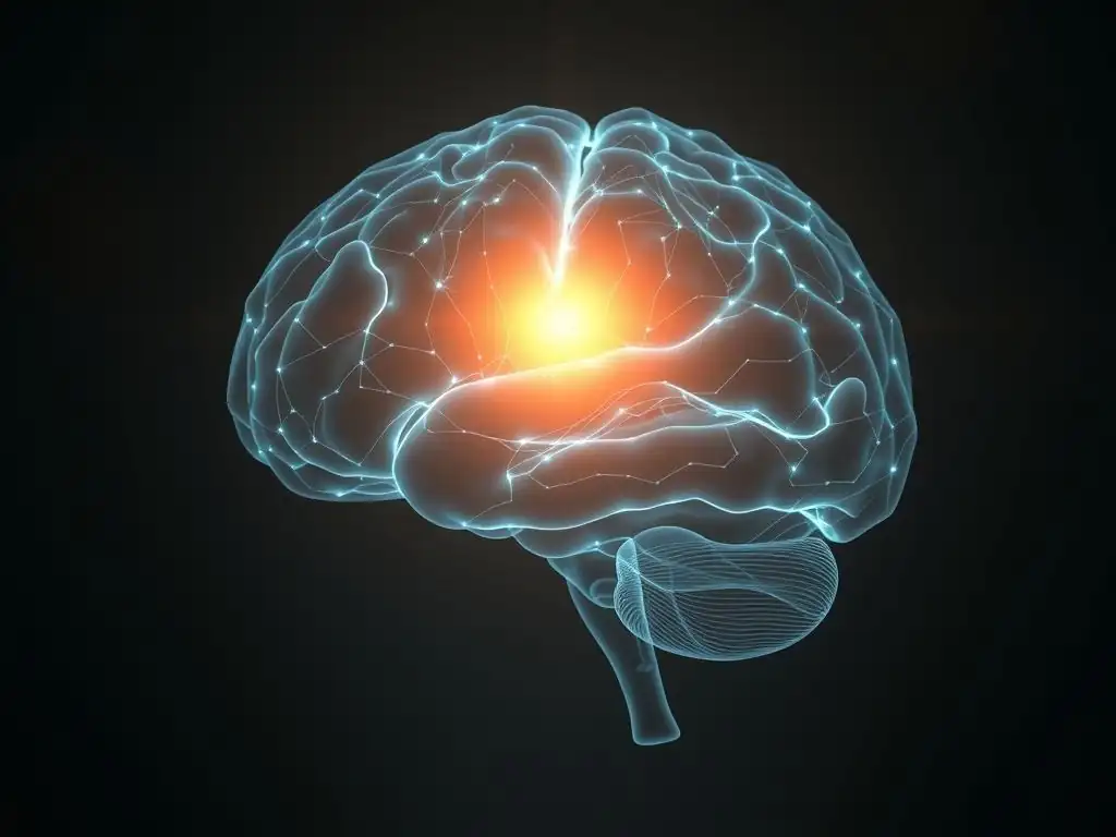
Successful therapy for fear, regardless of the specific approach, relies heavily on the principle of neuroplasticity – the brain's remarkable ability to reorganize itself by forming new neural connections throughout life. In the case of fear, therapy aims to facilitate fear extinction, which is not the erasure of the original fear memory, but rather the formation of a new memory that inhibits the original fear response in safe contexts. This process involves strengthening pathways between the prefrontal cortex (particularly the vmPFC) and the amygdala.
Brain scan studies using fMRI have provided compelling evidence of this neuroplasticity. For example, research on individuals with PTSD undergoing trauma-focused therapy often shows increased activity in the vmPFC and hippocampus, alongside decreased activity in the amygdala, during exposure to trauma-related cues post-treatment. These changes reflect the brain's rewiring, where the prefrontal cortex gains better control over the amygdala's fear signals, allowing for a more regulated and less intense fear response. This demonstrates how brain scan therapy for fear research can visualize the physical changes underlying psychological healing.
Cortical Reorganization and Cognitive Control Over Fear
Beyond the core fear circuit, successful therapy for fear also involves broader cortical reorganization, particularly in areas associated with cognitive control, attention, and self-referential processing. The dorsolateral prefrontal cortex (dlPFC) and anterior cingulate cortex (ACC) are critical in recruiting cognitive resources to regulate emotions, shift attention away from threats, and evaluate the salience of stimuli. Strengthening the function and connectivity of these regions is key to gaining conscious control over automatic fear responses.
Brain scan research has shown that effective fear therapy is often associated with increased activity or altered connectivity in these cortical areas. For instance, fMRI studies on individuals with phobias treated with CBT or exposure therapy have shown enhanced activation in the dlPFC and ACC when they are instructed to regulate their fear responses. This suggests that therapy helps individuals develop and utilize these cognitive control mechanisms more effectively, allowing them to override or modulate amygdala-driven reactions. This cortical shift is a vital component observed through brain scan therapy for fear studies.
Modulation of the Default Mode Network (DMN) in Fear Reduction
The Default Mode Network (DMN) is a network of brain regions that is typically most active when a person is at rest, not focused on the external world, and engaged in internal thought processes like mind-wandering, self-reflection, and thinking about the past or future. In individuals with anxiety disorders, the DMN can become hyperactive or abnormally connected, contributing to excessive worry, rumination, and heightened self-focus on internal states of fear.
Brain scan studies, particularly using resting-state fMRI, have explored how therapy impacts the DMN in individuals with fear-based conditions. Research suggests that successful treatment is sometimes associated with a normalization of DMN activity or altered connectivity between the DMN and other networks, such as the salience network (involved in detecting important stimuli).
For example, mindfulness-based interventions have been shown to decrease DMN activity and increase connectivity between the DMN and regions involved in present-moment awareness. Modulating the DMN is an emerging area where brain scan therapy for fear research is providing valuable insights into the broader impact of treatment on brain function.
Cognitive Behavioral Therapy (CBT) and Brain Scan Measured Outcomes
Cognitive Behavioral Therapy (CBT) is one of the most widely studied and effective treatments for anxiety disorders and phobias. CBT works by helping individuals identify and challenge negative thought patterns and develop more adaptive behaviors in the face of fear. Brain scan studies investigating the effects of CBT on fear-related brain activity have yielded consistent findings.
Research using fMRI often shows that successful CBT for conditions like social anxiety disorder or specific phobias is associated with decreased activity in the amygdala and insula when individuals are exposed to feared stimuli post-treatment. Simultaneously, studies frequently report increased activity in prefrontal cortical regions, such as the ventrolateral prefrontal cortex (vlPFC) and dorsomedial prefrontal cortex (dmPFC), which are involved in cognitive reappraisal and emotion regulation.
These changes, observable through brain scan therapy for fear research, reflect the neurobiological underpinnings of learning to manage anxious thoughts and reactions. For example, one meta-analysis of fMRI studies on CBT for anxiety disorders found a consistent pattern of reduced limbic (including amygdala) and increased prefrontal activation after treatment.
Exposure Therapy and Neural Changes in Fear Processing
Exposure therapy, often a component of CBT or used as a standalone treatment, involves gradually and systematically exposing individuals to their feared objects, situations, or memories in a safe and controlled environment. The goal is to facilitate fear extinction by learning that the feared stimulus does not lead to the predicted negative outcome. Brain scan studies have provided clear evidence of how exposure therapy achieves this neurobiologically.
fMRI studies on exposure therapy for specific phobias or PTSD consistently show a reduction in amygdala reactivity to the feared stimulus after successful treatment. Furthermore, these studies often reveal increased activity in the vmPFC, a key region for fear extinction learning, and strengthened connectivity between the vmPFC and the amygdala. This pattern reflects the brain's process of creating a new safety memory that inhibits the original fear response.
For instance, a study on spider phobia treatment showed decreased amygdala response to spider images after exposure therapy, correlating with symptom reduction. This provides direct brain scan therapy for fear evidence of the therapy's impact on core fear circuitry.
Mindfulness-Based Interventions and Amygdala Regulation
Mindfulness-based interventions (MBIs), such as Mindfulness-Based Stress Reduction (MBSR), teach individuals to cultivate present-moment awareness and accept their thoughts and feelings without judgment. While not directly targeting fear content like exposure therapy, MBIs can significantly impact emotion regulation and reduce anxiety. Brain scan research has explored how mindfulness practice influences the brain's fear network.
Studies using fMRI have shown that regular mindfulness practice can lead to structural and functional changes in brain regions involved in emotion regulation. Specifically, research suggests that mindfulness training can decrease amygdala reactivity to emotional stimuli and increase connectivity between the prefrontal cortex and the amygdala. This is thought to enhance the brain's ability to regulate emotional responses and reduce the automatic grip of fear. For example, an 8-week MBSR course has been shown to reduce amygdala size and functional connectivity to stress-related areas, demonstrating a potential mechanism through which this type of brain scan therapy for fear works.
Eye Movement Desensitization and Reprocessing (EMDR) Therapy and Associated Brain Activity
Eye Movement Desensitization and Reprocessing (EMDR) therapy is a treatment approach often used for trauma-related fear, like PTSD. It involves recalling distressing memories while simultaneously engaging in bilateral stimulation, typically eye movements. While the exact mechanisms are still debated, brain scan studies are beginning to shed light on EMDR's effects.
Preliminary brain scan research on EMDR has suggested that it may lead to decreased activity in the limbic system (including the amygdala) and increased activity in prefrontal regions during the processing of traumatic memories post-treatment. Some theories propose that the bilateral stimulation helps to facilitate the processing and integration of traumatic memories in a more adaptive way, potentially by reducing the vividness and emotional intensity of the memory. While more research is needed, these initial brain scan therapy for fear findings offer clues about how EMDR might help the brain reprocess distressing experiences.
Brain Scan Findings: Personalized Treatment Strategies for Fear
Identifying Individual Brain Patterns and Treatment Responses
One of the most promising applications of brain scan technology in mental health is the potential for personalized treatment. Just as individuals have different symptoms and life histories, their brains also exhibit unique patterns of activity and connectivity. These individual differences in neural circuitry may influence how a person experiences fear and how they respond to different types of brain scan therapy for fear.
Research is exploring whether specific baseline brain activity patterns, measured before therapy begins, can predict which individuals are most likely to benefit from a particular treatment approach. For instance, studies might investigate if the level of connectivity between the amygdala and vmPFC at baseline predicts response to exposure therapy, or if pre-treatment activity in the ACC predicts response to cognitive restructuring techniques in CBT. Identifying these neural markers could allow clinicians to move away from a one-size-fits-all approach towards more tailored interventions.
Tailoring Therapy Based on Brain Scan Data
The ultimate goal of identifying individual brain patterns is to tailor therapeutic interventions for better outcomes. If research can reliably link specific brain profiles to differential treatment responses, clinicians might someday use brain scan data to inform their therapeutic decisions. For example, a patient with unusually high amygdala reactivity and low prefrontal control might be guided towards therapies that specifically target emotion regulation skills or involve gradual exposure, potentially informed by brain scan therapy for fear insights.
Conversely, a patient whose fear seems more driven by cognitive distortions or rumination, perhaps reflected in specific DMN or cortical activity patterns, might benefit more from therapies focusing on changing thought processes or metacognitive skills. While still largely in the research phase, the potential to use brain scan data to match patients to the most effective therapy could significantly improve treatment success rates and reduce the burden of trial-and-error.
Predicting Therapy Outcomes with Pre-Treatment Brain Scans
Predicting who will respond well to therapy is a significant challenge in mental health care. Treatment non-response is common, and individuals may undergo months or years of therapy before finding an approach that works. Brain scan technology offers a potential avenue for developing biomarkers that can predict treatment outcomes before therapy even begins.
Numerous studies are investigating the predictive power of pre-treatment brain scans for fear disorders. For example, some research suggests that the strength of connectivity within certain brain networks (like the DMN or salience network) or the reactivity of the amygdala to emotional stimuli prior to therapy might predict the degree of symptom reduction achieved with CBT or medication. While these findings are still being validated and are not yet used routinely in clinical practice, they highlight the exciting possibility that brain scan therapy for fear research could one day provide tools to personalize treatment and improve prognosis prediction.
Case Studies: Illustrating Brain Scan-Guided Fear Therapy

Case Study 1: PTSD Treatment Guided by fMRI Feedback
Illustrative Example: Sarah, a 35-year-old veteran with chronic PTSD, experienced significant distress from intrusive memories and hypervigilance. Standard trauma-focused therapy had yielded limited results. As part of a research trial, she underwent fMRI scans. Her initial scan showed exceptionally high amygdala reactivity to trauma-related cues and low connectivity between her vmPFC and amygdala.
Based on these brain scan therapy for fear findings, the clinical team decided to incorporate neurofeedback using fMRI. Sarah was trained to consciously try to lower her amygdala activity while viewing trauma cues, receiving real-time feedback from her fMRI signal displayed on a screen. Over 10 sessions, her ability to reduce amygdala activity improved. Post-treatment fMRI showed a measurable decrease in amygdala reactivity and increased vmPFC-amygdala connectivity, correlating with a significant reduction in her PTSD symptoms and improved emotional regulation in daily life. This case illustrates the potential for brain scan feedback to enhance traditional therapy.
Illustrative Example: Mark, a 22-year-old student, suffered from severe social anxiety, experiencing intense fear and blushing in social situations. An initial EEG brain scan revealed heightened alpha wave activity in certain frontal brain regions, which has been correlated in some studies with excessive self-focus and worry in social contexts.
Mark began a course of CBT for social anxiety. Throughout his 12 weeks of therapy, he participated in follow-up EEG sessions every four weeks. The EEG monitoring, though not directly guiding each session, provided objective data points.
As Mark progressed through exposure exercises and cognitive restructuring, his EEG showed a gradual reduction in the problematic frontal alpha activity and shifts in ERPs related to processing social cues, becoming more typical of non-anxious individuals. By the end of therapy, his subjective report of reduced anxiety was supported by the normalization of his EEG patterns, providing a measurable correlate of his progress in this brain scan therapy for fear approach.
Case Study 3: Phobia Treatment Response Predicted by Pre-Treatment PET Scan
Illustrative Example: Emily, a 40-year-old with a severe specific phobia of heights, was considering treatment options. Before starting therapy, she participated in a research study involving a pre-treatment PET scan using a tracer that measures serotonin transporter (SERT) availability, as SERT levels have been implicated in anxiety disorders. Her PET scan showed relatively low SERT availability in key brain regions.
Based on previous research suggesting that individuals with lower SERT availability might respond better to exposure-based therapies than certain medications, and might have a particular neural profile related to fear processing, the researchers hypothesized Emily would be a good candidate for intensive exposure therapy. She completed a short, intensive course of exposure therapy.
A post-treatment assessment confirmed a significant reduction in her phobia symptoms, allowing her to comfortably engage in activities previously impossible. While preliminary, this case highlights how pre-treatment biological markers, assessed via a brain scan therapy for fear method like PET, might one day help predict responsiveness to specific treatments.
The Future of Brain Scan Enhanced Therapy for Fear
Advancements in Brain Scan Technology
The field of brain scan technology is continuously evolving, promising even more detailed and nuanced insights into the brain's workings. Future advancements may include higher-resolution fMRI, allowing for the study of activity in smaller brain regions or even cortical layers; faster EEG systems capable of capturing even more rapid neural dynamics; and new PET tracers that can target a wider range of neurotransmitter systems or specific cellular processes related to fear and anxiety.
Furthermore, the integration of different brain scan modalities (e.g., simultaneously collecting fMRI and EEG data) could provide a more complete spatio-temporal picture of brain activity during fear and therapy. Portable and more affordable brain imaging devices, perhaps based on technologies like fNIRS (functional Near-Infrared Spectroscopy) or advanced EEG, could also make brain scanning more accessible outside of specialized research centers, bringing brain scan therapy for fear insights closer to routine clinical use.
Integrating Brain Scan Data into Clinical Practice
Despite the valuable insights gained from research, the integration of brain scan data directly into routine clinical practice for diagnosing or guiding therapy for fear is still in its nascent stages. Current brain scans are primarily research tools due to their cost, accessibility, and the complexity of interpreting individual scan data in a clinical context. However, the trajectory of the field suggests increasing clinical relevance.
Future developments may involve creating standardized protocols for clinically relevant brain scans, developing user-friendly software for interpreting individual scan data in relation to normative databases, and identifying specific brain scan biomarkers that reliably predict treatment response or inform treatment selection. Bridging the gap between research findings and clinical application is a major focus, aiming to translate the power of brain scan therapy for fear research into tangible benefits for patients.
Ethical Considerations of Using Brain Scans in Mental Health Treatment
As brain scan technology becomes more powerful and potentially integrated into clinical practice, important ethical considerations arise. Issues around privacy and data security are paramount, given the sensitive nature of brain data. Ensuring equitable access to this potentially costly technology is another concern, to avoid creating disparities in mental health care.
Furthermore, there are ethical questions about the interpretation and communication of brain scan findings to patients. How should complex neural data be explained in a way that is informative but does not oversimplify or create undue anxiety or determinism? There's also the risk of misinterpreting group-level research findings when applied to an individual patient. As the field of brain scan therapy for fear evolves, ongoing dialogue among researchers, clinicians, ethicists, and the public is crucial to navigate these challenges responsibly.
Conclusion: Brain Scans as a Powerful Tool for Understanding and Treating Fear
Brain scan technology has moved our understanding of fear far beyond the initial focus on the amygdala. Techniques like fMRI, EEG, and PET scans provide powerful windows into the complex neural circuits and processes underlying fear responses, revealing the dynamic interplay between structures like the amygdala, prefrontal cortex, hippocampus, and insula. These tools have been instrumental in showing how therapy works at a biological level, illustrating the neuroplastic changes associated with fear extinction, cognitive control over fear, and the modulation of brain networks.
From demonstrating the impact of CBT and exposure therapy on the core fear circuit to exploring how mindfulness affects amygdala regulation or EMDR influences memory processing, brain scan therapy for fear research provides objective evidence for the effectiveness of diverse interventions. The potential to use brain scan data for personalizing treatment, predicting outcomes, and developing more targeted therapies represents an exciting frontier in mental health care.
While challenges related to clinical integration and ethics remain, the continued advancement and application of brain scan technology hold immense promise for improving our ability to understand, treat, and ultimately alleviate the burden of fear-based conditions.
Frequently Asked Questions
What is Brain Scan Therapy and how does it help with fear?
Brain scan therapy, often involving techniques like fMRI, provides visual insights into brain activity during fearful experiences. This allows therapists to tailor interventions to specifically target the neural pathways associated with fear.
Why is there "Brain Scan Therapy Fear" among patients?
The apprehension surrounding brain scan therapy often stems from concerns about radiation exposure, the claustrophobic nature of the scan environment, and the potential for discovering unexpected or unsettling findings about their brain.
Brain scans are highly accurate in identifying brain regions involved in fear responses. However, they are used in conjunction with other assessments to provide a comprehensive understanding and guide personalized treatment plans.
Are there alternatives to brain scan therapy for those experiencing "Brain Scan Therapy Fear"?
Yes, several alternatives exist, including cognitive behavioral therapy (CBT), exposure therapy, and medication. These therapies can effectively address fear without requiring a brain scan.
What are the potential benefits of overcoming "Brain Scan Therapy Fear" and undergoing the process?
Overcoming the fear can unlock deeper insights into the root causes of anxiety and phobias. This can lead to more effective and targeted therapeutic interventions, ultimately improving mental well-being and reducing fear-related symptoms.
Final Reflection: Unlocking the Fear Response Through Neuroscience
The insights gleaned from brain scan technology are revolutionizing our understanding of fear, offering a tangible path towards more effective therapeutic interventions. No longer are we reliant solely on subjective reports; we can now visualize the neural pathways involved in fear responses, paving the way for personalized treatments tailored to the individual's unique brain activity. This represents a significant leap forward in mental health care.
The ongoing exploration of the amygdala and its intricate connections within the brain is profoundly impacting how we approach anxiety and trauma. It's humbling to consider how our understanding of the brain's complex mechanisms is constantly evolving, leading to more compassionate and effective treatments. The brain is the most complex organ in the known universe. Understanding its intricacies is a journey, not a destination.
This quote encapsulates the ongoing nature of this research and the importance of continued investigation into the neural basis of fear. The implications extend beyond individual therapy, influencing our understanding of societal responses to fear-based narratives and the potential for preventative measures.
Consider the power you now hold. You've gained insight into the biological underpinnings of fear, a powerful emotion that shapes our lives. This knowledge isn't just for professionals; it empowers you to approach your own anxieties with a new level of understanding and compassion. Take time to reflect on your own experiences with fear. How might this new understanding inform your coping mechanisms? What steps can you take to foster resilience and manage fear more effectively? The journey towards understanding and overcoming fear is a personal one, but armed with this knowledge, you are better equipped to navigate it.
Unlock Your Fearless Potential: Next Steps to a Calmer You
Thanks for taking the time to explore the fascinating world of the amygdala and how brain scans are revolutionizing fear therapy! You've gained valuable insights into understanding and overcoming your anxieties. Now, it's time to put that knowledge into action.
- Start your personalized fear-reduction journey: Identify one specific fear you want to address and begin practicing the mindfulness techniques discussed in the article. Even five minutes a day can make a difference.
- Share your insights: Discuss this article with a friend or family member who might benefit from understanding the science behind fear and anxiety. Starting a conversation can be incredibly powerful!
- Explore further resources: Search online for guided meditations focused on amygdala regulation or look for local therapists specializing in brain-based therapy.
What small step will you take today to cultivate a more courageous and confident you?